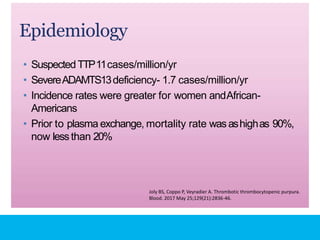
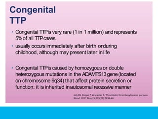
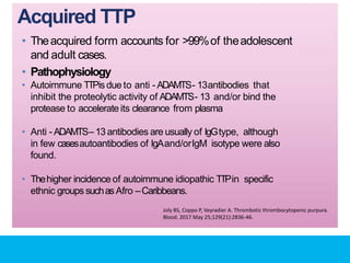
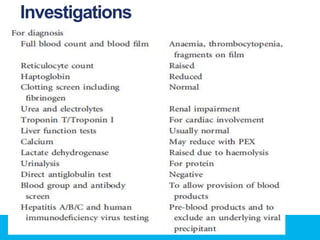
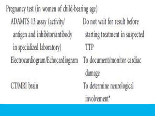
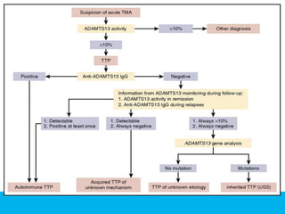
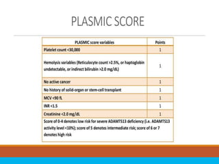
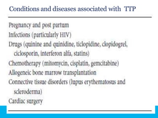
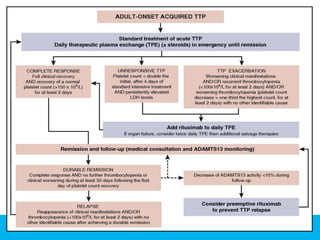
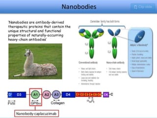
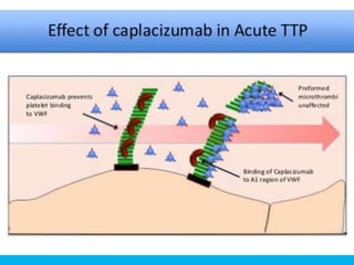
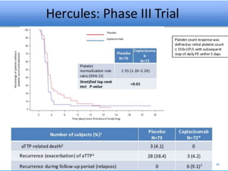
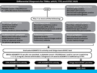
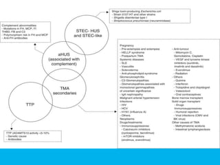
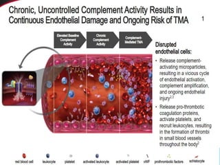
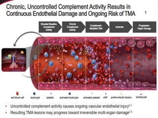
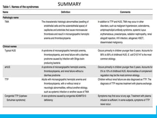
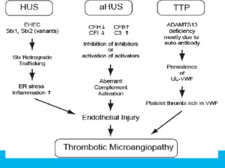
Dr. Darayus P.Gazder
PG R-1
For decades, we have used the term
“ (TTP/HUS)” for patients presenting with:
1) Acute renal failure,
2) Microangiopathic hemolysis
3) Thrombocytopenia, without diarrhea
prodrome.
Although, thrombotic thrombocytopenic purpura and atypical
hemolytic uremic syndrome are now known as 2 distinct diseases
“TTP/HUS” still can be seen in medical records and even in
medical journals today
Distinguishing Among the Major Thrombotic
Microangiopathies
TTP
Epidemiology
• Suspected TTP11cases/million/yr
• SevereADAMTS13deficiency- 1.7 cases/million/yr
• Incidence rates were greater for women andAfrican-
Americans
• Prior to plasma exchange, mortality rate wasashighas 90%,
now lessthan 20%
Joly BS, Coppo P, Veyradier A. Thrombotic thrombocytopenic purpura.
Blood. 2017 May 25;129(21):2836-46.
Definitions and
Diagnosis
• TheClassicPentad of TTP
• Microangiopathic hemolytic anemia
• Thrombocytopenia
• Renal insufficiency or abnormalities
• Neurologic abnormalities that can befluctuating
• Fever
• Most common symptoms at presentation arenonspecific
and include abdominal pain, nausea, vomiting and
weakness.
Joly BS, Coppo P, Veyradier A. Thrombotic thrombocytopenic purpura.
Blood. 2017 May 25;129(21):2836-46.
• MAHA
• nonimmune hemolysis (negative coombs) with
prominent red cell fragmentation (schistocytes) on
peripheral blood smear. Will exhibit increased LDH
and indirect bilirubin.
• Schistocytes
• in the appropriate clinical setting schistocyte count>1%
wasstrongly suggestive of TTP,ie 2 or moreschistos in
microscopic field at 100xmagnification.
• Neurologic symptoms
• most are subtle, suchastransient confusion orsevere
headache. Focal, objective abnormalities are less
common, but grand mall seizures and coma canoccur.
• Fever
• less frequent finding, but the presence of chills and high
spiking fever should suggest dx of sepsisorDIC.
• Cardiac involvement
• incidence is difficult to determine, but diffuse platelet
thrombi and associated hemorrhage in cardiac tissues
can lead to arrythmias, MIs, sudden death, shock, or
heart failure.
Types:
•
1. Congenital
2. Acquired
• Autoimmune forms, due to autoantibodiesagainst
ADAMTS– 13
• secondary to massive endothelial activation with
release of ultra - large VWFmultimers in large
amounts
Joly BS, Coppo P, Veyradier A. Thrombotic thrombocytopenic purpura.
Blood. 2017 May 25;129(21):2836-46.
Congenital
TTP
• Congenital TTPis very rare (1 in 1 million) andrepresents
5%of all TTPcases.
• usually occurs immediately after birth orduring
childhood, although may present later inlife
• Congenital TTPis caused by homozygous or double
heterozygous mutations in the ADAMTS13gene(located
on chromosome 9q34) that affect protein secretion or
function; it is inherited inautosomal recessive manner
Joly BS, Coppo P, Veyradier A. Thrombotic thrombocytopenic purpura.
Blood. 2017 May 25;129(21):2836-46.
Acquired TTP
• Theacquired form accounts for >99%of theadolescent
and adult cases.
• Pathophysiology
• Autoimmune TTPis due to anti - ADAMTS- 13antibodies that
inhibit the proteolytic activity of ADAMTS- 13 and/or bind the
protease to accelerate its clearance from plasma
• Anti - ADAMTS–13 antibodies are usually of IgGtype, although
in few casesautoantibodies of IgAand/orIgM isotype were also
found.
• Thehigher incidence of autoimmune idiopathic TTPin specific
ethnic groups suchas Afro –Caribbeans.
Joly BS, Coppo P, Veyradier A. Thrombotic thrombocytopenic purpura.
Blood. 2017 May 25;129(21):2836-46.
Investigations
Conditions and diseases associated with TTP
Joly BS, Coppo P, Veyradier A. Thrombotic thrombocytopenic purpura.
Blood. 2017 May 25;129(21):2836-46.
CLASSIFICATIONOFHUS ACCORDING TO
ETIOPATHOGENESIS
Typeof HUS Specific Cause
Infection related
Typical
Shiga toxin producing E.coli/Shigella
Pneumococcal infection
HIV
Other viral or bacterial infections
• Complement factorabnormality Factor H deficiency
Factor Ideficiency
• Miscellaneous Atypical
CTD
Drugs
Malignancy
SUMMARY
“Rectification of
names is a must”; we
shall no longer use
the term “TTP/HUS”!!

TTP HUS

  • 1.
  • 2.
    For decades, wehave used the term “ (TTP/HUS)” for patients presenting with: 1) Acute renal failure, 2) Microangiopathic hemolysis 3) Thrombocytopenia, without diarrhea prodrome. Although, thrombotic thrombocytopenic purpura and atypical hemolytic uremic syndrome are now known as 2 distinct diseases “TTP/HUS” still can be seen in medical records and even in medical journals today
  • 3.
    Distinguishing Among theMajor Thrombotic Microangiopathies
  • 4.
  • 5.
    Epidemiology • Suspected TTP11cases/million/yr •SevereADAMTS13deficiency- 1.7 cases/million/yr • Incidence rates were greater for women andAfrican- Americans • Prior to plasma exchange, mortality rate wasashighas 90%, now lessthan 20% Joly BS, Coppo P, Veyradier A. Thrombotic thrombocytopenic purpura. Blood. 2017 May 25;129(21):2836-46.
  • 6.
    Definitions and Diagnosis • TheClassicPentadof TTP • Microangiopathic hemolytic anemia • Thrombocytopenia • Renal insufficiency or abnormalities • Neurologic abnormalities that can befluctuating • Fever • Most common symptoms at presentation arenonspecific and include abdominal pain, nausea, vomiting and weakness. Joly BS, Coppo P, Veyradier A. Thrombotic thrombocytopenic purpura. Blood. 2017 May 25;129(21):2836-46.
  • 8.
    • MAHA • nonimmunehemolysis (negative coombs) with prominent red cell fragmentation (schistocytes) on peripheral blood smear. Will exhibit increased LDH and indirect bilirubin. • Schistocytes • in the appropriate clinical setting schistocyte count>1% wasstrongly suggestive of TTP,ie 2 or moreschistos in microscopic field at 100xmagnification.
  • 9.
    • Neurologic symptoms •most are subtle, suchastransient confusion orsevere headache. Focal, objective abnormalities are less common, but grand mall seizures and coma canoccur. • Fever • less frequent finding, but the presence of chills and high spiking fever should suggest dx of sepsisorDIC. • Cardiac involvement • incidence is difficult to determine, but diffuse platelet thrombi and associated hemorrhage in cardiac tissues can lead to arrythmias, MIs, sudden death, shock, or heart failure.
  • 10.
    Types: • 1. Congenital 2. Acquired •Autoimmune forms, due to autoantibodiesagainst ADAMTS– 13 • secondary to massive endothelial activation with release of ultra - large VWFmultimers in large amounts Joly BS, Coppo P, Veyradier A. Thrombotic thrombocytopenic purpura. Blood. 2017 May 25;129(21):2836-46.
  • 11.
    Congenital TTP • Congenital TTPisvery rare (1 in 1 million) andrepresents 5%of all TTPcases. • usually occurs immediately after birth orduring childhood, although may present later inlife • Congenital TTPis caused by homozygous or double heterozygous mutations in the ADAMTS13gene(located on chromosome 9q34) that affect protein secretion or function; it is inherited inautosomal recessive manner Joly BS, Coppo P, Veyradier A. Thrombotic thrombocytopenic purpura. Blood. 2017 May 25;129(21):2836-46.
  • 12.
    Acquired TTP • Theacquiredform accounts for >99%of theadolescent and adult cases. • Pathophysiology • Autoimmune TTPis due to anti - ADAMTS- 13antibodies that inhibit the proteolytic activity of ADAMTS- 13 and/or bind the protease to accelerate its clearance from plasma • Anti - ADAMTS–13 antibodies are usually of IgGtype, although in few casesautoantibodies of IgAand/orIgM isotype were also found. • Thehigher incidence of autoimmune idiopathic TTPin specific ethnic groups suchas Afro –Caribbeans. Joly BS, Coppo P, Veyradier A. Thrombotic thrombocytopenic purpura. Blood. 2017 May 25;129(21):2836-46.
  • 13.
  • 17.
    Conditions and diseasesassociated with TTP
  • 18.
    Joly BS, CoppoP, Veyradier A. Thrombotic thrombocytopenic purpura. Blood. 2017 May 25;129(21):2836-46.
  • 25.
    CLASSIFICATIONOFHUS ACCORDING TO ETIOPATHOGENESIS TypeofHUS Specific Cause Infection related Typical Shiga toxin producing E.coli/Shigella Pneumococcal infection HIV Other viral or bacterial infections • Complement factorabnormality Factor H deficiency Factor Ideficiency • Miscellaneous Atypical CTD Drugs Malignancy
  • 35.
  • 37.
    “Rectification of names isa must”; we shall no longer use the term “TTP/HUS”!!

Editor's Notes

  • #3 TTP/HUS” still can be seen in medical records and even in medical journals today Although, thrombotic thrombocytopenic purpura and atypical hemolytic uremic syndrome are now known as 2 distinct diseases
  • #9 Peripheral blood smear from a patient with a microangiopathic hemolytic anemia with marked red cell fragmentation. The smear shows multiple helmet cells (arrows) and other fragmented red cells (small arrowhead); microspherocytes are also seen (large arrowheads). The platelet number is reduced; the large platelet in the center (dashed arrow) suggests that the thrombocytopenia is due to enhanced destruction.
  • #10 Neurologic symptoms most are subtle, such as transient confusion or severe headache. Focal, objective abnormalities are less common, but grand mall seizures and coma can occur. Fever less frequent finding, but the presence of chills and high spiking fever should suggest dx of sepsis or DIC. Cardiac involvement incidence is difficult to determine, but diffuse platelet thrombi and associated hemorrhage in cardiac tissues can lead to arrythmias, MIs, sudden death, shock, or heart failure.
  • #12 To date, more than 80 mutations have been documented in patients with familial TTP. the majority cause severe ADAMTS - 13 deficiency by decreasing its biosynthesis, intacellular trafficing and secretion and/or proteolytic activity. While some patients have neonatal disease onset and multiple recurrent episodes and develop progressive organ failure, others may only experience a single episode that develops in adulthood and which is very responsive to plasma infusion,leaving no residual organ damage.
  • #13 Pathophysiology von Willebrand factor, a glycoprotein secreted from vascular endothelial cells in very large polymeric forms, supports platelet adhesion and aggregation at sites of vessel injury. ADAMTS13, a metalloprotease in plasma, cleaves von Willebrand factor when it is conformationally unfolded by shear stress. By cleaving vWF before it is fully activated by shear stress, ADAMTS13 prevents vWF-mediated platelet aggregation.
  • #17 predict the likelihood of ADAMTS13 activity <10 percent
  • #19 TPE with replacement of plasma remains the cornerstone of current management of TTP (Figure 5).17 According to previous studies,17,81 TPE (usually 1.5× plasma volume exchange for the first procedures, followed by 1.0× patient plasma volume thereafter) should be started as soon as the diagnosis of TTP is established or even suspected. TPE is performed daily until features related to organ involvement (cerebral manifestations, renal failure, increased troponin level, abdominal pain resulting from enteritis or pancreatitis) have resolved, the platelet count has stably recovered, and hemolysis has ceased. Some groups suggested progressively decreasing TPE sessions typically within 3 weeks to prevent severe exacerbations
  • #21 humanized monoclonal antibody-based fragment (a nanobody)
  • #23 Caplacizumab is a humanized monoclonal antibody-based fragment (a nanobody) that binds to VWF and blocks VWF interaction with platelet GPlb-IX-V. In a trial that randomly assigned 75 patients with acquired TTP to receive caplacizumab or placebo (all patients received standard plasma exchange [PEX]), caplacizumab was associated with a two-day reduction in the time to complete response (3 versus 4.9 days) [65]. Other endpoints also favored caplacizumab, including the frequency of complete remission (81 versus 46 percent) and the number of days of PEX (8 versus 12). Of interest, caplacizumab was associated with fewer TTP exacerbations during therapy but more exacerbations after therapy was stopped, suggesting a lack of effect on anti-ADAMTS13 antibody levels. There were no major adverse events. However, caplacizumab was associated with a greater risk of bleeding, consistent with its mechanism of blocking VWF-platelet binding. Bleeding was mostly minor and did not require administration of factor VIII or VWF. There were two deaths, both in the placebo group (one from refractory TTP and one from cerebral hemorrhage). Extended follow-up identified a lower rate of major thromboembolic events in the caplacizumab group compared with placebo (3 versus 19 percent); the majority of thromboembolic events occurred within the first month [66]. Caplacizumab is not yet available outside of a clinical trial 
  • #25 http://dev.nefro.elsevier.es/en-publicacion-nefrologia-imprimir-articulo-an-update-for-atypical-haemolytic-uraemic-syndrome-diagnosis-treatment-a-S2013251415000991 Fig. 2. Classification of the aetiologies of thrombotic microangiopathies. ADAMTS13: A Disintegrin And Metalloproteinase with a ThromboSpondin type 1 motif, member 13; aHUS: atypical haemolytic uraemic syndrome; CMV: cytomegalovirus; FB: complement factor B; FH: complement factor H; FI: complement factor I; HCV: hepatitis C virus; HELLP: Hemolysis, Elevated Liver enzymes, Low Platelet count; HIV: human immunodeficiency virus; HUS: haemolytic uraemic syndrome; MCP: membrane cofactor protein; mTOR: mammalian target of Rapamycin; SEL: systemic erythematous lupus; STEC: Shiga toxin-producing Escherichia coli; THBD: thrombomodulin; TMA: thrombotic microangiopathy; TTP: thrombocytopenic thrombotic purpura; VEGF: vascular endothelial growth factor.
  • #31 Treatment for aHUS has traditionally been supportive care, which includes platelet or blood transfusions in patients for whom this is necessary and dialysis for patients who suffer severe kidney injury.[6,25] Neither of these attack the root cause of the disease; they support the patient until the disease can burn itself out or otherwise be brought under control. Patients do receive kidney transplant to replace a severely damaged kidney, but those new kidneys are potentially subject to the same complement-mediated attack as the native kidneys. Because many of the complement factors, particularly factor H, are synthesized in the liver, liver transplant has been performed on a number of occasions for the treatment of factor H deficiency
  • #32 Consequences of complement activation. (A) The complement system can be activated via 3 pathways: classical, lectin, and alternative. All pathways lead to formation of powerful enzymes, the C3-convertases, followed by activation of the terminal cascade. The main effector functions of complement (promotion of opsonophagocytosis by opsonization and chemotaxis and formation of lytic membrane attack complexes) aim to destroy harmful agents such as microbes. Lysis of target cells can lead to damage-induced enhancement of complement activation. (B) Alternative pathway activation is based on continuous, low-level covalent deposition of C3b molecules onto practically all surfaces in contact with plasma. If the C3b molecule is allowed to form an enzyme (shown in red), new C3b deposits will be formed around the enzyme leading to rapid amplification of the activation. If the regulator factor H binds to C3b, the convertase enzyme is inactivated and no complement activation follows. The simultaneous interaction of factor H with both C3b and cell surface sialic acids (or possibly glycosaminoglycans [GAGs]) is essential for proper regulation on self red cells, platelets, and endothelial cells. If this fails, disbalance between activation and regulation may lead to pathogenesis of atypical HUS. iC3b, C3b molecule incapable of forming an enzyme with factor B.
  • #34 Treatment for aHUS has traditionally been supportive care, which includes platelet or blood transfusions in patients for whom this is necessary and dialysis for patients who suffer severe kidney injury.[6,25] Neither of these attack the root cause of the disease; they support the patient until the disease can burn itself out or otherwise be brought under control. Patients do receive kidney transplant to replace a severely damaged kidney, but those new kidneys are potentially subject to the same complement-mediated attack as the native kidneys. Because many of the complement factors, particularly factor H, are synthesized in the liver, liver transplant has been performed on a number of occasions for the treatment of factor H deficiency