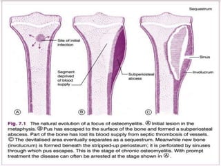
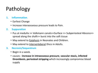
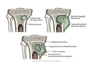
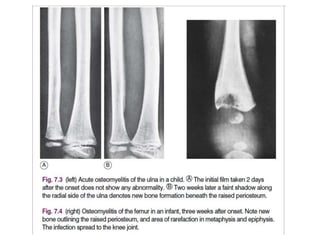
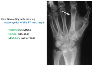
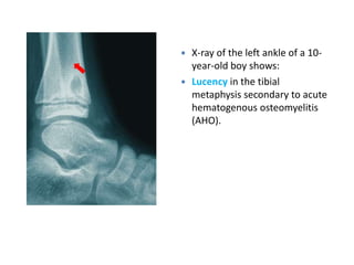
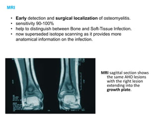
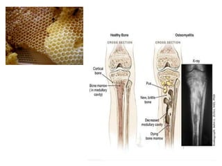
Osteomyelitis
Definition
• Osteomyelitis is the infection of the bone or BM which leads to a
subsequent Inflammatory process.
• Micro-Organisms may reach bones via the Bloodstream or by Direct
Invasion. (e.g : skin puncture, operation, open fracture)
• Factors which affects it’s development
– Virulence of the organism involved
– Host Factors (Age, Immunity, Diseases)
– Local factors (site of Involvement, damaged muscle presence of
foreign material , vascularity)
• It can be classified on the basis of the causative organism, the
route, duration and anatomic location of the infection.
• According to duration: acute, subacute, chronic
• In children, osteomyelitis most commonly affects the long bones of
the legs and upper arms.
• Adults are more likely to develop osteomyelitis in the vertebrae.
• Diabetic patients may develop osteomyelitis in their feet if they
have foot ulcers.
• Osteomyelitis usually begins as an acute infection, but it may evolve
into a chronic condition.
Acute Osteomyelitis
Types
1. Hematogenous Osteomyelitis
• Bacterial seeding from the blood.
• Seen primarily in Children.
• The most common site
• Metaphysis at the growing end of Long Bones in Children
• Vertebrae in Adults; involving two adjacent vertebrae with
intervertebral disk (may occur pelvis, long bones and clavicle)
2. Direct Inoculation Osteomyelitis
• Its osteomyelitis complicating open fracture or surgical
operation, in which organisms gain entry directly through the
wound.
• Tend to involve multiple organisms. but mainly S.Aureus
Acute Haematogenous Osteomyelitis
Causative Organisms
• Staph. aureus (Most common)
• Strep. pyogens or pneumoniae (Less).
• H.Influenzae (Young Children)
but still, the most common causative organism for osteomyelitis in young
children is staphylococcus aureus
H.flu infection has become less common due to vaccination
• Salmonella (Sickle-Cell)
still, the most common causative organism for osteomyelitis in sickle cell
anemia patient is staphylococcus aureus
Slamonella is the second most common infection in patients with sickle cell
disease
Pathology. Organisms reach the bone through the blood stream from a sep-
tic focus elsewhere in the body – for instance from a boil in the skin. In a rare
atypical form in adults infection reaches the vertebral column through the
spinal venous plexus from an infected intrapelvic lesion.
In the usual childhood manifestation, the infection begins in the metaphy-
sis of a long bone, which must be presumed to form a productive medium for
bacterial growth (Fig. 7.1A); thence it may spread to involve a large part of the bone. The
organisms induce an acute inflammatory reaction, but the marshalling of the body’s
defensive forces is greatly handicapped in bone because its rigid structure does not allow
swelling. Pus is formed and soon finds its way to the surface of the bone where it forms a
subperiosteal abscess (Fig. 7.1B); later the abscess may burst into the soft tissues and may
eventually reach the surface to form a sinus.
Often the blood supply to a part of the bone is cut off by septic thrombosis of
the vessels (Fig. 7.1B). The ischaemic bone dies and eventually separates from
the surrounding living bone as a sequestrum (Fig. 7.1C). Meanwhile new bone
is laid down beneath the stripped-up periosteum, forming an investing layer
known as the involucrum (Fig. 7.1C). The epiphysial cartilage plate is a barrier to the spread of
infection, but if the affected metaphysis lies partly within a joint cavity the joint is liable to
become infected (acute pyogenic arthritis). Metaphyses that lie wholly or partly within a joint
cavity include the upper metaphysis of the humerus, all the metaphyses at the elbow, and
the upper and lower metaphyses of the femur (Fig. 7.2). Even when the joint is not infected it
may swell from an effusion of clear fluid(sympathetic effusion).With efficient treatment, the
infection may be aborted in its earliest phase.But when it has progressed to the stage of
septic thrombosis and death of bone it almost inevitably passes into a state of chronic
osteomyelitis.
Pathology
1. Inflammation.
• Earliest Change
• Increase interaosseous pressure leads to Pain.
2. Suppuration
• Pus at medulla >> Volkmann canals>>Surface >> Subperiosteal Abscess>>
spread along the shaft>> burst into the soft tissue
• May extend to Epiphysis in Neonates and Children.
• May extend to Interverteberal Discs in Adults.
3. Necrosis/Sequestrum
• Begin in a week.
• causes : increase in intraosseous pressure, vascular stasis, infected
thrombosis, periosteal stripping which increasingly compromise blood
supply
4. New-bone formation
• New bone formation from the stripped surface of periosteum
• Bone thickens to form an involucrum enclosing the infected tissue.
5. Resolution
bone will heal if infection is controlled and intraosseous pressure is
released, though it may remain thickened. or progress to complications
Clinical Features
• Fever , chills and Malaise
• Pain
• Tenderness, Redness, Edema, Warmth (signs of inflammation)
• Restricted Joint Movement
History preceding Skin Lesion or Sore Throat.
Typically; child, boy. The bones most commonly
affected are the tibia, the femur and the humerus. The onset is rapid. The child
complains of feeling ill, and of severe pain over the affected bone. There may be
a history of recent boils or of a minor injury.
On examination there is pyrexia. Locallythere is exquisite tenderness over the affected bone.
The area of tenderness is clearly circumscribed; it is usually near the end of the bone in the
metaphysial region. The overlying skin is warmer than normal, and often the soft tissues are
indurated; later a fluctuant abscess may be present. The neighbouringjoint is sometimes
distended with clear fluid, but a good range of movementis retained unless the infection has
spread to the joint (septic arthritis)
Investigations
1. Lab studies
• CBC: leucocytosis
• Elevated CRP & ESR (nonspecific).
• Blood Culture
• Culture & sensitivity test; by aspiration from the subperiosteal abscess,
+ve in only 50% of patients with hematogenous osteomyelitis.
2. Radiological studies
• X-ray
• MRI
• Radionuclide bone scanning
• CT scan
• US
X-Ray
• 1st 10 days Show No Abnormality .
Only after two or three weeks do visible changes appear, and they may never do
so if efficient treatment is started very early.
• By the end of the 2nd Week signs of rarefaction of Metaphysis and
New Bone Formation. Then sigs of healing
• Soft-tissue edema at 3-5 days after infection.
• Bony changes are not evident for 14-21 days:
– Early radiographic signs of rarefraction (thining of bony tissue
sufficient to cause decreased density of bone) of the
metaphysis and new bone formation outlining the raised
periosteum
– Sclerosis and thickening of the bone cortex at healing
• Approximately 40-50% focal bone loss is necessary to cause
detectable lucency on plain films; a negative X-Ray does not
exclude osteomyelitis
Plain-film radiograph showing
osteomyelitis of the 2nd metacarpal
• Periosteal elevation
• Cortical disruption
• Medullary involvement.
 X-ray of the left ankle of a 10-
year-old boy shows:
 Lucency in the tibial
metaphysis secondary to acute
hematogenous osteomyelitis
(AHO).
MRI
• Early detection and surgical localization of osteomyelitis.
• sensitivity 90-100%
• help to distinguish between Bone and Soft-Tissue Infection.
• now superseded isotope scanning as it provides more
anatomical information on the infection.
MRI sagittal section shows
the same AHO lesions
with the right lesion
extending into the
growth plate.
Radionuclide bone scanning
• A 3-phase bone scan with technetium 99m is probably
the initial imaging modality of choice. VS reserved for
the diagnosis of bone infection in the less clinically
accessible sites such as the hip, pelvis and spine.
• Show increase activity (non specific sign of
inflamation).
Accumulation of isotope depends upon the rate of bone
turnover and its vascuarity, so that in the early stages
of disease inadequate blood supply may result in a
‘cold’ lesion. More commonly, within a few hours or
days of the onset of symptoms there is an increased
uptake of isotope, giving a ‘hot’ scan at the site of the
bone lesion.
A. Anterior view B. lateral view
• Both showing the accumulation of radioactive tracer at the
right ankle (arrow). This focal accumulation is characteristic
of osteomyelitis.
CT scan
• Spinal vertebral lesions
• Complex anatomy (pelvis, sternum & calcaneus)
Ultrasound
• In children with acute osteomyelitis.
• May demonstrate early changes, 1-2 days after onset of
symptoms.
• Shows soft tissue abscess, fluid collection & periosteal
elevation.
• Ultrasonography allows for ultrasound-guided aspiration.
• It does not allow for evaluation of bone cortex.
Acute osteomyelitis is to be distinguished from pyogenic arthritis of the
adjacent joint by the following features:
1. the point of greatest tenderness is over the bone rather than the joint
2. a good range of joint movement is retained
3. although the joint may be distended with fluid it does not contain pus
(this may be confirmed by aspiration).
Diagnosis
• Criteria (2 of 4):
1. Localized classic physical findings (tenderness, erythema
or edema).
2. Purulent material on aspiration of affected bone.
3. Positive findings of bone tissue or blood culture.
4. Positive radiological imaging study.
Treatment
1. Analgesia
2. Rest of the affected part
3. Antibiotic treatment.
– IV antibiotics for 1-2 weeks then oral for 3-6 weeks.
– Cultures & sensitivity test.
– systemic antibiotic therapy started intravenously to ensure high blood levels. Initially, it is
recommended that broad-spectrum antibiotics with good anti-Staphylococcus activity are used,
such as a third-generation cephalosporin combined with a synthetic penicillin, but as soon as the
causative organism has been identified the antibiotic to which it is most sensitive should be
ordered. In cases where a multiple-resistant Staphylococcus aureus (MRSA) is suspected it may be
appropriate to use vancomycin instead of the penicillin. Antibiotics should be continued for at least
4 weeks, even when the response has been rapid.
4. Surgery
– Debridement
– Drainage of subperiosteal abscess
– Operation may be unnecessary if effective antibiotic treatment can be begun within 24 hours of
the onset of symptoms, but in practice diagnosis is not always so prompt, and in that event it
seems wiser to undertake early operation, in order to release pus and to relieve pain, which is
often severe. This should definitely be performed if there has not been a marked improvement to
the antibiotic treatment within 48 hours.
Complications
The important complications are:
1. septicaemia or pyaemia
2. extension of infection to the adjacent joint with consequent
pyogenic arthritis
3. retardation of growth from damage to the epiphysial cartilage
Prevention
• Improve immunity.
• Post-traumatic infection (regular wound dressing for established
infection):
• Debridement of open fractures.
• Stabilization of fractures.
• Antibiotics.
• Closure of exposed bone surfaces.
• Postoperative infection:
• Cleanest possible surgical environment.
• Careful haemostasis.
• Suction drainage.
• Prophylactic antibiotics in high risk surgeries.
Osteomyelitis Complicating Open
Fracture Or Surgical Operation
• When acute infection complicates open fracture or surgical operation the
organisms are introduced directly through the wound. Any part of the
bone may be affected, depending upon the site of injury.
• Suppuration and necrosis occur as in haematogenous osteomyelitis, but
the pus discharges through the primary wound rather than collecting
under the periosteum.
• The infection often becomes chronic.
• Clinical features: This type of osteomyelitis may occur in children or in
adults.
The temperature fails to settle after the primary treatment of the wound
or rises a few days later. Pain is not a prominent feature because pus is not
contained under pressure. Re-examination of the wound reveals a
purulent discharge.
• Radiographic features. In the early stages radiographs do not help
significantly. Later there may be local rarefaction, and eventually
sequestrum formation may be evident.
• Treatment. The main principle of treatment is to secure free drainage
through the wound, which may be enlarged if necessary for the purpose.
Appropriate antibacterial drugs should be ordered. Later, any bone
fragment that has sequestrated should be removed.
Chronic Osteomyelitis
• It is nearly always a sequel to acute osteomyelitis, generally secondary to
open fractures, bacteremia and contiguous soft tissue infection. It is
difficult to treat and is characterized with frequent relapses
• Occasionally infection is subacute or chronic from the beginning.
• Cause. As with acute osteomyelitis, the Staphylococcus is the usual
causative organism, but streptococci, pneumococci, typhoid bacilli, or other
bacteria may be responsible.
• Chronic osteomyelitis from contiguous soft tissue infection is becoming
more common due to increasing prevalence of diabetic foot infection and
peripheral vascular disease
Pathology
• It is commonest in the long bones. It is often confined to one end of
the bone, but it may affect the whole length. The bone is thickened
and generally denser than normal, though often honeycombed with
granulation tissue, fibrous tissue, or pus. Sequestra (Fragment of dead
bone) are commonly present within cavities in the bone. Often a sinus
tract leads to the skin surface: the sinus tends to heal and break down
recurrently, but if a sequestrum is present it never heals permanently.
Clinical features
Non-specific and difficult to recognize. Raise suspicion :
• Persistent sinus tract
• Exposed bone
• Tissue necrosis overlying bone
• Chronic wound overlying surgical hardware
• Chronic wound overlying fracture
• May also present as recurrent condition with periods of quiescence, with
exacerbation, patient complaining increasing pain at affected site
• a purulent discharge (the main symptom is usually a purulent discharge from a sinus
over the affected bone. Discharge of pus may be continuous or intermittent)
• Pain may be the predominant feature
• ‘flare-up’, or ‘flare’, of infection (Reappearance of a sinus that has been healed for
some time and it is heralded by local pain, pyrexia, and the formation of an abscess)
• On examination the bone is palpably thickened, and there are nearly always a
number of overlying scars or sinuses.
Imaging
• Radiographic examination
– The bone is often thickened and shows irregular and patchy sclerosis which
may give a honeycombed appearance. If a sequestrum is present it is seen
as a dense loose fragment, with irregular but sharply demarcated edges,
lying within a cavity in the bone
• Radioisotope scanning
– increased uptake in the vicinity of the lesion
• MRI and CT scanning
– localisation of abscess cavities and sequestra in diffuse disease, thus
allowing accurate planning of operative treatment.
Complications
1. Pathological fracture
2. Rarely, amyloid disease
3. Rarely, squamous celled carcinoma in a sinus
Treatment
• Surgical debridement and appropriate antibiotic therapy is usually indicated
• rest and antibiotics to subsides an acute flare-up of chronic osteomyelitis .
• If an abscess forms outside the bone it must be drained.
• If there is a persistent and profuse discharge of pus a more extensive
operation is advised. to remove fragments of infected dead bone (sequestra)
and to open up or ‘saucerise’ abscess cavities by chiselling away the overlying
bone. and to obliterate the cavity.
• The principles of treatment are (1) remove dead and foreign material, (2)
obliterate dead space, (3) if necessary, stabilise the skeleton, (4) obtain soft
tissue cover, (5) if necessary, reconstruction of the bone defect, (6) possible
appropriate antibiotic cover.
Subacute Osteomyelitis
• Results from
– Less virulent Microorganism
– Patient with an elevated resistance.
• Occurs Mostly at the Distal Femur or Proximal Tibia
• X-Ray we See Brodie’s Abcess:
– Small and Oval in shape
– It is surrounded by sclerotic bone
– May be mistaken for Ostieoid Osteoma
Brodie’s abscess
(chronic bone abscess)
• This is a special form of chronic osteomyelitis which arises insidiously,
without a preceding acute attack. There is a localised abscess within the
bone, often near the site of the metaphysis. A deep ‘boring’ pain is the
predominant symptom.
• Imaging; Radiographically, the lesion is seen as a circular or oval cavity
surrounded by a zone of sclerosis ,but the site and extent of the lesion can
be shown more accurately on an MRI scan. The rest of the bone is normal.
• Treatment is by operation. The cavity is de-roofed and the pus evacuated.
Whenever possible the cavity should be filled with a muscle flap to
obliterate the dead space.
Post Operative Infections
• Not Uncommon, about a 5% incidence.
• Prophylaxis is KEY in prevention
• Predisposed by:
1. Debility
2. Chronic disease
3. Previous Infection
4. Tight Dressing
5. Corticosteroid Treatment
6. Long Surgery
7. Hematoma
8. Foreign Material Implants

The secrets and ideas behind osteomyelitis.ppt

  • 1.
  • 2.
    Definition • Osteomyelitis isthe infection of the bone or BM which leads to a subsequent Inflammatory process. • Micro-Organisms may reach bones via the Bloodstream or by Direct Invasion. (e.g : skin puncture, operation, open fracture) • Factors which affects it’s development – Virulence of the organism involved – Host Factors (Age, Immunity, Diseases) – Local factors (site of Involvement, damaged muscle presence of foreign material , vascularity)
  • 3.
    • It canbe classified on the basis of the causative organism, the route, duration and anatomic location of the infection. • According to duration: acute, subacute, chronic • In children, osteomyelitis most commonly affects the long bones of the legs and upper arms. • Adults are more likely to develop osteomyelitis in the vertebrae. • Diabetic patients may develop osteomyelitis in their feet if they have foot ulcers. • Osteomyelitis usually begins as an acute infection, but it may evolve into a chronic condition.
  • 4.
  • 5.
    Types 1. Hematogenous Osteomyelitis •Bacterial seeding from the blood. • Seen primarily in Children. • The most common site • Metaphysis at the growing end of Long Bones in Children • Vertebrae in Adults; involving two adjacent vertebrae with intervertebral disk (may occur pelvis, long bones and clavicle) 2. Direct Inoculation Osteomyelitis • Its osteomyelitis complicating open fracture or surgical operation, in which organisms gain entry directly through the wound. • Tend to involve multiple organisms. but mainly S.Aureus
  • 6.
  • 7.
    Causative Organisms • Staph.aureus (Most common) • Strep. pyogens or pneumoniae (Less). • H.Influenzae (Young Children) but still, the most common causative organism for osteomyelitis in young children is staphylococcus aureus H.flu infection has become less common due to vaccination • Salmonella (Sickle-Cell) still, the most common causative organism for osteomyelitis in sickle cell anemia patient is staphylococcus aureus Slamonella is the second most common infection in patients with sickle cell disease
  • 8.
    Pathology. Organisms reachthe bone through the blood stream from a sep- tic focus elsewhere in the body – for instance from a boil in the skin. In a rare atypical form in adults infection reaches the vertebral column through the spinal venous plexus from an infected intrapelvic lesion. In the usual childhood manifestation, the infection begins in the metaphy- sis of a long bone, which must be presumed to form a productive medium for bacterial growth (Fig. 7.1A); thence it may spread to involve a large part of the bone. The organisms induce an acute inflammatory reaction, but the marshalling of the body’s defensive forces is greatly handicapped in bone because its rigid structure does not allow swelling. Pus is formed and soon finds its way to the surface of the bone where it forms a subperiosteal abscess (Fig. 7.1B); later the abscess may burst into the soft tissues and may eventually reach the surface to form a sinus. Often the blood supply to a part of the bone is cut off by septic thrombosis of the vessels (Fig. 7.1B). The ischaemic bone dies and eventually separates from the surrounding living bone as a sequestrum (Fig. 7.1C). Meanwhile new bone is laid down beneath the stripped-up periosteum, forming an investing layer known as the involucrum (Fig. 7.1C). The epiphysial cartilage plate is a barrier to the spread of infection, but if the affected metaphysis lies partly within a joint cavity the joint is liable to become infected (acute pyogenic arthritis). Metaphyses that lie wholly or partly within a joint cavity include the upper metaphysis of the humerus, all the metaphyses at the elbow, and the upper and lower metaphyses of the femur (Fig. 7.2). Even when the joint is not infected it may swell from an effusion of clear fluid(sympathetic effusion).With efficient treatment, the infection may be aborted in its earliest phase.But when it has progressed to the stage of septic thrombosis and death of bone it almost inevitably passes into a state of chronic osteomyelitis.
  • 10.
    Pathology 1. Inflammation. • EarliestChange • Increase interaosseous pressure leads to Pain. 2. Suppuration • Pus at medulla >> Volkmann canals>>Surface >> Subperiosteal Abscess>> spread along the shaft>> burst into the soft tissue • May extend to Epiphysis in Neonates and Children. • May extend to Interverteberal Discs in Adults. 3. Necrosis/Sequestrum • Begin in a week. • causes : increase in intraosseous pressure, vascular stasis, infected thrombosis, periosteal stripping which increasingly compromise blood supply
  • 11.
    4. New-bone formation •New bone formation from the stripped surface of periosteum • Bone thickens to form an involucrum enclosing the infected tissue. 5. Resolution bone will heal if infection is controlled and intraosseous pressure is released, though it may remain thickened. or progress to complications
  • 13.
    Clinical Features • Fever, chills and Malaise • Pain • Tenderness, Redness, Edema, Warmth (signs of inflammation) • Restricted Joint Movement History preceding Skin Lesion or Sore Throat. Typically; child, boy. The bones most commonly affected are the tibia, the femur and the humerus. The onset is rapid. The child complains of feeling ill, and of severe pain over the affected bone. There may be a history of recent boils or of a minor injury. On examination there is pyrexia. Locallythere is exquisite tenderness over the affected bone. The area of tenderness is clearly circumscribed; it is usually near the end of the bone in the metaphysial region. The overlying skin is warmer than normal, and often the soft tissues are indurated; later a fluctuant abscess may be present. The neighbouringjoint is sometimes distended with clear fluid, but a good range of movementis retained unless the infection has spread to the joint (septic arthritis)
  • 14.
    Investigations 1. Lab studies •CBC: leucocytosis • Elevated CRP & ESR (nonspecific). • Blood Culture • Culture & sensitivity test; by aspiration from the subperiosteal abscess, +ve in only 50% of patients with hematogenous osteomyelitis. 2. Radiological studies • X-ray • MRI • Radionuclide bone scanning • CT scan • US
  • 15.
    X-Ray • 1st 10days Show No Abnormality . Only after two or three weeks do visible changes appear, and they may never do so if efficient treatment is started very early. • By the end of the 2nd Week signs of rarefaction of Metaphysis and New Bone Formation. Then sigs of healing • Soft-tissue edema at 3-5 days after infection. • Bony changes are not evident for 14-21 days: – Early radiographic signs of rarefraction (thining of bony tissue sufficient to cause decreased density of bone) of the metaphysis and new bone formation outlining the raised periosteum – Sclerosis and thickening of the bone cortex at healing • Approximately 40-50% focal bone loss is necessary to cause detectable lucency on plain films; a negative X-Ray does not exclude osteomyelitis
  • 17.
    Plain-film radiograph showing osteomyelitisof the 2nd metacarpal • Periosteal elevation • Cortical disruption • Medullary involvement.
  • 18.
     X-ray ofthe left ankle of a 10- year-old boy shows:  Lucency in the tibial metaphysis secondary to acute hematogenous osteomyelitis (AHO).
  • 19.
    MRI • Early detectionand surgical localization of osteomyelitis. • sensitivity 90-100% • help to distinguish between Bone and Soft-Tissue Infection. • now superseded isotope scanning as it provides more anatomical information on the infection. MRI sagittal section shows the same AHO lesions with the right lesion extending into the growth plate.
  • 20.
    Radionuclide bone scanning •A 3-phase bone scan with technetium 99m is probably the initial imaging modality of choice. VS reserved for the diagnosis of bone infection in the less clinically accessible sites such as the hip, pelvis and spine. • Show increase activity (non specific sign of inflamation). Accumulation of isotope depends upon the rate of bone turnover and its vascuarity, so that in the early stages of disease inadequate blood supply may result in a ‘cold’ lesion. More commonly, within a few hours or days of the onset of symptoms there is an increased uptake of isotope, giving a ‘hot’ scan at the site of the bone lesion.
  • 21.
    A. Anterior viewB. lateral view • Both showing the accumulation of radioactive tracer at the right ankle (arrow). This focal accumulation is characteristic of osteomyelitis.
  • 22.
    CT scan • Spinalvertebral lesions • Complex anatomy (pelvis, sternum & calcaneus) Ultrasound • In children with acute osteomyelitis. • May demonstrate early changes, 1-2 days after onset of symptoms. • Shows soft tissue abscess, fluid collection & periosteal elevation. • Ultrasonography allows for ultrasound-guided aspiration. • It does not allow for evaluation of bone cortex.
  • 23.
    Acute osteomyelitis isto be distinguished from pyogenic arthritis of the adjacent joint by the following features: 1. the point of greatest tenderness is over the bone rather than the joint 2. a good range of joint movement is retained 3. although the joint may be distended with fluid it does not contain pus (this may be confirmed by aspiration).
  • 24.
    Diagnosis • Criteria (2of 4): 1. Localized classic physical findings (tenderness, erythema or edema). 2. Purulent material on aspiration of affected bone. 3. Positive findings of bone tissue or blood culture. 4. Positive radiological imaging study.
  • 25.
    Treatment 1. Analgesia 2. Restof the affected part 3. Antibiotic treatment. – IV antibiotics for 1-2 weeks then oral for 3-6 weeks. – Cultures & sensitivity test. – systemic antibiotic therapy started intravenously to ensure high blood levels. Initially, it is recommended that broad-spectrum antibiotics with good anti-Staphylococcus activity are used, such as a third-generation cephalosporin combined with a synthetic penicillin, but as soon as the causative organism has been identified the antibiotic to which it is most sensitive should be ordered. In cases where a multiple-resistant Staphylococcus aureus (MRSA) is suspected it may be appropriate to use vancomycin instead of the penicillin. Antibiotics should be continued for at least 4 weeks, even when the response has been rapid. 4. Surgery – Debridement – Drainage of subperiosteal abscess – Operation may be unnecessary if effective antibiotic treatment can be begun within 24 hours of the onset of symptoms, but in practice diagnosis is not always so prompt, and in that event it seems wiser to undertake early operation, in order to release pus and to relieve pain, which is often severe. This should definitely be performed if there has not been a marked improvement to the antibiotic treatment within 48 hours.
  • 26.
    Complications The important complicationsare: 1. septicaemia or pyaemia 2. extension of infection to the adjacent joint with consequent pyogenic arthritis 3. retardation of growth from damage to the epiphysial cartilage
  • 27.
    Prevention • Improve immunity. •Post-traumatic infection (regular wound dressing for established infection): • Debridement of open fractures. • Stabilization of fractures. • Antibiotics. • Closure of exposed bone surfaces. • Postoperative infection: • Cleanest possible surgical environment. • Careful haemostasis. • Suction drainage. • Prophylactic antibiotics in high risk surgeries.
  • 28.
  • 29.
    • When acuteinfection complicates open fracture or surgical operation the organisms are introduced directly through the wound. Any part of the bone may be affected, depending upon the site of injury. • Suppuration and necrosis occur as in haematogenous osteomyelitis, but the pus discharges through the primary wound rather than collecting under the periosteum. • The infection often becomes chronic. • Clinical features: This type of osteomyelitis may occur in children or in adults. The temperature fails to settle after the primary treatment of the wound or rises a few days later. Pain is not a prominent feature because pus is not contained under pressure. Re-examination of the wound reveals a purulent discharge.
  • 30.
    • Radiographic features.In the early stages radiographs do not help significantly. Later there may be local rarefaction, and eventually sequestrum formation may be evident. • Treatment. The main principle of treatment is to secure free drainage through the wound, which may be enlarged if necessary for the purpose. Appropriate antibacterial drugs should be ordered. Later, any bone fragment that has sequestrated should be removed.
  • 31.
  • 32.
    • It isnearly always a sequel to acute osteomyelitis, generally secondary to open fractures, bacteremia and contiguous soft tissue infection. It is difficult to treat and is characterized with frequent relapses • Occasionally infection is subacute or chronic from the beginning. • Cause. As with acute osteomyelitis, the Staphylococcus is the usual causative organism, but streptococci, pneumococci, typhoid bacilli, or other bacteria may be responsible. • Chronic osteomyelitis from contiguous soft tissue infection is becoming more common due to increasing prevalence of diabetic foot infection and peripheral vascular disease
  • 33.
    Pathology • It iscommonest in the long bones. It is often confined to one end of the bone, but it may affect the whole length. The bone is thickened and generally denser than normal, though often honeycombed with granulation tissue, fibrous tissue, or pus. Sequestra (Fragment of dead bone) are commonly present within cavities in the bone. Often a sinus tract leads to the skin surface: the sinus tends to heal and break down recurrently, but if a sequestrum is present it never heals permanently.
  • 35.
    Clinical features Non-specific anddifficult to recognize. Raise suspicion : • Persistent sinus tract • Exposed bone • Tissue necrosis overlying bone • Chronic wound overlying surgical hardware • Chronic wound overlying fracture • May also present as recurrent condition with periods of quiescence, with exacerbation, patient complaining increasing pain at affected site • a purulent discharge (the main symptom is usually a purulent discharge from a sinus over the affected bone. Discharge of pus may be continuous or intermittent) • Pain may be the predominant feature • ‘flare-up’, or ‘flare’, of infection (Reappearance of a sinus that has been healed for some time and it is heralded by local pain, pyrexia, and the formation of an abscess) • On examination the bone is palpably thickened, and there are nearly always a number of overlying scars or sinuses.
  • 37.
    Imaging • Radiographic examination –The bone is often thickened and shows irregular and patchy sclerosis which may give a honeycombed appearance. If a sequestrum is present it is seen as a dense loose fragment, with irregular but sharply demarcated edges, lying within a cavity in the bone • Radioisotope scanning – increased uptake in the vicinity of the lesion • MRI and CT scanning – localisation of abscess cavities and sequestra in diffuse disease, thus allowing accurate planning of operative treatment.
  • 39.
    Complications 1. Pathological fracture 2.Rarely, amyloid disease 3. Rarely, squamous celled carcinoma in a sinus
  • 40.
    Treatment • Surgical debridementand appropriate antibiotic therapy is usually indicated • rest and antibiotics to subsides an acute flare-up of chronic osteomyelitis . • If an abscess forms outside the bone it must be drained. • If there is a persistent and profuse discharge of pus a more extensive operation is advised. to remove fragments of infected dead bone (sequestra) and to open up or ‘saucerise’ abscess cavities by chiselling away the overlying bone. and to obliterate the cavity. • The principles of treatment are (1) remove dead and foreign material, (2) obliterate dead space, (3) if necessary, stabilise the skeleton, (4) obtain soft tissue cover, (5) if necessary, reconstruction of the bone defect, (6) possible appropriate antibiotic cover.
  • 41.
  • 42.
    • Results from –Less virulent Microorganism – Patient with an elevated resistance. • Occurs Mostly at the Distal Femur or Proximal Tibia • X-Ray we See Brodie’s Abcess: – Small and Oval in shape – It is surrounded by sclerotic bone – May be mistaken for Ostieoid Osteoma
  • 44.
  • 45.
    • This isa special form of chronic osteomyelitis which arises insidiously, without a preceding acute attack. There is a localised abscess within the bone, often near the site of the metaphysis. A deep ‘boring’ pain is the predominant symptom. • Imaging; Radiographically, the lesion is seen as a circular or oval cavity surrounded by a zone of sclerosis ,but the site and extent of the lesion can be shown more accurately on an MRI scan. The rest of the bone is normal. • Treatment is by operation. The cavity is de-roofed and the pus evacuated. Whenever possible the cavity should be filled with a muscle flap to obliterate the dead space.
  • 47.
  • 48.
    Post Operative Infections •Not Uncommon, about a 5% incidence. • Prophylaxis is KEY in prevention • Predisposed by: 1. Debility 2. Chronic disease 3. Previous Infection 4. Tight Dressing 5. Corticosteroid Treatment 6. Long Surgery 7. Hematoma 8. Foreign Material Implants