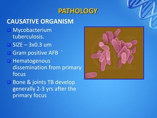
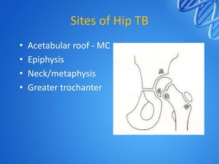
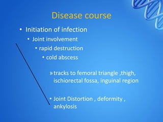
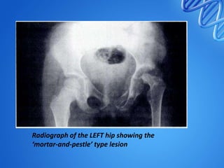
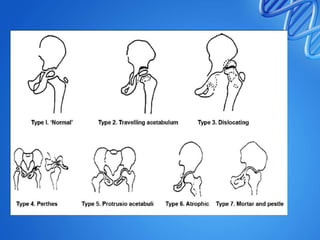
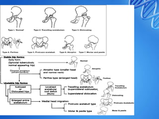
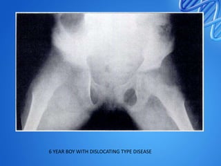
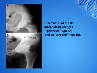
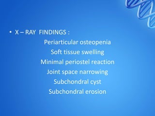
TUBERCULOSIS OF HIP
DR. HARDIK PAWAR
CARE HOSPITALS
INTRODUCTION
•
•
•
•
•
•
•

Oldest disease of human beings
Chronic granulomatous infectious disease
EPTB – 10-15 %
Bone and joint – 1-3 %
Hip joint 2nd most common
M>F
Age group – 20 -30 years
HISTORICAL ASPECTS



Robert Koch discovered
Mycobacterium
tuberculosis in 1882.
PATHOLOGY
CAUSATIVE ORGANISM
Mycobacterium
tuberculosis.
 SIZE – 3x0.3 um
 Gram positive AFB `
 Hematogenous
dissemination from primary
focus
 Bone & joints TB develop
generally 2-3 yrs after the
primary focus

PATHOLOGY
1. TB bacilli phagocytosed
by mononuclear cells

2. Epitheloid cell formation

3.Langhans giant cell formed by fusion
epitheliod cells. Formed only if caseation
necrosis has occurred
PATHOLOGY
4.Lymphocytes form a
ring around the lesion

5.Tubercle formation




Epitheloid cells-characteristic of TB.
Presence of caseation necrosis almost diagnostic of TB
PATHOGENESIS
Caseous exudative type
• more in children
• more destruction
• more exudation
• abscess formation.

Granular proliferative type
• more in adults
• less destructive
• insidious onset/course
• Cellular proliferation
Sites of Hip TB
•
•
•
•

Acetabular roof - MC
Epiphysis
Neck/metaphysis
Greater trochanter
Disease course
• Initiation of infection
• Joint involvement
• rapid destruction
• cold abscess
»tracks to femoral triangle ,thigh,
ischiorectal fossa, inguinal region

• Joint Distortion , deformity ,
ankylosis
Clinical features
• Insidious onset
• Chronic course
• Commonly associated with constitutional
symptoms
• Limp or lameness – earliest and commonest
sign , antalgic gait
• Pain - absent in early stage
• Night cries
• Decreased ROM
Cont.
•
•
•
•
•

Fullness around the hip – cold abscess
Deformity - depending on the stage
Wasting of the thigh and gluteal muscle
Limb length discrepency
Due to fixed deformity Secondary chages –
lordosis , scoliosis ,
Clinicoradiological staging
1. Stage of synovitis :
• Irritable hip , painful movements
• Flexion , abduction , external rotation .
(FABER ) apparent lenghening.
• X-RAY – soft tissue swelling , haziness of
articular margins & rarefaction
• USG – soft tissue swelling
• MRI – synovial effusion
• Biopsy – can be done for confirmation
•
•
•
•

2. Stage of early arthritis :
Destruction of articular cartilage
Spasm of adductors , flexors + wasting
Flexion , adduction , internal roatation
(FADIR) , Apparent shortening
• ↓sed ROM
• X-RAY – osteopenia , erosion of articular
margins , ↓ joint space
• MRI - synovial effusion , edema , minimal
bone destruction
•
•
•
•
•
•

3. Stage of advanced arthritis :
Further destruction of joint
True shortening > 1 cm
Muscle wasting
Decrease in ROM
X RAY - further decrease in joint space.
• 4. Advanced arthritis with subluxation /
dislocation :
• Furhter destruction of acetabulum , head ,
capsule and ligaments.
• Gross restriction of ROM
• Head – upwards and posteriorly
• Wandering / migrating acetabulum
• Mortle & pestle appearance
• Reduced joint space
Radiograph showing a ‘travelling’ or
‘wandering’ right hip
Radiograph of the LEFT hip showing the
‘mortar-and-pestle’ type lesion
CLINICO RADIO LOGICAL
CLASSIFICATION
6 YEAR BOY WITH DISLOCATING TYPE DISEASE
Tuberculous of the Hip.
Morphologic changes
“protrusio” type (A)
and an “atrophic” type (B).
2 YEARS BOY WITH PERTHES TYPE DISEASE
COXA MAGNA
MANAGEMENT
INVESTIGATIONS










CBC
ESR
MANTOUX TEST
TB ELISA
IMAGING : X RAY – HIP
SYNOVIAL BIOPSY
SMEAR , CULTURE AND GUINEA PIG INOCULATION
MRI - EFFUSION , SYNOVIAL THICKENING , JUXTRA
ARTICULAR OSTEOPENIA
PCR – RT PCR , NESTED PCR
• X – RAY FINDINGS :
Periarticular osteopenia
Soft tissue swelling
Minimal periostel reaction
Joint space narrowing
Subchondral cyst
Subchondral erosion
INVESTIGATIONS
 Sputum examination :

for AFB
INVESTIGATIONS


Aspiration of abscess :
smears & culture of pus
CULTURE=GOLD STANDARD

SOLID MEDIA: 3-8 weeks
Lowenstein Jensen (egg Based)
Middlebrook 7H11(agar based)

LIQUID BROTH:
1-3 weeks
INVESTIGATIONS


BACTEC media: for faster culture (within 1-2wks)

Bactec 460

Bactec9000MB

Bactec mgit960b
AIM OF THE MANAGEMENT
PAINLESS
SYMPTOM FREE
STABLE
FREELY MOBLE
NORMAL GAIT
NO DEFORMITY
NO LLD
•
•
•
•

GENERAL
CHEMOTHERAPY
LOCAL TREATMENT
ROLE OF SURGERY
GENERAL TREATMENT
GOOD DIET
FRESH AIR
SUNLIGHT
EDUCATION
OCCUPATION
CHEMOTHERAPY
• 6-9 MONTH DURATION
• 12 MONTH FOR PEDIATRIC AGE GROUP
• 1st line - HRZES
• 2ND Line Capreomycin
Kanamycin
Ethionamide
Cycloserine
PAS
• Newer drugs : rifapentin , rifabutin ,
gatifloxacin , moxifloxacin
Bactericidal drugs

1.Isoniazid

2. Rifampicin

Dose

5mg/kg

10-15 mg/kg

3. Streptomycin

20mg/kg

4. Pyrazinamide

20-25 mg/kg

Bacteriostatic drugs
1. Ethambutol

Dose
25mg/kg (x 2mnths)
Then 15mg/kg
LOCAL TREATMENT
• A) STAGE OF SYNOVITIS AND EARLY
ARTHRITIS :
ATT
TRACTION
ASPIRATION OF COLD ABSCESS
• TRACTION :
Relieves muscle spasm
To give rest to joint
Prevents deformity
Maintain joint space
Minimizes chances of developing migrtaing
acetabulum
• PROTOCOL :
• B ) STAGE OF ADVANCED ARTHRITIS :
Traction
Usually gross fibrous ankylosis is out come
Role of surgery
• To establish diagnosis
• Therapeutic
• Failure of conservative treatment
•
•
•
•

SYNOVECTOMY
DEBRIDEMENT OF JOINT
ARTHRODESIS
ARTHROPLASTY
ARTHRODESIS
• Surgical fusion of joint
• INDICATIONS :
Failure of conservative Mx
Relapse
Destructive lesion in head
Painful fibrous ankylosis
Ankylosis with severe deformity
• TYPES :

INTRAARTICULAR
EXTRAARTICULAR
COMBINED
INTRAARTICULAR
• Active disease
• Painful fibrous ankylosis
• Lower rate of fusion

EXTRAARTICULAR
• ILIOFEMORAL - HIBBS
• ISHIO FEMORAL BRITTAIN’S
• ABOTT LUCAS TECHNIQUE :
2 STAGE PROCEDURE
1ST – Arthrodesis in wide abduction
2nd – subtrochantric osteotomy to repostion
limb
• BEST POSITION :
30 DEGREES OF FLEXION
5-10 DEGREES OF ER
NO ADDUCTION
EXCISION ARTHROPLASTY
HEAD , NECK , RPOXIMAL PART OF TROCHANTER
AND ACETABULAR RIM EXCISED
PAINLESS
MOBILE HIP
PERMITS SQUATTING
BUT
INSTABILITY
SHORTENING
REPLACEMENT ARTHROPLASTY
• EARLY CEMENTLESS THR IN ACTIVE TB
PROVIDED PATIENT IS ADMINISTERED ATT
AND THOROUGH DEBRIDEMENT Of DISEASED
TISSUE IS DONE.
• ( ACCORDING TO RECENT AUTHORS )
• Cemented
- Hardinge et al. 1979
- Kim et al. 1988
• Uncemented - Eskola et al. 1988
- Caparros et al. 1999
- Yoon et al. 2001
•

Normal hip type tuberculosis lesion of left hip (a, b). Triple chemotherapy for
12 months and bed rest was given. The tuberculosis lesion had healed
without joint damage and sequelae (c)
Perthes type hip
epiphysio-metaphyseal tuberculosis of the left proximal femur (a, b).
After joint debridement, the child was immobilized with cast for 6 weeks under
cover of triple chemotherapy (12 months).
Tuberculosis healed without joint destruction,
though residual coxa magna deformity was present (c)
Atrophic type tuberculosis of hip in
a 11-year-old girl. There is slight joint space narrowing of left hip (a).
After synovectomy chemotherapy for 12 months was given (b, c, d).
Tuberculosis healed (3 years followup) though atrophic head and neck are seen (d)
REFERENCES
1.

Campbell JA, Hoffman EB. Tuberculosis of the hip in children.
J Bone Joint Surg Br 1995;77:319-26.

2. Shanmugasundaram TK. A clinicoradiological classification of
tuberculosis of the hip. In: Shanmugasundaram TK (editor).
Current concepts in bone and joint tuberculosis. Madras,
India: Proceedings of Combined Congress of International
Bone and Joint Tuberculosis Club and the Indian Orthop
Assoc;1983. P. 60.

3. Moon MS, Kim SS, Lee SR, Moon YW, Moon JL, Moon SI.
Tuberculosis of the hip in children: A retrospective analysis. Ind J
Orthop 2012;46: 91-99.
• 4.

Eskola A, Santavirta S, Konttinen YT, Tallroth K, Hoikka V,
Lindholm ST. Cementless total replacement for old
tuberculosis of the hip. J Bone Joint Surg (Br) 1988;70:603–
606.

• 5.

Yoon TR, Rowe SM, Santosa SB, Jung ST, Seon JK.
Immediate cementless total hip arthroplasty for the
treatment of active tuberculosis. J Arthroplasty. 2005;20:923–
926. doi: 10.1016/j.arth.2004.08.002.
THANK YOU

Tuberculosis of hip

  • 1.
    TUBERCULOSIS OF HIP DR.HARDIK PAWAR CARE HOSPITALS
  • 2.
    INTRODUCTION • • • • • • • Oldest disease ofhuman beings Chronic granulomatous infectious disease EPTB – 10-15 % Bone and joint – 1-3 % Hip joint 2nd most common M>F Age group – 20 -30 years
  • 3.
    HISTORICAL ASPECTS  Robert Kochdiscovered Mycobacterium tuberculosis in 1882.
  • 4.
    PATHOLOGY CAUSATIVE ORGANISM Mycobacterium tuberculosis.  SIZE– 3x0.3 um  Gram positive AFB `  Hematogenous dissemination from primary focus  Bone & joints TB develop generally 2-3 yrs after the primary focus 
  • 5.
    PATHOLOGY 1. TB bacilliphagocytosed by mononuclear cells 2. Epitheloid cell formation 3.Langhans giant cell formed by fusion epitheliod cells. Formed only if caseation necrosis has occurred
  • 6.
    PATHOLOGY 4.Lymphocytes form a ringaround the lesion 5.Tubercle formation   Epitheloid cells-characteristic of TB. Presence of caseation necrosis almost diagnostic of TB
  • 7.
    PATHOGENESIS Caseous exudative type •more in children • more destruction • more exudation • abscess formation. Granular proliferative type • more in adults • less destructive • insidious onset/course • Cellular proliferation
  • 8.
    Sites of HipTB • • • • Acetabular roof - MC Epiphysis Neck/metaphysis Greater trochanter
  • 9.
    Disease course • Initiationof infection • Joint involvement • rapid destruction • cold abscess »tracks to femoral triangle ,thigh, ischiorectal fossa, inguinal region • Joint Distortion , deformity , ankylosis
  • 10.
    Clinical features • Insidiousonset • Chronic course • Commonly associated with constitutional symptoms • Limp or lameness – earliest and commonest sign , antalgic gait • Pain - absent in early stage • Night cries • Decreased ROM
  • 11.
    Cont. • • • • • Fullness around thehip – cold abscess Deformity - depending on the stage Wasting of the thigh and gluteal muscle Limb length discrepency Due to fixed deformity Secondary chages – lordosis , scoliosis ,
  • 12.
    Clinicoradiological staging 1. Stageof synovitis : • Irritable hip , painful movements • Flexion , abduction , external rotation . (FABER ) apparent lenghening. • X-RAY – soft tissue swelling , haziness of articular margins & rarefaction • USG – soft tissue swelling • MRI – synovial effusion • Biopsy – can be done for confirmation
  • 14.
    • • • • 2. Stage ofearly arthritis : Destruction of articular cartilage Spasm of adductors , flexors + wasting Flexion , adduction , internal roatation (FADIR) , Apparent shortening • ↓sed ROM • X-RAY – osteopenia , erosion of articular margins , ↓ joint space • MRI - synovial effusion , edema , minimal bone destruction
  • 16.
    • • • • • • 3. Stage ofadvanced arthritis : Further destruction of joint True shortening > 1 cm Muscle wasting Decrease in ROM X RAY - further decrease in joint space.
  • 18.
    • 4. Advancedarthritis with subluxation / dislocation : • Furhter destruction of acetabulum , head , capsule and ligaments. • Gross restriction of ROM • Head – upwards and posteriorly • Wandering / migrating acetabulum • Mortle & pestle appearance • Reduced joint space
  • 19.
    Radiograph showing a‘travelling’ or ‘wandering’ right hip
  • 20.
    Radiograph of theLEFT hip showing the ‘mortar-and-pestle’ type lesion
  • 21.
  • 25.
    6 YEAR BOYWITH DISLOCATING TYPE DISEASE
  • 26.
    Tuberculous of theHip. Morphologic changes “protrusio” type (A) and an “atrophic” type (B).
  • 27.
    2 YEARS BOYWITH PERTHES TYPE DISEASE
  • 28.
  • 29.
  • 30.
    INVESTIGATIONS          CBC ESR MANTOUX TEST TB ELISA IMAGING: X RAY – HIP SYNOVIAL BIOPSY SMEAR , CULTURE AND GUINEA PIG INOCULATION MRI - EFFUSION , SYNOVIAL THICKENING , JUXTRA ARTICULAR OSTEOPENIA PCR – RT PCR , NESTED PCR
  • 31.
    • X –RAY FINDINGS : Periarticular osteopenia Soft tissue swelling Minimal periostel reaction Joint space narrowing Subchondral cyst Subchondral erosion
  • 32.
  • 33.
    INVESTIGATIONS  Aspiration of abscess: smears & culture of pus CULTURE=GOLD STANDARD SOLID MEDIA: 3-8 weeks Lowenstein Jensen (egg Based) Middlebrook 7H11(agar based) LIQUID BROTH: 1-3 weeks
  • 34.
    INVESTIGATIONS  BACTEC media: forfaster culture (within 1-2wks) Bactec 460 Bactec9000MB Bactec mgit960b
  • 35.
    AIM OF THEMANAGEMENT PAINLESS SYMPTOM FREE STABLE FREELY MOBLE NORMAL GAIT NO DEFORMITY NO LLD
  • 36.
  • 37.
    GENERAL TREATMENT GOOD DIET FRESHAIR SUNLIGHT EDUCATION OCCUPATION
  • 38.
    CHEMOTHERAPY • 6-9 MONTHDURATION • 12 MONTH FOR PEDIATRIC AGE GROUP
  • 39.
    • 1st line- HRZES • 2ND Line Capreomycin Kanamycin Ethionamide Cycloserine PAS • Newer drugs : rifapentin , rifabutin , gatifloxacin , moxifloxacin
  • 40.
    Bactericidal drugs 1.Isoniazid 2. Rifampicin Dose 5mg/kg 10-15mg/kg 3. Streptomycin 20mg/kg 4. Pyrazinamide 20-25 mg/kg Bacteriostatic drugs 1. Ethambutol Dose 25mg/kg (x 2mnths) Then 15mg/kg
  • 41.
    LOCAL TREATMENT • A)STAGE OF SYNOVITIS AND EARLY ARTHRITIS : ATT TRACTION ASPIRATION OF COLD ABSCESS
  • 42.
    • TRACTION : Relievesmuscle spasm To give rest to joint Prevents deformity Maintain joint space Minimizes chances of developing migrtaing acetabulum • PROTOCOL :
  • 43.
    • B )STAGE OF ADVANCED ARTHRITIS : Traction Usually gross fibrous ankylosis is out come
  • 44.
    Role of surgery •To establish diagnosis • Therapeutic • Failure of conservative treatment
  • 45.
  • 46.
    ARTHRODESIS • Surgical fusionof joint • INDICATIONS : Failure of conservative Mx Relapse Destructive lesion in head Painful fibrous ankylosis Ankylosis with severe deformity
  • 47.
  • 48.
    INTRAARTICULAR • Active disease •Painful fibrous ankylosis • Lower rate of fusion EXTRAARTICULAR • ILIOFEMORAL - HIBBS • ISHIO FEMORAL BRITTAIN’S
  • 50.
    • ABOTT LUCASTECHNIQUE : 2 STAGE PROCEDURE 1ST – Arthrodesis in wide abduction 2nd – subtrochantric osteotomy to repostion limb
  • 51.
    • BEST POSITION: 30 DEGREES OF FLEXION 5-10 DEGREES OF ER NO ADDUCTION
  • 52.
    EXCISION ARTHROPLASTY HEAD ,NECK , RPOXIMAL PART OF TROCHANTER AND ACETABULAR RIM EXCISED PAINLESS MOBILE HIP PERMITS SQUATTING BUT INSTABILITY SHORTENING
  • 54.
    REPLACEMENT ARTHROPLASTY • EARLYCEMENTLESS THR IN ACTIVE TB PROVIDED PATIENT IS ADMINISTERED ATT AND THOROUGH DEBRIDEMENT Of DISEASED TISSUE IS DONE. • ( ACCORDING TO RECENT AUTHORS ) • Cemented - Hardinge et al. 1979 - Kim et al. 1988 • Uncemented - Eskola et al. 1988 - Caparros et al. 1999 - Yoon et al. 2001
  • 55.
    • Normal hip typetuberculosis lesion of left hip (a, b). Triple chemotherapy for 12 months and bed rest was given. The tuberculosis lesion had healed without joint damage and sequelae (c)
  • 56.
    Perthes type hip epiphysio-metaphysealtuberculosis of the left proximal femur (a, b). After joint debridement, the child was immobilized with cast for 6 weeks under cover of triple chemotherapy (12 months). Tuberculosis healed without joint destruction, though residual coxa magna deformity was present (c)
  • 57.
    Atrophic type tuberculosisof hip in a 11-year-old girl. There is slight joint space narrowing of left hip (a). After synovectomy chemotherapy for 12 months was given (b, c, d). Tuberculosis healed (3 years followup) though atrophic head and neck are seen (d)
  • 58.
    REFERENCES 1. Campbell JA, HoffmanEB. Tuberculosis of the hip in children. J Bone Joint Surg Br 1995;77:319-26. 2. Shanmugasundaram TK. A clinicoradiological classification of tuberculosis of the hip. In: Shanmugasundaram TK (editor). Current concepts in bone and joint tuberculosis. Madras, India: Proceedings of Combined Congress of International Bone and Joint Tuberculosis Club and the Indian Orthop Assoc;1983. P. 60. 3. Moon MS, Kim SS, Lee SR, Moon YW, Moon JL, Moon SI. Tuberculosis of the hip in children: A retrospective analysis. Ind J Orthop 2012;46: 91-99.
  • 59.
    • 4. Eskola A,Santavirta S, Konttinen YT, Tallroth K, Hoikka V, Lindholm ST. Cementless total replacement for old tuberculosis of the hip. J Bone Joint Surg (Br) 1988;70:603– 606. • 5. Yoon TR, Rowe SM, Santosa SB, Jung ST, Seon JK. Immediate cementless total hip arthroplasty for the treatment of active tuberculosis. J Arthroplasty. 2005;20:923– 926. doi: 10.1016/j.arth.2004.08.002.
  • 60.

Editor's Notes

  • #23 % - PROGNOSIS , C- CHILDREN , A - adult NORMAL 92% , C - Joint space normal Rx – skin traction , ATT TRAVELLING ACETABULUM – 29% , CA – Lesion in roof of aceta. , decreased joint space .50% C – HIP JOINT DISLOCATED POSt. , destruction of surface and cystic lesion in the acetabulum and head80 % . C – Capital epiphysis fragmented , dense , flattened , neck widened , heals by fibrous ankylosis Protrusio
  • #24 % - PROGNOSIS , C- CHILDREN , A - adult NORMAL 92% , C - Joint space normal Rx – skin traction , ATT TRAVELLING ACETABULUM – 29% , CA – Lesion in roof of aceta. , decreased joint space .50% C – HIP JOINT DISLOCATED POSt. , destruction of surface and cystic lesion in the acetabulum and head80 % . C – Capital epiphysis fragmented , dense , flattened , neck widened , heals by fibrous ankylosis Protrusio – CA -
  • #36 TO OBTAIN
  • #43 Active asisted movements should be started as soon as pain is aubsidedHip mobilzation exercises With traction exercise – sittin , After 4-6 month – ambulation wid crutches , 12 wks – non wt bearing , next 12 wks – partial wt bearing After total 12 mnth – full wt bearing
  • #49 ADVANTAGES - permits curratge ,to obtain tissue for HPE, exploration of joint , excision of diseased tissue Iliofemoral – easy in ABDUCTION Ishiofemoral – EASY IN ADDUCTION
  • #51 Indication – extensive femoral head destruction – ABSENCE HEAD, deficient bone stock , Strong , fused , painless hip , BOSWORTH TECH ,.- FEMORO ISCHIAL ARTHRODESIS