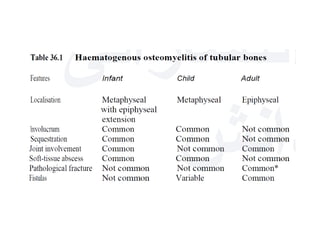
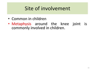
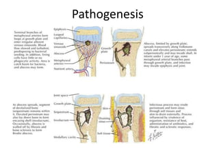
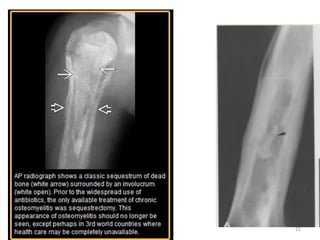
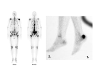
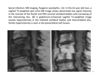
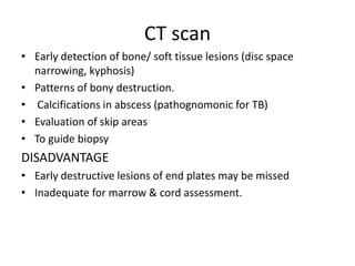
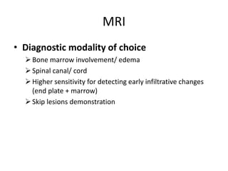
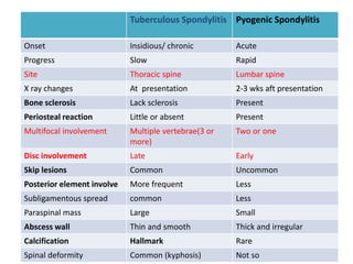
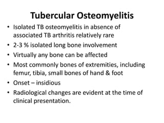
about basics of cartilage imaging.
how does normal cartilage look , how does diseased cartilage look.
what are advanced techniques in cartilage imaging
about basics of cartilage imaging.
how does normal cartilage look , how does diseased cartilage look.
what are advanced techniques in cartilage imaging
Imaging features of acute and chronic osteomyelitis are described in this PPT. Infective arthritis along with fungal infections of soft tissue are also covered very well. Special emphasis is given on tubercular infection of bone.
Title: Sense of Smell
Presenter: Dr. Faiza, Assistant Professor of Physiology
Qualifications:
MBBS (Best Graduate, AIMC Lahore)
FCPS Physiology
ICMT, CHPE, DHPE (STMU)
MPH (GC University, Faisalabad)
MBA (Virtual University of Pakistan)
Learning Objectives:
Describe the primary categories of smells and the concept of odor blindness.
Explain the structure and location of the olfactory membrane and mucosa, including the types and roles of cells involved in olfaction.
Describe the pathway and mechanisms of olfactory signal transmission from the olfactory receptors to the brain.
Illustrate the biochemical cascade triggered by odorant binding to olfactory receptors, including the role of G-proteins and second messengers in generating an action potential.
Identify different types of olfactory disorders such as anosmia, hyposmia, hyperosmia, and dysosmia, including their potential causes.
Key Topics:
Olfactory Genes:
3% of the human genome accounts for olfactory genes.
400 genes for odorant receptors.
Olfactory Membrane:
Located in the superior part of the nasal cavity.
Medially: Folds downward along the superior septum.
Laterally: Folds over the superior turbinate and upper surface of the middle turbinate.
Total surface area: 5-10 square centimeters.
Olfactory Mucosa:
Olfactory Cells: Bipolar nerve cells derived from the CNS (100 million), with 4-25 olfactory cilia per cell.
Sustentacular Cells: Produce mucus and maintain ionic and molecular environment.
Basal Cells: Replace worn-out olfactory cells with an average lifespan of 1-2 months.
Bowman’s Gland: Secretes mucus.
Stimulation of Olfactory Cells:
Odorant dissolves in mucus and attaches to receptors on olfactory cilia.
Involves a cascade effect through G-proteins and second messengers, leading to depolarization and action potential generation in the olfactory nerve.
Quality of a Good Odorant:
Small (3-20 Carbon atoms), volatile, water-soluble, and lipid-soluble.
Facilitated by odorant-binding proteins in mucus.
Membrane Potential and Action Potential:
Resting membrane potential: -55mV.
Action potential frequency in the olfactory nerve increases with odorant strength.
Adaptation Towards the Sense of Smell:
Rapid adaptation within the first second, with further slow adaptation.
Psychological adaptation greater than receptor adaptation, involving feedback inhibition from the central nervous system.
Primary Sensations of Smell:
Camphoraceous, Musky, Floral, Pepperminty, Ethereal, Pungent, Putrid.
Odor Detection Threshold:
Examples: Hydrogen sulfide (0.0005 ppm), Methyl-mercaptan (0.002 ppm).
Some toxic substances are odorless at lethal concentrations.
Characteristics of Smell:
Odor blindness for single substances due to lack of appropriate receptor protein.
Behavioral and emotional influences of smell.
Transmission of Olfactory Signals:
From olfactory cells to glomeruli in the olfactory bulb, involving lateral inhibition.
Primitive, less old, and new olfactory systems with different path
MANAGEMENT OF ATRIOVENTRICULAR CONDUCTION BLOCK.pdfJim Jacob Roy
Cardiac conduction defects can occur due to various causes.
Atrioventricular conduction blocks ( AV blocks ) are classified into 3 types.
This document describes the acute management of AV block.
Ozempic: Preoperative Management of Patients on GLP-1 Receptor Agonists Saeid Safari
Preoperative Management of Patients on GLP-1 Receptor Agonists like Ozempic and Semiglutide
ASA GUIDELINE
NYSORA Guideline
2 Case Reports of Gastric Ultrasound
ARTIFICIAL INTELLIGENCE IN HEALTHCARE.pdfAnujkumaranit
Artificial intelligence (AI) refers to the simulation of human intelligence processes by machines, especially computer systems. It encompasses tasks such as learning, reasoning, problem-solving, perception, and language understanding. AI technologies are revolutionizing various fields, from healthcare to finance, by enabling machines to perform tasks that typically require human intelligence.
Explore natural remedies for syphilis treatment in Singapore. Discover alternative therapies, herbal remedies, and lifestyle changes that may complement conventional treatments. Learn about holistic approaches to managing syphilis symptoms and supporting overall health.
Lung Cancer: Artificial Intelligence, Synergetics, Complex System Analysis, S...Oleg Kshivets
RESULTS: Overall life span (LS) was 2252.1±1742.5 days and cumulative 5-year survival (5YS) reached 73.2%, 10 years – 64.8%, 20 years – 42.5%. 513 LCP lived more than 5 years (LS=3124.6±1525.6 days), 148 LCP – more than 10 years (LS=5054.4±1504.1 days).199 LCP died because of LC (LS=562.7±374.5 days). 5YS of LCP after bi/lobectomies was significantly superior in comparison with LCP after pneumonectomies (78.1% vs.63.7%, P=0.00001 by log-rank test). AT significantly improved 5YS (66.3% vs. 34.8%) (P=0.00000 by log-rank test) only for LCP with N1-2. Cox modeling displayed that 5YS of LCP significantly depended on: phase transition (PT) early-invasive LC in terms of synergetics, PT N0—N12, cell ratio factors (ratio between cancer cells- CC and blood cells subpopulations), G1-3, histology, glucose, AT, blood cell circuit, prothrombin index, heparin tolerance, recalcification time (P=0.000-0.038). Neural networks, genetic algorithm selection and bootstrap simulation revealed relationships between 5YS and PT early-invasive LC (rank=1), PT N0—N12 (rank=2), thrombocytes/CC (3), erythrocytes/CC (4), eosinophils/CC (5), healthy cells/CC (6), lymphocytes/CC (7), segmented neutrophils/CC (8), stick neutrophils/CC (9), monocytes/CC (10); leucocytes/CC (11). Correct prediction of 5YS was 100% by neural networks computing (area under ROC curve=1.0; error=0.0).
CONCLUSIONS: 5YS of LCP after radical procedures significantly depended on: 1) PT early-invasive cancer; 2) PT N0--N12; 3) cell ratio factors; 4) blood cell circuit; 5) biochemical factors; 6) hemostasis system; 7) AT; 8) LC characteristics; 9) LC cell dynamics; 10) surgery type: lobectomy/pneumonectomy; 11) anthropometric data. Optimal diagnosis and treatment strategies for LC are: 1) screening and early detection of LC; 2) availability of experienced thoracic surgeons because of complexity of radical procedures; 3) aggressive en block surgery and adequate lymph node dissection for completeness; 4) precise prediction; 5) adjuvant chemoimmunoradiotherapy for LCP with unfavorable prognosis.
Pulmonary Thromboembolism - etilogy, types, medical- Surgical and nursing man...VarunMahajani
Disruption of blood supply to lung alveoli due to blockage of one or more pulmonary blood vessels is called as Pulmonary thromboembolism. In this presentation we will discuss its causes, types and its management in depth.
Title: Sense of Taste
Presenter: Dr. Faiza, Assistant Professor of Physiology
Qualifications:
MBBS (Best Graduate, AIMC Lahore)
FCPS Physiology
ICMT, CHPE, DHPE (STMU)
MPH (GC University, Faisalabad)
MBA (Virtual University of Pakistan)
Learning Objectives:
Describe the structure and function of taste buds.
Describe the relationship between the taste threshold and taste index of common substances.
Explain the chemical basis and signal transduction of taste perception for each type of primary taste sensation.
Recognize different abnormalities of taste perception and their causes.
Key Topics:
Significance of Taste Sensation:
Differentiation between pleasant and harmful food
Influence on behavior
Selection of food based on metabolic needs
Receptors of Taste:
Taste buds on the tongue
Influence of sense of smell, texture of food, and pain stimulation (e.g., by pepper)
Primary and Secondary Taste Sensations:
Primary taste sensations: Sweet, Sour, Salty, Bitter, Umami
Chemical basis and signal transduction mechanisms for each taste
Taste Threshold and Index:
Taste threshold values for Sweet (sucrose), Salty (NaCl), Sour (HCl), and Bitter (Quinine)
Taste index relationship: Inversely proportional to taste threshold
Taste Blindness:
Inability to taste certain substances, particularly thiourea compounds
Example: Phenylthiocarbamide
Structure and Function of Taste Buds:
Composition: Epithelial cells, Sustentacular/Supporting cells, Taste cells, Basal cells
Features: Taste pores, Taste hairs/microvilli, and Taste nerve fibers
Location of Taste Buds:
Found in papillae of the tongue (Fungiform, Circumvallate, Foliate)
Also present on the palate, tonsillar pillars, epiglottis, and proximal esophagus
Mechanism of Taste Stimulation:
Interaction of taste substances with receptors on microvilli
Signal transduction pathways for Umami, Sweet, Bitter, Sour, and Salty tastes
Taste Sensitivity and Adaptation:
Decrease in sensitivity with age
Rapid adaptation of taste sensation
Role of Saliva in Taste:
Dissolution of tastants to reach receptors
Washing away the stimulus
Taste Preferences and Aversions:
Mechanisms behind taste preference and aversion
Influence of receptors and neural pathways
Impact of Sensory Nerve Damage:
Degeneration of taste buds if the sensory nerve fiber is cut
Abnormalities of Taste Detection:
Conditions: Ageusia, Hypogeusia, Dysgeusia (parageusia)
Causes: Nerve damage, neurological disorders, infections, poor oral hygiene, adverse drug effects, deficiencies, aging, tobacco use, altered neurotransmitter levels
Neurotransmitters and Taste Threshold:
Effects of serotonin (5-HT) and norepinephrine (NE) on taste sensitivity
Supertasters:
25% of the population with heightened sensitivity to taste, especially bitterness
Increased number of fungiform papillae
The prostate is an exocrine gland of the male mammalian reproductive system
It is a walnut-sized gland that forms part of the male reproductive system and is located in front of the rectum and just below the urinary bladder
Function is to store and secrete a clear, slightly alkaline fluid that constitutes 10-30% of the volume of the seminal fluid that along with the spermatozoa, constitutes semen
A healthy human prostate measures (4cm-vertical, by 3cm-horizontal, 2cm ant-post ).
It surrounds the urethra just below the urinary bladder. It has anterior, median, posterior and two lateral lobes
It’s work is regulated by androgens which are responsible for male sex characteristics
Generalised disease of the prostate due to hormonal derangement which leads to non malignant enlargement of the gland (increase in the number of epithelial cells and stromal tissue)to cause compression of the urethra leading to symptoms (LUTS
Ethanol (CH3CH2OH), or beverage alcohol, is a two-carbon alcohol
that is rapidly distributed in the body and brain. Ethanol alters many
neurochemical systems and has rewarding and addictive properties. It
is the oldest recreational drug and likely contributes to more morbidity,
mortality, and public health costs than all illicit drugs combined. The
5th edition of the Diagnostic and Statistical Manual of Mental Disorders
(DSM-5) integrates alcohol abuse and alcohol dependence into a single
disorder called alcohol use disorder (AUD), with mild, moderate,
and severe subclassifications (American Psychiatric Association, 2013).
In the DSM-5, all types of substance abuse and dependence have been
combined into a single substance use disorder (SUD) on a continuum
from mild to severe. A diagnosis of AUD requires that at least two of
the 11 DSM-5 behaviors be present within a 12-month period (mild
AUD: 2–3 criteria; moderate AUD: 4–5 criteria; severe AUD: 6–11 criteria).
The four main behavioral effects of AUD are impaired control over
drinking, negative social consequences, risky use, and altered physiological
effects (tolerance, withdrawal). This chapter presents an overview
of the prevalence and harmful consequences of AUD in the U.S.,
the systemic nature of the disease, neurocircuitry and stages of AUD,
comorbidities, fetal alcohol spectrum disorders, genetic risk factors, and
pharmacotherapies for AUD.
Report Back from SGO 2024: What’s the Latest in Cervical Cancer?bkling
Are you curious about what’s new in cervical cancer research or unsure what the findings mean? Join Dr. Emily Ko, a gynecologic oncologist at Penn Medicine, to learn about the latest updates from the Society of Gynecologic Oncology (SGO) 2024 Annual Meeting on Women’s Cancer. Dr. Ko will discuss what the research presented at the conference means for you and answer your questions about the new developments.
HOT NEW PRODUCT! BIG SALES FAST SHIPPING NOW FROM CHINA!! EU KU DB BK substit...GL Anaacs
Contact us if you are interested:
Email / Skype : kefaya1771@gmail.com
Threema: PXHY5PDH
New BATCH Ku !!! MUCH IN DEMAND FAST SALE EVERY BATCH HAPPY GOOD EFFECT BIG BATCH !
Contact me on Threema or skype to start big business!!
Hot-sale products:
NEW HOT EUTYLONE WHITE CRYSTAL!!
5cl-adba precursor (semi finished )
5cl-adba raw materials
ADBB precursor (semi finished )
ADBB raw materials
APVP powder
5fadb/4f-adb
Jwh018 / Jwh210
Eutylone crystal
Protonitazene (hydrochloride) CAS: 119276-01-6
Flubrotizolam CAS: 57801-95-3
Metonitazene CAS: 14680-51-4
Payment terms: Western Union,MoneyGram,Bitcoin or USDT.
Deliver Time: Usually 7-15days
Shipping method: FedEx, TNT, DHL,UPS etc.Our deliveries are 100% safe, fast, reliable and discreet.
Samples will be sent for your evaluation!If you are interested in, please contact me, let's talk details.
We specializes in exporting high quality Research chemical, medical intermediate, Pharmaceutical chemicals and so on. Products are exported to USA, Canada, France, Korea, Japan,Russia, Southeast Asia and other countries.
3. • The term osteomyelitis implies an infection of bone
and marrow. (osteo-myelo-itis)
• Osteomyelitis most commonly results from bacterial
infections, although fungi, parasites, and viruses can
infect the bone and the marrow
3
Introduction
4. Classification
• Acute- diagnosed within 2 weeks of onset of
symptoms
• Subacute-Symptoms exceed 2 weeks duration
• Chronic- diagnosed months after the onset of
symptoms
• All cases of osteomyelitis don’t progress
through each of these phases as well.
4
15. • A sequestrum:
– represents a segment of necrotic bone that is
separated from living bone by granulation tissue.
– Sequestra- nidus for infection.
– As sequestra are devitalized, they remain denser than
surrounding vital bone which become demineralized
due to hyperemia and immobilization.
• An involucrum:
– denotes a layer of living bone that has formed about
the dead bone.
• Cloaca-an opening in an involucrum which allows
drainage of purulent and necrotic material out of the
dead bone.
16
16. CLINICAL PRESENTATION
• Common in childhood
• High fever, a toxic state, and local signs of inflammation,
although this presentation is certainly not uniform.
• The adult form of hematogenous osteomyelitis may have a
more insidious onset with a relatively longer period between
the appearance of symptoms and signs and accurate
diagnosis.
17
18. Plain radiography
• In the early stage of
osteomyelitis, the findings on
conventional radiography may
be normal 5 to 7 days in
children and 10 to 14 days in
adults after the onset of
infection
• A 30% to 50% loss of bone
density must occur before a
radiograph becomes abnormal.
• Soft-tissue changes are the
earliest manifestations
– non specific
– Swelling, with edema and
blurring of fat planes is
seen
19
19. • Localized osteoporosis and bone resorption caused by
infection in the medullary space with hyperemia, edema
or abscess formation, and trabecular destruction occur.
• Later, there is increased lysis and cortical lucency as
infection spreads into the haversian and Volkmann's
canals of the cortex.
• Next are periostitis and involucrum formation, which are
caused by subperiosteal abscess formation with lifting of
the periosteum and bone formation .
20
Bone Changes:
20. • Bony changes –
- 10-14 days required
– Local osteopenia (at the site of infection,
usually metaphysis) is the earliest bony
change.
– Periosteal reaction follows
– An involucrum is usually visualized after 3
weeks
– Sequestrum is seen as dense, irregular bone
surrounded by lucency (representing
granulation tissue).
– Cloacae (fistula tract) are seen as lucent
areas which are created by external
migration of dead pieces of bone with
breakdown of skin and subcutaneous tissue.
21
22. Radionuclide scanning:
• Should precede plain film examination in suspected
bone infection.
• Diagnosis can be confirmed as early as 48 hours after
the onset of the disease, even if clinical signs are
equivocal.
• This may help to initiate early aggressive treatment
to prevent gross bone destruction.
23
23. Radionuclides used:
• Tc 99m labelled phosphonates: most
commonly used
• Ga - 67 labelled citrate scan
• Indium-111 labelled WBC scan
24
24. a) Plain film shows no abnormality;
b )blood flow phase and c)
late phase shows increased
bone tracer activity
25
25. Tc - 99m scan
• 3 phase scan:
– Vascular flow phase (2-5 sec)
– Blood pool phase (5 min)
– Delayed phase (at 3-4 hours)
• In osteomyelitis, there is increased uptake noted in all the
three phases in the affected area.
• In cellulitis, increased uptake is seen in first two phases.
26
26. • Limitations:
– Non specific (tumors, fractures etc give similar
appearance)
– False negative if blood vessels are occluded in the infective
process by tamponade or thrombus (in neonates, up to
30% of scans may be negative because of this).
27.
28. CT scanning:
• Less value in the diagnosis in the acute infection
• Useful in subacute and chronic Osteomyelitis.
• Better evaluation of cortical bone, periosteum.
• CT – guided biopsy can be used to obtain material for
culture.
• Demonstrate abnormalities earlier than conventional
radiographs.
• It is superior to MRI for visualizing bony destruction,
and a bony sequestration.
29
29. Chronic osteomyelitis of tibia in a 57-year-old man.
A and B, Axial CTs of tibia-fibula show hypertrophic expanded new bone (involucrum)
around the central dead bones (sequestra).
- cortical break anteriorly at fistulous tract (cloaca).
30
30. CT
• Early findings of osteomyelitis may include intramedullary
gas or increased marrow density (>+20 HU difference to
healthy side).
• Chronic osteomyelitis shows sclerosis, demineralization,
periosteal reaction and sequestra.
• Sequestra are seen as dense or high attenuation spicules
of bone lying in areas of osteolysis.
• allows ready identification of these bony fragments, their
position within the medullary cavity, and their relationship
to the overlying cortex.
• Cloacae, periostitis & local soft tissue masses are shown.
These may enhance in CECT.
31
31. Chronic osteomyelitis: CT. {A} On this transaxial scan,
osteomyelitis has resulted in erosion of the anterior cortex of the
tibia (arrow) and a sinus tract (arrowheads).
--{B} In a second patient, transverse CT reveals bone destruction
and a sequestrum in the calcaneus that have resulted from a pin
tract infection.
32
32. MRI
• Modality of choice in the diagnosis of
musculoskeletal infection:
– Marrow abnormality is more sensitive indicator of
disease than lytic changes seen in radiography, and
appears much earlier in the course of the disease.
– Demonstrates osteomyelitis as early as isotope
scanning
– Ischemia and destruction of cortex and marrow edema
may be identified
– Subsequent soft tissue extension of pus through
cloacae and para-osseous abscesses may be seen.
33
33. Signal changes with different weightings
T1WI T2WI STIR
Normal Cortex Low signal Low signal Low signal
Normal
Medulla
Very bright Less bright Low signal
Edematous
cortex
Low signal Bright Very Bright
Edematous
medulla
Low signal;
Enhancement
with Gd
Bright Very Bright
34
34.
35. MR imaging features of osteomyelitis
generally include the following
1. Marrow edema with or without associated
contrast enhancement at early stages
2. Focal cortical destruction and associated periosteal
elevation
3. Associated intraosseous or juxtacortical soft
tissue abscesses or edema
4. Cloaca (marrow to periosteum) or sinus tract
formation
5. Sequestrum or involucrum formation
36
36. Ultrasonography
• In acute osteomyelitis, US changes are demonstrable 1 or
2 days after the onset of symptoms.
• Ultrasound excels as a fast and cheap examination of the
soft tissues.
• USG guided aspiration of subperiosteal collection for
bacterial culture.
• It has little direct role in the assessment of osteomyelitis,
as it is unable to visualise within bone
• It does, however, have a role to play in the assessment
of soft tissues and joints adjacent to infected bone, able
to visualise soft tissue abscesses, cellulitis,
subperiosteal collections and joint effusion
38
37. Ultrasound study in a patient with acute osteomyelitis of the right tibia 3 weeks
after the onset of symptoms.
Transverse and sagittal high-resolution real-time ultrasound scans showing
periosteal elevation as a thick echogenic line (arrow) and anechoic subperiosteal
pus accumulation in the antero-medial aspect of the tibial shaft.
39
39. Bone Biopsy
• Aim:
– To confirm the presence of infection and exclude tumor or other
causes of radioloical lesion
– To distinguish organism
– To allow correct antibiotic treatment after appropriate sensitivities
have been established
• Blood cultures are positive in approximately 50 per cent of
patients with acute hematogenous osteomyelitis.
• Spine is the area more frequently biopsied.
41
40. • Brodie abscess is an intraosseous abscess related to a
focus of subacute pyogenic osteomyelitis.
• They are found characteristically in subacute pyogenic
osteomyelitis, usually of staphylococcal origin, but
cultures are often negative.
• common in children, more typically boys.
• Appear mainly in the metaphysis, particularly that of the
distal or proximal portions of the tibia.
42
Brodie's Abscess
41. Plain radiograph
• lytic lesion often in an oval configuration that is
oriented along the long axis of the bone
• surrounded by a thick dense rim of reactive
sclerosis
• lucent tortuous channel extending toward growth
plate prior to physeal closure-tunneling
(pathognomonic)
• periosteal new-bone formation +/- adjacent soft-
tissue swelling
• may persist for many months
42.
43. • CT
• central intramedullary hypodense cystic lesion
with thick rim ossification
• extensive thick well-circumscribed periosteal
reaction and bone sclerosis around the lesion
could be seen
• MRI
• The “penumbra sign” - a rim lining of an abscess
cavity with higher signal intensity than that of the
main abscess on T1-weighted images
45. • A sclerotic nonpurulent form of osteomyelitis,
also known as STERILE OSTEOMYELITIS.
• There is intense proliferation of the periosteum
leading to bony deposition - focal bulge of
thickened cortex (sclerosing periosteal reaction).
• no necrosis or purulent exudate and little
granulation tissue are present.
• It is rare, typically results from Staphylococcus
aureus infection
• Seen most commonly in the mandible
47
Garre's sclerosing osteomyelitis
46. Chronic sclerosing osteomyelitis of Garre in a 39-year-old
AP (A) and lateral (B) radiographs of the tibia show osseous expansion
and sclerosis of the midshaft.
C- CT scan (bone window) shows osseous expansion and sclerosis,
cortical thickening, and serpinginous cortical lucency. 48
47. Complications
• Severe Osteolysis
• Soft tissue abscess
• Septic arthritis- extension into joint
• Severe deformity
• Epiphyseal Growth Disturbance
• Neoplasms: Epithelioma of tract, Osteosarcoma
• Secondary amyloidosis
• Pathological fractures
49
48. Pyogenic spondylitis/Infective discitis
• infection involving the intervertebral disc and
adjacent vertebra
• paediatric age group- infection often starts in
the intervertebral disc itself
• Adults infection-infection starts at the
vertebral body endplate, extending into the
intervertebral disc space and then into the
adjacent vertebral body endplate.
49. Spinal infection: Sequential stages. {A} An anterior subchondral focus in the vertebral body
is typical. {B} Infection then may perforate the vertebral surface, reaching the intervertebral
disc space. {C} With further spread of infection, contamination of the adjacent vertebral
body and narrowing of the intervertebral disc space are recognizable. {D} With continued
dissemination, infection may spread in a subligamentous fashion, eroding the anterior
surface of the vertebral body (arrowhead), or perforating the anterior ligamentous
structures (arrow).
53
50. Spinal infection: MR imaging. Pyogenic spondylitis. { A} In this 61 year old man, a
sagittal T1-weighted spin echo MR image shows abnormally low signal intensity
in the marrow of the fourth and fifth cervical vertebral bodies with narrowing of
the intervening disc. {B} A gadolinium-enhanced sagittal T1-weighted image
reveals hyperintensity in the infected vertebral bodies and intervertebral disc.
Similar hyperintensity is seen in the prevertebral soft tissues.
54
51. Aetiology
• Staphylococcus aureus (most common; 60%)
• Streptococcus viridans
(IVDU, immunocompromised)
• gram-negative organisms, e.g. Enterobacter spp,
E. coli
• Location-
• most commonly lumbar region
• In most patients only two bodies are involved.
and only rarely is the infection confined to one
vertebral body.
52. Investigations
• Plain radiograph
• Usually normal appearances up to 2-4 weeks.
Thereafter disc space narrowing and
irregularity or ill definition of the vertebral
endplates can be seen.
• CT
• CT findings are similar to plain film but are
more sensitive to earlier changes.
53. • MRI
– MRI is the imaging modality of choice
– Fluid, bone marrow edema, vertebral endplate
and soft tissues involvement
– Bone scan
Increased uptake
54. Septic arthritis
• Can occur at any age, common in children
• Organisms:
– Staphylococcus - most common
– Streptococcus
– Pseudomonas
• Usually monoarticular
• Polyarticular in immunocompromised/ Steroid
therapy
58
55. • Joint may be contaminated by:
– Direct intervention – following Surgery, aspiration
or perforating injury
– Spread from adjacent bone
– Hematogenous Spread – direct infection of
Synovium by Septic emboli
59
56. Radiologic features:
• Initially, Synovial thickening and effusion distend the joint
• Fat planes are displaced but may be blurred by edema
• Demineralization occurs due to hyperemia and
immobilization.
• Following cartilage destruction, joint Space Is narrowed.
• Articular Surface Is blurred initially and Is then eroded, both
peripherally and centrally.
• Severe cases may cause massive destruction, Separation of
bone ends, Subluxation and dislocation
• Bony ankylosis can occur
61
57.
58. • Ultrasonography is a useful technique to detect joint
effusion and in aspiration.
• Isotope scan will be positive at an early stage
• CT can be used for biopsy
• MRI is the investigation of choice in the early
diagnosis of septic arthritis, where effusion and bone
edema are clearly seen.
64
59. Arthritis of hip in infants
• Also called Tom Smith arthritis
• Infection of hip joint is common in infants
• Streptococcus is common in neonates
• In infants, because of lax muscles around the hip and the
cartilaginous nature of acetabulum, an effusion may dislocate
the hip.
• Gross metaphyseal destruction is soon apparent with cortical
and medullary erosions.
• Gross sequestration rapidly occurs and involucrum may
involve the entire femur.
• Femoral neck and head may be totally destroyed, though
shaft usually heals.
• Deformity and shortening result
66
64. Tuberculosis of Spine (Pott’s spine)
• Comprises 50% of skeletal TB
• 3 or more vertebra typically affected
• Vertebral body (anterior predilection) more commonly
involved than posterior elements,
65.
66. Paradiscal type
• Most common site
• Infective focus is in anterior
aspect of vertebral body adjacent
to subchondral bone plate.
• Disk space narrowing is caused
either by destruction of
subchondral bone with
subsequent herniation of the disc
into the vertebral body or by
direct involvement of the disc.
67. Central type
• Centred on vertebral body.
• Disc not involved.
• Vertebral collapse can occur, producing a
vertebra plana appearance.
• MR: signal abnormality of vertebral body
with preservation of disc.
68. Anterior type
• Subperiosteal lesion under ALL.
• Pus spreads over multiple vertebral
segments, stripping periosteum & ALL from
ant surface of vertebral bodies.
• Periosteal stripping renders vertebrae
avascular & susceptible to infection.
• Both pressure and ischemia combine to
produce anterior scalloping.
• Collapse of the vertebral body & diminution
of disc space usually minimal & occurs late.
• Relatively more common in thoracic spine
and in children.
69. Appendicial type
• Uncommon lesion (< 5%).
• Isolated infection of pedicles & laminae (neural arch),
transverse processes & spinous process.
• May occur in conjunction with typical paradiscal variant
• Radiographically, they appear as erosive lesions,
paravertebral shadows with intact disc space.
• Rarely, present as synovitis of facet joints.
70. Paraspinal abscess
• With collapse of vertebral body, tubercular
granulation tissue, caseous matter, and necrotic
bone accumulate beneath ALL; rarely
posteriorly to epidural space (significant
morbidity)
• Gravitate along fascial planes & present
externally at some distance from site of original
lesion aka Burrowing abscesses
In lumbar region, appear as paraspinal soft
tissue opacity and gravitate along psoas fascial
sheath & points into groin just below inguinal
ligament.
71. In thoracic region, longitudinal
ligaments limit abscess, which is
seen as a fusiform radiopaque
shadow at or just below level of
involved vertebra (as posterior
mediastinal masses.) or may reach
ant chest wall in parasternal area by
tracking via intercostal vessels.
Paravertebral abscesses can
penetrate lung, then esophagus,
bronchus, mediastinum, liver,
intestine
• A healed psoas abscess may calcify bilateral tuberculous psoas
abscesses with peripheral
calcification
72. Imaging modalities
• X Ray
• CT scan
• MRI
• Myelography
• Nuclear medicine scintigraphy
73. PLAIN RADIOGRAPH
• >50% of bone to be destroyed before a
lesion can be seen on X-ray. This
process takes approximately six
months.
• A reduction in vertebral height is often
seen with the irregularity of the
anterosuperior end plate being
relatively early and subtle sign.
• Due to the subligamentous extension
there may be some irregularity of the
anterior vertebral margin. This is a
classical appearance with TB
spondylitis.
obliteration of disk space with
destruction of adjacent end
plates and anterior wedging.
75. • Typical tubercular spondylitic features
in long standing paraspinal
abscesses
a} produce concave erosions
around ant margins of vertebral
bodies producing scalloped
appearance called Aneurysmal
phenomenon.
b} bilateral fusiform paraspinal soft
tissue shadow (out of proportion to
degree of osseous/discal
destruction) with calcifications.
• Skip lesions as involvement of non
contiguous vertebrae (7 – 10 %
cases).
76. DEFORMITIES:
• Knuckle/Gibbus/Kyphosis: degree of
angulation varies with site & extent of
disease
– More acute in Dorsal spine
– More severe in 1-2 vertebral involvement
– Despite striking deformity, spinal canal diameter
not sign altered
• Scoliosis: asymmetric or Unilateral
destruction of body or disc
• Anterior wedging
• Vertebra plana - single collapsed vertebr
• Ivory vertebra: healing phase
77. CT scan
• Early detection of bone/ soft tissue lesions (disc space
narrowing, kyphosis)
• Patterns of bony destruction.
• Calcifications in abscess (pathognomonic for TB)
• Evaluation of skip areas
• To guide biopsy
DISADVANTAGE
• Early destructive lesions of end plates may be missed
• Inadequate for marrow & cord assessment.
78. MRI
• Diagnostic modality of choice
Bone marrow involvement/ edema
Spinal canal/ cord
Higher sensitivity for detecting early infiltrative changes
(end plate + marrow)
Skip lesions demonstration
79. Tuberculous Spondylitis Pyogenic Spondylitis
Onset Insidious/ chronic Acute
Progress Slow Rapid
Site Thoracic spine Lumbar spine
X ray changes At presentation 2-3 wks aft presentation
Bone sclerosis Lack sclerosis Present
Periosteal reaction Little or absent Present
Multifocal involvement Multiple vertebrae(3 or
more)
Two or one
Disc involvement Late Early
Skip lesions Common Uncommon
Posterior element involve More frequent Less
Subligamentous spread common Less
Paraspinal mass Large Small
Abscess wall Thin and smooth Thick and irregular
Calcification Hallmark Rare
Spinal deformity Common (kyphosis) Not so
80. Tubercular Osteomyelitis
• Isolated TB osteomyelitis in absence of
associated TB arthritis relatively rare
• 2-3 % isolated long bone involvement
• Virtually any bone can be affected
• Most commonly bones of extremities, including
femur, tibia, small bones of hand & foot
• Onset – insidious
• Radiological changes are evident at the time of
clinical presentation.
81. Pathogenesis
• Hematogenous spread
• Primary focus- metaphysis
• Tubercle -> granulation tissue: intense
reactive hyperemia
• Enlarges -> breach of cortex &
periosteum, formation of soft tissue
exudates (abscess/ sinus)
• Growth plate is no barrier to spread,
In children, metaphyseal foci can
involve the growth plate. This feature
differentiates tuberculosis from
pyogenic infection.
• Metaphysis-epiphysis-joint involved
82. X-ray appearances
• Earliest lesion: eccentric osteolytic
focus in metaphysis.
• Sharply defined/ imperceptibly
faded into surrounding bone
• No surrounding sclerosis
• Little periosteal reaction
• Growth plate is no barrier to the
spread, thus transphyseal lesion
are more common and usually
epiphysis also involved along with
metaphysis at presentation.
• Uncommonly– arises in epiphysis
Well
circumscribed
epi and
metaphyseal
lesion.
83.
84. Tubercular Arthritis
• Two theories of origin
– Invasion from adjacent
bony lesion
– hematogenous (Synovium)
• Usually monoarticular
• A monoarticular process must
be regarded as infective until
proved otherwise
• Lower limb > upper limb
• Any joints, but mainly large
weight bearing joints as hip &
knee
85. • By radiological features, tubercular arthropathy can be divided into
early and late stages:
• early stages (stage of synovitis and arthritis)
– periarticular demineralisation
– joint space widening (due to joint effusion)
– mild subchondral marginal erosion
• late stages (stage of erosion and destruction)
– gradual narrowing of joint space (there is involvement of articular
cartilage)
– severe subchondral erosion and destruction, Occasionally, wedge-
shaped areas of necrosis (kissing sequestra)
– pathological subluxation and dislocation
– fibrous ankylosis -
– atrophic changes in bones may occur and lead to atrophic arthropathy
(seen in shoulder joint as carries sicca)
90. Differences between tubercular and
pyogenic arthritis
Tuberculous arthritis Pyogenic arthritis
Progression Slow Rapid
Bone erosion Peripheral Central
Joint space loss Gradual Early
Bony proliferation
(sclerosis, periostitis)
more Less
Ankylosis fibrous bony
91. Brucellosis
• Transmission is from unpasteurised milk or by
direct contact with affected animals.
• Organisms-Brucellus melitensis , Br abortus
• Bones affected in 10 percent and mostly spine-
lumbar is affected.
• Spinal lesions-disc and adjacent vertebra
rapidly destroyed
Joints and bone poor blood supply compared to soft tissues
more likely to become chronic and recurrent if they are not adequately treated in the early stages.
Difficult to treat infection characterized by the progressive inflammatory destruction and new apposition of bone.
Coagulase negative staphs
Single or multiple bones can be infected;
In the younger age group, the long tubular bones of the extremities (femur, humerus, tibia) are especially vulnerable;
In adults, hematogenous osteomyelitis is encountered more frequently in the axial skeleton.
In infants, group B streptococcus typically involves a single bone, particularly the humerus.
Destruction of >60% of trabecular bone is necessary before osteomyelitis can be detected reliably.
Transepiphyseal growth in TB osteomyelitis
Plain film and Tc-99m-MDP in a 16-year-old patient with acute osteomyelitis of the left distal tibial metaphysis 10 days after the onset of symptoms.
Ewing’s sarcoma:
Peak age 15 yrs, severe localised pain, soft tissue mass, fever, leukocytosis
Location: most commonly in femur (metadiaphyseal), can also be in flat bones.
Mostly lytic lesion with mottle moth-eaten destructive permeative lesion
Penetration into soft tissue with preservation of tissue plane (DDx: osteomyelitis with diffuse soft tissue swelling)
Fusiform lamellated onion-skin periosteal reaction.
DDx: osteomyelitis – duration of pain <2 wks
Osteosarcoma:
Young adults
Aggressive periosteal reaction
Eosinophilic granuloma:
Usually flat bones (skull and axial skeleton)
Mostly diaphyseal location in tubular bones
Expansile lytic lesion with ill-defined/sclerotic edges
Lesion respect joint space and growth plate
Infection starts from center of vertebral body; reaches there through Batson’s venous plexus or through posterior vertebral artery.
Tuberculous spondylitis. Axial CT scan demonstrates lytic destruction of the vertebral body (black arrow) with an adjoining soft-tissue abscess (white arrow). (25) Iliopsoas abscess. Axial CT scan demonstrates large, multiloculated iliopsoas abscesses bilaterally (arrowheads). Note also the presacral abscess (solid arrow) accompanied by erosion of the anterior sacrum (open arrow)
Tuberculous arthritis of the knee joint. Ra polyarticular early and significant loss of joint space Frontal radiograph demonstrates periarticular osteopenia (black arrow), peripheral osseous erosions (white arrow), and relative preservation of the joint space.
(Grossly destroyed, collapsed head & neck of femur in an enlarged acetabulum)