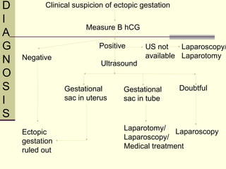
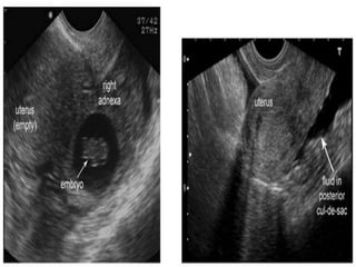
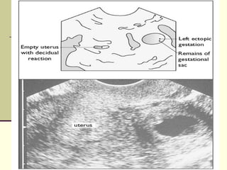
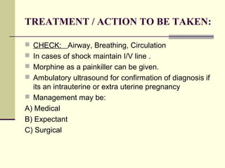
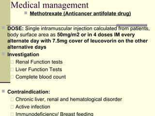
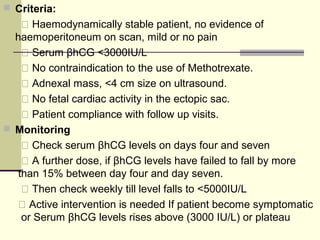
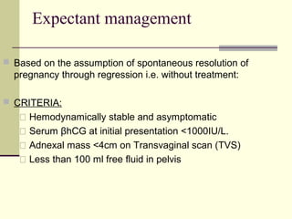
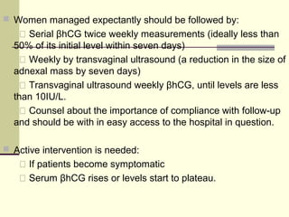
ECTOPIC
PREGNANCY
By :Darayus P.Gazder (DPG)
Roll Number: 18
Ectopic Pregnancy
 Definition: It is defined as an implantation of a
conceptus outside the normal uterine cavity
Or
 In theory, any mechanical or functional factors that
prevent or interfere with the passage of the fertilized
egg to the uterine cavity may be aetiological
factors for an ectopic pregnancy.
If not diagnosed on time, it may be life
threatening!!
 Incidence (11.1/1000 pregnancies)
Sites of ectopic pregnancy
ETIOLOGY ,INCIDENCE
 Mostly unknown
 Premature implantation (infection –damage to tubes-
delay of passage of fertilized ovum along tubes)
 One in 90 pregnancies is ectopic .
 A combined intra uterine and extra uterine pregnancy
is very rare and occurs in 1;40000 spontaneous
pregnancies and 1;1000 IVF pregnancies.
“ Heterotopic pregnancy ”
RISK FACTORS
 Congenital anomalies of the fallopian tubes
 Scarring due to a ruptured appendix
 Previous ectopic pregnancy
 Infections of the female genital organs
 History of PID
 Maternal age more than 35 yrs
 IUCD
 IVF- fertility treatment
 Tubal ligation/ reversal of tubal ligation
 Smoking
 Progesterone only contraceptive pills
OUTCOME
 Ectopic gestation mostly terminates between 6-10 weeks:
1) TUBAL ABORTION- 65 % of cases, usually in the fimbrial and
ampullary implantations. Repeated small hemorrhages from
the invaded area of the tubal wall detach the ovum, which dies
and :
1. Is absorbed completely
2. Is aborted completely through the tubal ostium into the
peritoneal cavity
3. Is absorbed incompletely with the result that the clot covered
conceptus distends the ostium
4. Forms a tubal blood mole
Outcome
2) TUBAL RUPTURE- 35 % of the cases more
common when the implantation is in the
isthmus. This may occur earlier then the 6th
week.
The trophoblast burrows deeply and erodes
the serosal coat of the tube, causing it to break
and rupture.
If this is seen on the mesenteric side of the
tube a broad ligament hematoma will form
Outcome
3) SECONDARY ABDOMINAL PREGNANCY
– very rarely the extruded ovum continues to
grow and attaches to the abdominal organs. A
few advance to term, the fetus dies and is
converted to a lithopaedion
Clinical Presentation:
 Unruptured ectopic: (TRIAD)
1) Lower abdominal pain, mild vaginal bleeding,
amenorrhea
 Ruptured ectopic:
1. Epigastric pain, severe lower abdominal pain,
shoulder tip pain, shock
CLINICAL PATTERNS:
1) Subacute presentation
 Mild lower abdominal pain
 Occasionally sharp pain and faintness
 Slight vaginal bleeding
 On examination lower abdominal tenderness, vaginal
examination may show a tender fornix or vague
mass.
 Acute collapse ( incase of ruptured or incomplete
tubal abortion)
 Cessation of symptoms (incase of complete abortion
with or without a pelvic hematocoele)
2) Acute presentation
 Sudden collapse (especially in isthmal tubal
pregnancy)
 Acute lower abdominal pain
 Fainting
 Signs of Internal haemorrhage (leading to pallor,
collapse, falling blood pressure, rapid weak pulse )
 Pain maybe either epigastric or shoulder tip pain
(referred)
 Abdominal examination may reveal tenderness with
some fullness and muscle guarding.
 Vaginal examination will reveal extreme cervical
tenderness.
 Vitals:
 BP, Pulse, Temperature, R/R
Laboratory Investigations:
 CBC(complete blood count),
 Blood Grouping and Cross Matching
 Beta hCG ( Levels double every 48 hrs a rise <66%=EP )
 Progesterone (Viable Preg: >79nmol/L, EP: <15.9 nmol/L)
 Ultrasound
 TVS (Transvaginal U/S Scan)
• Can detect 75-80% on initial scan and further 25% on follow
up.
• Transabdominal U/S has a limited role.
 Laparoscopy
(Advantage: Diagnostic and Therapeutic)
Clinical suspicion of ectopic gestation
Measure B hCG
Negative
Ectopic
gestation
ruled out
Positive
Gestational
sac in uterus
Gestational
sac in tube
Doubtful
Laparotomy/
Laparoscopy/
Medical treatment
Laparoscopy
Laparoscopy/
Laparotomy
US not
available
Ultrasound
D
I
A
G
N
O
S
I
S
TREATMENT / ACTION TO BE TAKEN:
 CHECK: Airway, Breathing, Circulation
 In cases of shock maintain I/V line .
 Morphine as a painkiller can be given.
 Ambulatory ultrasound for confirmation of diagnosis if
its an intrauterine or extra uterine pregnancy
 Management may be:
A) Medical
B) Expectant
C) Surgical
Medical management
 Methotrexate (Anticancer antifolate drug)
 DOSE: Single intramuscular injection calculated from patients,
body surface area as 50mg/m2 or in 4 doses IM every
alternate day with 7.5mg cover of leucovorin on the other
alternative days
 Investigation
 Renal Function tests
 Liver Function Tests
 Complete blood count
 Contraindication:
 Chronic liver, renal and hematological disorder
 Active infection
 Immunodeficiency/ Breast feeding
 Criteria:
 Haemodynamically stable patient, no evidence of
haemoperitoneum on scan, mild or no pain
 Serum βhCG <3000IU/L
 No contraindication to the use of Methotrexate.
 Adnexal mass, <4 cm size on ultrasound.
 No fetal cardiac activity in the ectopic sac.
 Patient compliance with follow up visits.
 Monitoring
 Check serum βhCG levels on days four and seven
 A further dose, if βhCG levels have failed to fall by more
than 15% between day four and day seven.
 Then check weekly till level falls to <5000IU/L
 Active intervention is needed If patient become symptomatic
or Serum βhCG levels rises above (3000 IU/L) or plateau
Expectant management
 Based on the assumption of spontaneous resolution of
pregnancy through regression i.e. without treatment:
 CRITERIA:
 Hemodynamically stable and asymptomatic
 Serum βhCG at initial presentation <1000IU/L.
 Adnexal mass <4cm on Transvaginal scan (TVS)
 Less than 100 ml free fluid in pelvis
 Women managed expectantly should be followed by:
 Serial βhCG twice weekly measurements (ideally less than
50% of its initial level within seven days)
 Weekly by transvaginal ultrasound (a reduction in the size of
adnexal mass by seven days)
 Transvaginal ultrasound weekly βhCG, until levels are less
than 10IU/L.
 Counsel about the importance of compliance with follow-up
and should be with in easy access to the hospital in question.
 Active intervention is needed:
 If patients become symptomatic
 Serum βhCG rises or levels start to plateau.
Surgical management
 If Serum βhCG is >3000IU/L
 Patient is symptomatic(or hemodynamically unstable)
Salpingotomy: Removal of conceptus with conservation of
tubes
Done in women with diseased contralateral tube and fertility
desired
Salpingectomy: Removal of tube in the presence of healthy
contra lateral tube
Can be done by two methods;
 Laparoscopy
 Laparotomy
CRITERIA
Laparoscopy
 Pt hemodynamically
stable
 No previous surgeries
 Unruptured ectopic
 Adnexal mass<4cm
Laparotomy
 Hemodynamically
unstable
 Previous surgeries
 Ruptured ectopic
 Expertise for laparoscopy
is not available
Prognosis:
 10-20 % are at risk of having a second
gestation with ectopic.
 60 % will become pregnant again with a
normal intrauterine gestation.
 Early careful monitoring is required in women
who become pregnant with previous history
of ectopic gestation. It is recommended to get
a TVS between 6-10 weeks .

Ectopic pregnancy

  • 1.
  • 2.
    Ectopic Pregnancy  Definition:It is defined as an implantation of a conceptus outside the normal uterine cavity Or  In theory, any mechanical or functional factors that prevent or interfere with the passage of the fertilized egg to the uterine cavity may be aetiological factors for an ectopic pregnancy. If not diagnosed on time, it may be life threatening!!  Incidence (11.1/1000 pregnancies)
  • 4.
  • 5.
    ETIOLOGY ,INCIDENCE  Mostlyunknown  Premature implantation (infection –damage to tubes- delay of passage of fertilized ovum along tubes)  One in 90 pregnancies is ectopic .  A combined intra uterine and extra uterine pregnancy is very rare and occurs in 1;40000 spontaneous pregnancies and 1;1000 IVF pregnancies. “ Heterotopic pregnancy ”
  • 7.
    RISK FACTORS  Congenitalanomalies of the fallopian tubes  Scarring due to a ruptured appendix  Previous ectopic pregnancy  Infections of the female genital organs  History of PID  Maternal age more than 35 yrs  IUCD  IVF- fertility treatment  Tubal ligation/ reversal of tubal ligation  Smoking  Progesterone only contraceptive pills
  • 8.
    OUTCOME  Ectopic gestationmostly terminates between 6-10 weeks: 1) TUBAL ABORTION- 65 % of cases, usually in the fimbrial and ampullary implantations. Repeated small hemorrhages from the invaded area of the tubal wall detach the ovum, which dies and : 1. Is absorbed completely 2. Is aborted completely through the tubal ostium into the peritoneal cavity 3. Is absorbed incompletely with the result that the clot covered conceptus distends the ostium 4. Forms a tubal blood mole
  • 10.
    Outcome 2) TUBAL RUPTURE-35 % of the cases more common when the implantation is in the isthmus. This may occur earlier then the 6th week. The trophoblast burrows deeply and erodes the serosal coat of the tube, causing it to break and rupture. If this is seen on the mesenteric side of the tube a broad ligament hematoma will form
  • 12.
    Outcome 3) SECONDARY ABDOMINALPREGNANCY – very rarely the extruded ovum continues to grow and attaches to the abdominal organs. A few advance to term, the fetus dies and is converted to a lithopaedion
  • 13.
    Clinical Presentation:  Unrupturedectopic: (TRIAD) 1) Lower abdominal pain, mild vaginal bleeding, amenorrhea  Ruptured ectopic: 1. Epigastric pain, severe lower abdominal pain, shoulder tip pain, shock
  • 14.
    CLINICAL PATTERNS: 1) Subacutepresentation  Mild lower abdominal pain  Occasionally sharp pain and faintness  Slight vaginal bleeding  On examination lower abdominal tenderness, vaginal examination may show a tender fornix or vague mass.  Acute collapse ( incase of ruptured or incomplete tubal abortion)  Cessation of symptoms (incase of complete abortion with or without a pelvic hematocoele)
  • 15.
    2) Acute presentation Sudden collapse (especially in isthmal tubal pregnancy)  Acute lower abdominal pain  Fainting  Signs of Internal haemorrhage (leading to pallor, collapse, falling blood pressure, rapid weak pulse )  Pain maybe either epigastric or shoulder tip pain (referred)  Abdominal examination may reveal tenderness with some fullness and muscle guarding.  Vaginal examination will reveal extreme cervical tenderness.
  • 16.
     Vitals:  BP,Pulse, Temperature, R/R Laboratory Investigations:  CBC(complete blood count),  Blood Grouping and Cross Matching  Beta hCG ( Levels double every 48 hrs a rise <66%=EP )  Progesterone (Viable Preg: >79nmol/L, EP: <15.9 nmol/L)  Ultrasound  TVS (Transvaginal U/S Scan) • Can detect 75-80% on initial scan and further 25% on follow up. • Transabdominal U/S has a limited role.  Laparoscopy (Advantage: Diagnostic and Therapeutic)
  • 17.
    Clinical suspicion ofectopic gestation Measure B hCG Negative Ectopic gestation ruled out Positive Gestational sac in uterus Gestational sac in tube Doubtful Laparotomy/ Laparoscopy/ Medical treatment Laparoscopy Laparoscopy/ Laparotomy US not available Ultrasound D I A G N O S I S
  • 20.
    TREATMENT / ACTIONTO BE TAKEN:  CHECK: Airway, Breathing, Circulation  In cases of shock maintain I/V line .  Morphine as a painkiller can be given.  Ambulatory ultrasound for confirmation of diagnosis if its an intrauterine or extra uterine pregnancy  Management may be: A) Medical B) Expectant C) Surgical
  • 21.
    Medical management  Methotrexate(Anticancer antifolate drug)  DOSE: Single intramuscular injection calculated from patients, body surface area as 50mg/m2 or in 4 doses IM every alternate day with 7.5mg cover of leucovorin on the other alternative days  Investigation  Renal Function tests  Liver Function Tests  Complete blood count  Contraindication:  Chronic liver, renal and hematological disorder  Active infection  Immunodeficiency/ Breast feeding
  • 22.
     Criteria:  Haemodynamicallystable patient, no evidence of haemoperitoneum on scan, mild or no pain  Serum βhCG <3000IU/L  No contraindication to the use of Methotrexate.  Adnexal mass, <4 cm size on ultrasound.  No fetal cardiac activity in the ectopic sac.  Patient compliance with follow up visits.  Monitoring  Check serum βhCG levels on days four and seven  A further dose, if βhCG levels have failed to fall by more than 15% between day four and day seven.  Then check weekly till level falls to <5000IU/L  Active intervention is needed If patient become symptomatic or Serum βhCG levels rises above (3000 IU/L) or plateau
  • 23.
    Expectant management  Basedon the assumption of spontaneous resolution of pregnancy through regression i.e. without treatment:  CRITERIA:  Hemodynamically stable and asymptomatic  Serum βhCG at initial presentation <1000IU/L.  Adnexal mass <4cm on Transvaginal scan (TVS)  Less than 100 ml free fluid in pelvis
  • 24.
     Women managedexpectantly should be followed by:  Serial βhCG twice weekly measurements (ideally less than 50% of its initial level within seven days)  Weekly by transvaginal ultrasound (a reduction in the size of adnexal mass by seven days)  Transvaginal ultrasound weekly βhCG, until levels are less than 10IU/L.  Counsel about the importance of compliance with follow-up and should be with in easy access to the hospital in question.  Active intervention is needed:  If patients become symptomatic  Serum βhCG rises or levels start to plateau.
  • 25.
    Surgical management  IfSerum βhCG is >3000IU/L  Patient is symptomatic(or hemodynamically unstable) Salpingotomy: Removal of conceptus with conservation of tubes Done in women with diseased contralateral tube and fertility desired Salpingectomy: Removal of tube in the presence of healthy contra lateral tube Can be done by two methods;  Laparoscopy  Laparotomy
  • 26.
    CRITERIA Laparoscopy  Pt hemodynamically stable No previous surgeries  Unruptured ectopic  Adnexal mass<4cm Laparotomy  Hemodynamically unstable  Previous surgeries  Ruptured ectopic  Expertise for laparoscopy is not available
  • 27.
    Prognosis:  10-20 %are at risk of having a second gestation with ectopic.  60 % will become pregnant again with a normal intrauterine gestation.  Early careful monitoring is required in women who become pregnant with previous history of ectopic gestation. It is recommended to get a TVS between 6-10 weeks .

Editor's Notes

  • #5 http://books.google.com.pk/books?id=zlB4T2ER4msC&amp;pg=PA82&amp;dq=risk+factors+of+ectopic+pregnancy&amp;hl=en&amp;sa=X&amp;ei=CrSCU_3ZN4mZO43SgfAK&amp;ved=0CEAQ6AEwBA#v=onepage&amp;q=risk%20factors%20of%20ectopic%20pregnancy&amp;f=false http://books.google.com.pk/books?id=KZLubBxJEwEC&amp;pg=PT2152&amp;dq=risk+factors+of+ectopic+pregnancy&amp;hl=en&amp;sa=X&amp;ei=CrSCU_3ZN4mZO43SgfAK&amp;ved=0CC8Q6AEwAQ#v=onepage&amp;q=risk%20factors%20of%20ectopic%20pregnancy&amp;f=false
  • #8 Pelvic inflammatory disease (PID) is a bacterial infection of the female reproductive system. Most cases of PID are caused by an infection in the vagina or the neck of the womb (cervix) that has spread to the reproductive organs higher up.Many different types of bacteria can cause PID, but most cases are the result of a chlamydia infection, which is a type of sexually transmitted infection that can be spread during unprotected sex. Chlamydia often exhibits no noticeable symptoms, so women may be unaware they are infected. But the bacteria can cause inflammation of the fallopian tubes, which is known as salpingitis. Salpingitis leads to a four-fold increase in the risk of having an ectopic pregnancy. 2)Having a previous history of ectopic pregnancy means that you have an increased risk of having one in the future compared to other women. Depending on the underlying factors, the risk of having another ectopic pregnancy is somewhere between 1 in 10 and 1 in 4. 3) If you have ever had surgery that involved your fallopian tubes, you have an increased risk of having an ectopic pregnancy. Types of surgery known to increase your risk include: female sterilisation (specifically a type of surgery known as tubal ligation or ‘tying the tubes’) – in around 1 in 200 cases the surgery fails, the woman becomes pregnant and it can result in an ectopic pregnancy earlier surgery to remove a previous ectopic pregnancy 4) Taking medication to stimulate ovulation (the release of an egg) can increase the risk of ectopic pregnancy by around four-fold. In addition, the type of fertility treatment known as in-vitro fertilisation (IVF) is not always successful and can accidentally result in an ectopic pregnancy. This occurs in around 1 in 22 cases of IVF. 5) The intrauterine device (IUD) and the intrauterine system (IUS) types of contraception are usually very effective in preventing pregnancy – estimated to be effective in around 99 out of 100 cases. But if a pregnancy does occur when using these types of contraception, it is more likely to be an ectopic pregnancy than a normal pregnancy.There is also a risk that if you take emergency contraception and it fails to work, any subsequent pregnancy could be an ectopic pregnancy.
  • #19  An extrauterine gestational sac containing an embryo or yolk sac is also diagnostic and is seen in a significant percentage of ectopic pregnancies (Figure 13).8 Free fluid in the posterior pelvic cul-de-sac or in other intraperitoneal sites is highly suggestive of ectopic pregnancy (Figure 14).88