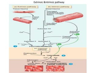
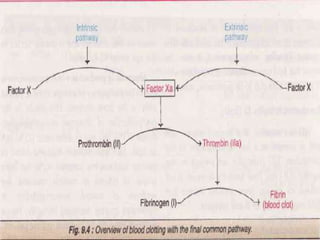
Blood coagulation cascade
Introduction
Hemostasis:
• Hemostasis is a sequence of responses that
stops bleeding. When blood vessels are
damaged or ruptured.
• The hemostatic response must be quick
localized to the region of damage, and carefully
controlled in order to be effective.
• Three mechanisms reduce blood loss:
(1)vascular spasm, (2) platelet plug formation,
and (3) blood clotting.
Vascular Spasm: When arteries or arterioles are
damaged, the circularly arranged smooth muscle
in their walls contracts immediately, a reaction
called vascular spasm.
Platelet Plug Formation:Considering their small
size, platelets store an impressive array of
chemicals. Within many vesicles are clotting
factors,ADP, ATP, Ca2, and serotonin. Also present
are enzymes that produce thromboxane A2, a
prostaglandin; fibrin-stabilizing factor, which
helps to strengthen a blood clot.
Blood clotting
• Normally, blood remains in its liquid form as long as it stays within
its vessels.
• If it is drawn from the body, however,it thickens and forms a gel.
• The gel separates from the liquid.The straw-colored liquid, called
serum, is simply blood plasma minus the clotting proteins. The
gel is called a clot.
• It consists of a network of insoluble protein fibers called fibrin in
which the formed elements of blood are trapped,The process of
gel formation, called clotting or coagulation.
• The series of chemical reactions that culminates in formation of
fibrin threads. If blood clots too easily, the result can be
thrombosis—clotting in an undamaged blood vessel. If the blood
takes too long to clot.
• Clotting involves several substances known as
clotting factors.
• These factors include calcium ions (Ca2),several
inactive enzymes that are synthesized by
hepatocytes(liver cells) and released into the
bloodstream, and various molecules associated with
platelets or released by damaged tissues.
• Clotting is a complex cascade of enzymatic reactions
in which each clotting factor activates many
molecules of the next one in a fixed sequence.
Finally, a large quantity of product (the insoluble
protein fibrin) is formed.
Clotting can be divided in 3 stages
• The two pathways called the extrinsic pathways and
the intrinsic pathway
• These two pathways lead to the formation of
prothrombinase.
• Once prothrombinase is formed the steps involved in
the next two stages of clotting are same for both
extrinsic & intrinsic pathway.
• In 2nd stage- prothrombinase converts prothrombin
in to thrombin.
• Thrombin converts soluble fibrinogen into fibrin.
• Fibrin forms the threads of the blood clotting.
Extrinsic &intrinsic pathway
The extrinsic pathway
• The extrinsic pathway of blood clotting has fewer
steps than the intrinsic pathway.
• It occurs rapidly—within a matter of seconds if
trauma is severe. It is so named because a
tissue,protein called tissue factor (TF), also known as
thromboplastin.
• TF is a complex mixture of lipoproteins and
phospholipids released from the surfaces of
damaged cells. In the presence of Ca2, TF begins a
sequence of reactions that ultimately activates
clotting factor X.
• Once factor X is activated, it combines with factor V
in the presence of Ca2+ to form the active enzyme
prothrombinase, completing the extrinsic pathway.
The intrinsic pathway
• The intrinsic pathway of blood clotting is more
complex than the extrinsic pathway.
• It occurs more slowly, usually requiring several
minutes.
• The intrinsic pathway is so named because its
activators are either in direct contact with blood.
• If endothelial cells become or damaged blood
can come in contact with collagen fibers
• The endothelium cell of the blood vessel. In
addition,trauma to endothelial cells causes
damage to platelets, resulting in the release of
phospholipids by the platelets Contact with
collagen fibers.
• Activates clotting factor XII
• Which begins a sequence of reactions that
eventually activates clotting factor X.
• Platelet phospholipids and Ca2+ can also
participate in the activation of factor X.
• Once factor X is activated, it combines with
factor V to form the active enzyme
prothrombinase completing the intrinsic
pathway.
Anticoagulants
• In some thromboembolic conditions, it is desirable to
delay the coagulation process. Various
anticoagulants have been developed for this
purpose. The ones most useful clinically are heparin
and the coumarins.
• Heparin :Commercial heparin is extracted from
several different animal tissues and prepared in
almost pure form.
• Injection of relatively small quantities, about 0.5 to1
mg/kg of body weight, causes the blood-clotting
time to increase from a normal of about 6 minutes to
30 or more minutes
• The action of heparin lasts about 1.5 to 4 hours.The injected
heparin is destroyed by an enzyme in the blood known as
heparinase.
• Coumarins:When a coumarin, such as warfarin, is given to a
patient, the plasma levels of prothrombin and Factors VII, IX, and X,
all formed by the liver, begin to fall, indicating that warfarin has a
potent depressant effect on liver formation of these compounds.
Warfarin causes this effect by competing with vitamin K for reactive
sites in the enzymatic processes for formation of prothrombin and
the other three clotting factors, thereby blocking the action of
vitamin K.
• After administration of an effective dose of warfarin, the coagulant
activity of the blood decreases to about 50 per cent of normal by
the end of 12 hours and to about 20 per cent of normal by the end
of 24 hours.In other words, the coagulation process is not blocked
immediately but must await the natural consumption of the
prothrombin and the other affected coagulation factors already
present in the plasma. Normal coagulation usually returns 1 to 3
days after discontinuing coumarin therapy.
Blood coagulation cascade

Blood coagulation cascade

  • 1.
  • 2.
    Introduction Hemostasis: • Hemostasis isa sequence of responses that stops bleeding. When blood vessels are damaged or ruptured. • The hemostatic response must be quick localized to the region of damage, and carefully controlled in order to be effective. • Three mechanisms reduce blood loss: (1)vascular spasm, (2) platelet plug formation, and (3) blood clotting.
  • 3.
    Vascular Spasm: Whenarteries or arterioles are damaged, the circularly arranged smooth muscle in their walls contracts immediately, a reaction called vascular spasm. Platelet Plug Formation:Considering their small size, platelets store an impressive array of chemicals. Within many vesicles are clotting factors,ADP, ATP, Ca2, and serotonin. Also present are enzymes that produce thromboxane A2, a prostaglandin; fibrin-stabilizing factor, which helps to strengthen a blood clot.
  • 4.
    Blood clotting • Normally,blood remains in its liquid form as long as it stays within its vessels. • If it is drawn from the body, however,it thickens and forms a gel. • The gel separates from the liquid.The straw-colored liquid, called serum, is simply blood plasma minus the clotting proteins. The gel is called a clot. • It consists of a network of insoluble protein fibers called fibrin in which the formed elements of blood are trapped,The process of gel formation, called clotting or coagulation. • The series of chemical reactions that culminates in formation of fibrin threads. If blood clots too easily, the result can be thrombosis—clotting in an undamaged blood vessel. If the blood takes too long to clot.
  • 6.
    • Clotting involvesseveral substances known as clotting factors. • These factors include calcium ions (Ca2),several inactive enzymes that are synthesized by hepatocytes(liver cells) and released into the bloodstream, and various molecules associated with platelets or released by damaged tissues. • Clotting is a complex cascade of enzymatic reactions in which each clotting factor activates many molecules of the next one in a fixed sequence. Finally, a large quantity of product (the insoluble protein fibrin) is formed.
  • 7.
    Clotting can bedivided in 3 stages • The two pathways called the extrinsic pathways and the intrinsic pathway • These two pathways lead to the formation of prothrombinase. • Once prothrombinase is formed the steps involved in the next two stages of clotting are same for both extrinsic & intrinsic pathway. • In 2nd stage- prothrombinase converts prothrombin in to thrombin. • Thrombin converts soluble fibrinogen into fibrin. • Fibrin forms the threads of the blood clotting.
  • 8.
  • 10.
    The extrinsic pathway •The extrinsic pathway of blood clotting has fewer steps than the intrinsic pathway. • It occurs rapidly—within a matter of seconds if trauma is severe. It is so named because a tissue,protein called tissue factor (TF), also known as thromboplastin. • TF is a complex mixture of lipoproteins and phospholipids released from the surfaces of damaged cells. In the presence of Ca2, TF begins a sequence of reactions that ultimately activates clotting factor X. • Once factor X is activated, it combines with factor V in the presence of Ca2+ to form the active enzyme prothrombinase, completing the extrinsic pathway.
  • 11.
    The intrinsic pathway •The intrinsic pathway of blood clotting is more complex than the extrinsic pathway. • It occurs more slowly, usually requiring several minutes. • The intrinsic pathway is so named because its activators are either in direct contact with blood. • If endothelial cells become or damaged blood can come in contact with collagen fibers • The endothelium cell of the blood vessel. In addition,trauma to endothelial cells causes damage to platelets, resulting in the release of phospholipids by the platelets Contact with collagen fibers.
  • 12.
    • Activates clottingfactor XII • Which begins a sequence of reactions that eventually activates clotting factor X. • Platelet phospholipids and Ca2+ can also participate in the activation of factor X. • Once factor X is activated, it combines with factor V to form the active enzyme prothrombinase completing the intrinsic pathway.
  • 13.
    Anticoagulants • In somethromboembolic conditions, it is desirable to delay the coagulation process. Various anticoagulants have been developed for this purpose. The ones most useful clinically are heparin and the coumarins. • Heparin :Commercial heparin is extracted from several different animal tissues and prepared in almost pure form. • Injection of relatively small quantities, about 0.5 to1 mg/kg of body weight, causes the blood-clotting time to increase from a normal of about 6 minutes to 30 or more minutes
  • 14.
    • The actionof heparin lasts about 1.5 to 4 hours.The injected heparin is destroyed by an enzyme in the blood known as heparinase. • Coumarins:When a coumarin, such as warfarin, is given to a patient, the plasma levels of prothrombin and Factors VII, IX, and X, all formed by the liver, begin to fall, indicating that warfarin has a potent depressant effect on liver formation of these compounds. Warfarin causes this effect by competing with vitamin K for reactive sites in the enzymatic processes for formation of prothrombin and the other three clotting factors, thereby blocking the action of vitamin K. • After administration of an effective dose of warfarin, the coagulant activity of the blood decreases to about 50 per cent of normal by the end of 12 hours and to about 20 per cent of normal by the end of 24 hours.In other words, the coagulation process is not blocked immediately but must await the natural consumption of the prothrombin and the other affected coagulation factors already present in the plasma. Normal coagulation usually returns 1 to 3 days after discontinuing coumarin therapy.