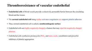
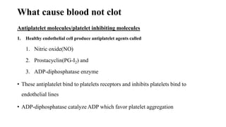
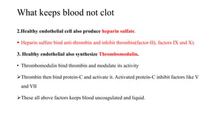
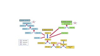
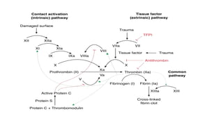
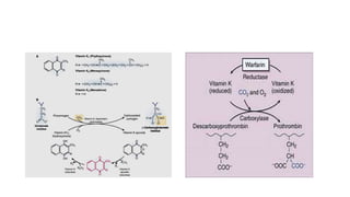
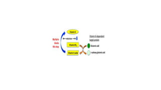
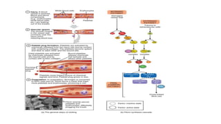
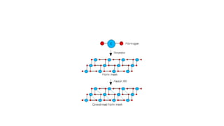
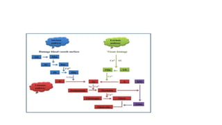
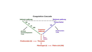
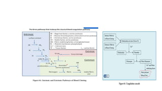
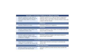
Coagulation pathway
Thromboresistance of vascular endothelial
• Endothelial cells of blood vessels provide a selectively permeable barrier between the circulating
blood and the tissues.
• The normal endothelial cell lining neither activates coagulation nor supports platelet adhesion
• Thus, normal endothelial cell is called a nonthrombogenic surface
• Endothelial cells are highly negatively charged, a feature that may repel the negatively charged
platelets.
• Endothelial cells synthesize prostacyclin (PGI2) and nitric oxide, vasodilators and powerful
inhibitors of platelet aggregation.
What cause blood not clot
Antiplatelet molecules/platelet inhibiting molecules
1. Healthy endothelial cell produce antiplatelet agents called
1. Nitric oxide(NO)
2. Prostacyclin(PG-I2) and
3. ADP-diphosphatase enzyme
• These antiplatelet bind to platelets receptors and inhibits platelets bind to
endothelial lines
• ADP-diphosphatase catalyze ADP which favor platelet aggregation
What keeps blood not clot
2.Healthy endothelial cell also produce heparin sulfate.
 Heparin sulfate bind anti-thrombin and inhibit thrombin(factor-II), factors IX and X).
3. Healthy endothelial also synthesize Thrombomodulin.
• Thrombomodulin bind thrombin and modulate its activity
Thrombin then bind protein-C and activate it. Activated protein-C inhibit factors like V
and VII
These all above factors keeps blood uncoagulated and liquid.
What make blood not clot
4.Healthy endothelium produce tissue plasminogen activator(T.P.A)
• Tissue plasminogen activator bind plasminogen released by liver and convert it to plasmin.
• Plasmin take the circulating fibrin and degrade it to inhibit coagulation
Coagulation initiation
• When there is a damage to endothelial cell the antiplatelet or anticoagulation
properties of endothelial cell will be inhibited
• Damaged endothelial cells start to produce von Willebrand factor(VWF) that stick with
underline collagen
• VWB bind with platelet through its receptors and form process called platelet adherence
• Once vwf bind to platelet, the platelet activated, and its PLC cleaves membrane
phospholipid to arachidonic then TXA2 by cyclooxygenase.
• TXA2 has two function (vasoconstrictor and platelet aggregator)
Coagulation initiation
• Ca2+ used to sticks coagulating factors to each others and into phospholipid
membrane.
• Because both coagulation factors and phospholipid membranes are negatively
charged that can repeal each other and can't form aggregation.
• Aspirin inhibit COX and prevent platelet aggregation.
• alpha granules of platelet release PDGF that stimulate collagen synthesis to seal
damage
• Platelet aggregation is loose, and it is primary platelet formation
Hemostasis
• Haemostasis is the body's normal physiological response for the prevention and
stopping of bleeding.
• It is a complex series of events involving both cellular and plasma components
• The actual mechanism of haemostasis can be divided into two main components.
• primary haemostasis
• secondary haemostasis
Components of Hemostasis
1.Primary haemostasis – platelet aggregation at the site of injury to form a
temporary platelet plug.
• There are four phases involved in primary haemostasis:
1. vasoconstriction,
2. platelet adhesion,
3. platelet activation,
4. platelet aggregation.
2. Secondary haemostasis – formation of stable fibrin clot
Coagulation
• Is a complex physiological process by which soluble fibrinogen convert to
insoluble fibrin
• Clotting can prevent us from bleeding to death and protect us from the entry of
bacteria and viruses.
• The coagulation factors are present in circulation as inactive zymogen forms.
• They are converted to their active forms only when the clotting process is
initiated.
• This would prevent unnecessary intravascular coagulation.
Coagulation cascade
• The coagulation cascade is often classified into three pathways—
• the extrinsic pathway,
• the intrinsic pathway, and
• the common pathway
Intrinsic pathway
• It's "intrinsic" because it's initiated by a factor inside the blood vessel.
• It initiated when inactive factor XII contact with negatively charged collagen of exposed
endothelial cell
• Endothelial collagen is only exposed when endothelial damage occurs.
• Factor XIIA acts as a catalyst to activate factor XI to Factor XIA
• Factor XIA then goes on to activate factor IX to factor IXA.
• Factor IXA goes on to serve as a catalyst for turning factor X into factor Xa
• This process is known as a cascade.
• The intrinsic pathway is clinically measured as the partial thromboplastin time (PTT).
Extrinsic pathway
• The extrinsic pathway is triggered by a chemical called tissue factor that is
released by damaged cells.
• This pathway is "extrinsic" because it's initiated by a factor outside the blood
vessels.
• Tissue factor(TF) called factor III released from damaged endothelial .
• The source of TF is not circulating components that is why called extrinsic
• This TF activate factor VIIb to active form VIIa
Extrinsic pathway
• Activated factor VII activate factor X and form the rest of common pathway
• The extrinsic pathway is clinically measured as the prothrombin time (PT
• The activated factor XA, activate prothrombin (IIB) to thrombin(IIA)
• Activated factor II (thrombin) activate factor V, then activated factor V back
activate factor II
• Thrombin activate fibrinogen to fibrin(Ib-Ia)
• Common pathways: XI,II,V,I and XIII
Intrinsic vs extrinsic
• The intrinsic pathway consists of factors I, II, IX, X, XI, and XII. Respectively,
each one is named, fibrinogen, prothrombin, Christmas factor, Stuart-Prower
factor, plasma thromboplastin, and Hageman factor
• Extrinsic pathway consists of factors I, II, VII, and X.
• The common pathway consists of factors I, II, V, VIII, X
• The following are serine proteases: factors II, VII, IX, X, XI and XII.
• These are not serine proteases: factors V, VIII, XIII
• The factors circulate through the bloodstream as zymogens and are activated into
serine proteases.
• These serine proteases act as a catalyst to cleave the next zymogen into more
serine proteases and ultimately activate fibrinogen.
Negative Feedback
• To prevent over-coagulation, which causes thrombosis, there are certain processes to keep
the coagulation cascade in check.
• As thrombin acts as a procoagulant, it also acts as a negative feedback by activating
plasminogen to plasmin and stimulating the production of antithrombin (AT).
• Plasmin acts directly on the fibrin mesh and breaks it down. AT decreases the production
of thrombin from prothrombin and decreases the amount of activated factor X.
• Protein C and S also act to prevent coagulation, mainly by inactivating factors V and VIII
Factor Complexes
• Factors VII, IX, X, and prothrombin contain a domain in which one or more glutamate residues are
carboxylated to γ-carboxyglutamate in a reaction that requires vitamin K
• Prothrombin and factor X both contain 10 or more γ-carboxyglutamate residues that bind Ca2+.
• The Ca2+ forms a coordination complex with the negatively charged platelet membrane
phospholipids and the γ-carboxyglutamates.
• This reduce the repeal b/n carboxylated and membrane of platelet, then localize the complex
assembly and thrombin formation to the platelet surface
Vitamin K Requirement for Blood Coagulation
• The formation of the γ-carboxyglutamate residues on blood coagulation factors takes place in the
hepatocyte before release of the protein
• Within the hepatocyte, vitamin K is reduced to form vitamin KH2 by a microsomal quinone
reductase
• Vitamin KH2 is a cofactor for carboxylases that add a carboxyl group to the appropriate glutamate
residues in the proenzyme to form the carboxylated proenzyme
• In the same reaction, vitamin K is converted to vitamin K epoxide.
• To recover active vitamin KH2, vitamin K is first reduced to the quinone form by vitamin K
epoxide reductase, and then to the active hydroquinone form.
Warfarin as anticoagulant
• Warfarin (Coumadin) is a slow- and long-acting blood anticoagulant with a structure resembling
that of vitamin K.
• The structural similarity allows the compound to compete with vitamin K and prevent γ-
carboxylation of glutamate residues in factors II, VII, IX, and X and proteins C and S.
• The noncarboxylated blood clotting protein precursors increase in both the blood and plasma, but
they are unable to promote blood coagulation because they cannot bind calcium
• and thus cannot bind to their phospholipid membrane sites of activation.
• Cofactor Va contains a binding site for both factor Xa and prothrombin, the zymogen
substrate of factor Xa.
• Upon binding to the factor Va–platelet complex, prothrombin undergoes a conformational
change, rendering it more susceptible to enzymatic cleavage.
• Binding of factor Xa to the factor Va–prothrombin–platelet complex allows the
prothrombin-to-thrombin conversion
• Complex assembly accelerates the rate of this conversion 10,000- to 15,000-fold as
compared with non–complex formation
Coagulation pathway.pptx

Coagulation pathway.pptx

  • 1.
  • 2.
    Thromboresistance of vascularendothelial • Endothelial cells of blood vessels provide a selectively permeable barrier between the circulating blood and the tissues. • The normal endothelial cell lining neither activates coagulation nor supports platelet adhesion • Thus, normal endothelial cell is called a nonthrombogenic surface • Endothelial cells are highly negatively charged, a feature that may repel the negatively charged platelets. • Endothelial cells synthesize prostacyclin (PGI2) and nitric oxide, vasodilators and powerful inhibitors of platelet aggregation.
  • 3.
    What cause bloodnot clot Antiplatelet molecules/platelet inhibiting molecules 1. Healthy endothelial cell produce antiplatelet agents called 1. Nitric oxide(NO) 2. Prostacyclin(PG-I2) and 3. ADP-diphosphatase enzyme • These antiplatelet bind to platelets receptors and inhibits platelets bind to endothelial lines • ADP-diphosphatase catalyze ADP which favor platelet aggregation
  • 4.
    What keeps bloodnot clot 2.Healthy endothelial cell also produce heparin sulfate.  Heparin sulfate bind anti-thrombin and inhibit thrombin(factor-II), factors IX and X). 3. Healthy endothelial also synthesize Thrombomodulin. • Thrombomodulin bind thrombin and modulate its activity Thrombin then bind protein-C and activate it. Activated protein-C inhibit factors like V and VII These all above factors keeps blood uncoagulated and liquid.
  • 5.
    What make bloodnot clot 4.Healthy endothelium produce tissue plasminogen activator(T.P.A) • Tissue plasminogen activator bind plasminogen released by liver and convert it to plasmin. • Plasmin take the circulating fibrin and degrade it to inhibit coagulation
  • 6.
    Coagulation initiation • Whenthere is a damage to endothelial cell the antiplatelet or anticoagulation properties of endothelial cell will be inhibited • Damaged endothelial cells start to produce von Willebrand factor(VWF) that stick with underline collagen • VWB bind with platelet through its receptors and form process called platelet adherence • Once vwf bind to platelet, the platelet activated, and its PLC cleaves membrane phospholipid to arachidonic then TXA2 by cyclooxygenase. • TXA2 has two function (vasoconstrictor and platelet aggregator)
  • 7.
    Coagulation initiation • Ca2+used to sticks coagulating factors to each others and into phospholipid membrane. • Because both coagulation factors and phospholipid membranes are negatively charged that can repeal each other and can't form aggregation. • Aspirin inhibit COX and prevent platelet aggregation. • alpha granules of platelet release PDGF that stimulate collagen synthesis to seal damage • Platelet aggregation is loose, and it is primary platelet formation
  • 8.
    Hemostasis • Haemostasis isthe body's normal physiological response for the prevention and stopping of bleeding. • It is a complex series of events involving both cellular and plasma components • The actual mechanism of haemostasis can be divided into two main components. • primary haemostasis • secondary haemostasis
  • 9.
    Components of Hemostasis 1.Primaryhaemostasis – platelet aggregation at the site of injury to form a temporary platelet plug. • There are four phases involved in primary haemostasis: 1. vasoconstriction, 2. platelet adhesion, 3. platelet activation, 4. platelet aggregation. 2. Secondary haemostasis – formation of stable fibrin clot
  • 10.
    Coagulation • Is acomplex physiological process by which soluble fibrinogen convert to insoluble fibrin • Clotting can prevent us from bleeding to death and protect us from the entry of bacteria and viruses. • The coagulation factors are present in circulation as inactive zymogen forms. • They are converted to their active forms only when the clotting process is initiated. • This would prevent unnecessary intravascular coagulation.
  • 11.
    Coagulation cascade • Thecoagulation cascade is often classified into three pathways— • the extrinsic pathway, • the intrinsic pathway, and • the common pathway
  • 12.
    Intrinsic pathway • It's"intrinsic" because it's initiated by a factor inside the blood vessel. • It initiated when inactive factor XII contact with negatively charged collagen of exposed endothelial cell • Endothelial collagen is only exposed when endothelial damage occurs. • Factor XIIA acts as a catalyst to activate factor XI to Factor XIA • Factor XIA then goes on to activate factor IX to factor IXA. • Factor IXA goes on to serve as a catalyst for turning factor X into factor Xa • This process is known as a cascade. • The intrinsic pathway is clinically measured as the partial thromboplastin time (PTT).
  • 14.
    Extrinsic pathway • Theextrinsic pathway is triggered by a chemical called tissue factor that is released by damaged cells. • This pathway is "extrinsic" because it's initiated by a factor outside the blood vessels. • Tissue factor(TF) called factor III released from damaged endothelial . • The source of TF is not circulating components that is why called extrinsic • This TF activate factor VIIb to active form VIIa
  • 15.
    Extrinsic pathway • Activatedfactor VII activate factor X and form the rest of common pathway • The extrinsic pathway is clinically measured as the prothrombin time (PT • The activated factor XA, activate prothrombin (IIB) to thrombin(IIA) • Activated factor II (thrombin) activate factor V, then activated factor V back activate factor II • Thrombin activate fibrinogen to fibrin(Ib-Ia) • Common pathways: XI,II,V,I and XIII
  • 16.
    Intrinsic vs extrinsic •The intrinsic pathway consists of factors I, II, IX, X, XI, and XII. Respectively, each one is named, fibrinogen, prothrombin, Christmas factor, Stuart-Prower factor, plasma thromboplastin, and Hageman factor • Extrinsic pathway consists of factors I, II, VII, and X. • The common pathway consists of factors I, II, V, VIII, X • The following are serine proteases: factors II, VII, IX, X, XI and XII. • These are not serine proteases: factors V, VIII, XIII • The factors circulate through the bloodstream as zymogens and are activated into serine proteases. • These serine proteases act as a catalyst to cleave the next zymogen into more serine proteases and ultimately activate fibrinogen.
  • 18.
    Negative Feedback • Toprevent over-coagulation, which causes thrombosis, there are certain processes to keep the coagulation cascade in check. • As thrombin acts as a procoagulant, it also acts as a negative feedback by activating plasminogen to plasmin and stimulating the production of antithrombin (AT). • Plasmin acts directly on the fibrin mesh and breaks it down. AT decreases the production of thrombin from prothrombin and decreases the amount of activated factor X. • Protein C and S also act to prevent coagulation, mainly by inactivating factors V and VIII
  • 19.
    Factor Complexes • FactorsVII, IX, X, and prothrombin contain a domain in which one or more glutamate residues are carboxylated to γ-carboxyglutamate in a reaction that requires vitamin K • Prothrombin and factor X both contain 10 or more γ-carboxyglutamate residues that bind Ca2+. • The Ca2+ forms a coordination complex with the negatively charged platelet membrane phospholipids and the γ-carboxyglutamates. • This reduce the repeal b/n carboxylated and membrane of platelet, then localize the complex assembly and thrombin formation to the platelet surface
  • 20.
    Vitamin K Requirementfor Blood Coagulation • The formation of the γ-carboxyglutamate residues on blood coagulation factors takes place in the hepatocyte before release of the protein • Within the hepatocyte, vitamin K is reduced to form vitamin KH2 by a microsomal quinone reductase • Vitamin KH2 is a cofactor for carboxylases that add a carboxyl group to the appropriate glutamate residues in the proenzyme to form the carboxylated proenzyme • In the same reaction, vitamin K is converted to vitamin K epoxide. • To recover active vitamin KH2, vitamin K is first reduced to the quinone form by vitamin K epoxide reductase, and then to the active hydroquinone form.
  • 22.
    Warfarin as anticoagulant •Warfarin (Coumadin) is a slow- and long-acting blood anticoagulant with a structure resembling that of vitamin K. • The structural similarity allows the compound to compete with vitamin K and prevent γ- carboxylation of glutamate residues in factors II, VII, IX, and X and proteins C and S. • The noncarboxylated blood clotting protein precursors increase in both the blood and plasma, but they are unable to promote blood coagulation because they cannot bind calcium • and thus cannot bind to their phospholipid membrane sites of activation.
  • 24.
    • Cofactor Vacontains a binding site for both factor Xa and prothrombin, the zymogen substrate of factor Xa. • Upon binding to the factor Va–platelet complex, prothrombin undergoes a conformational change, rendering it more susceptible to enzymatic cleavage. • Binding of factor Xa to the factor Va–prothrombin–platelet complex allows the prothrombin-to-thrombin conversion • Complex assembly accelerates the rate of this conversion 10,000- to 15,000-fold as compared with non–complex formation