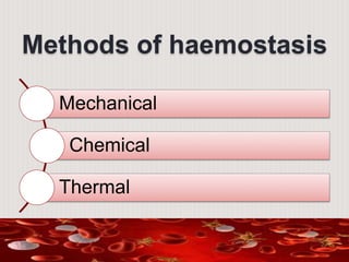
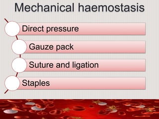
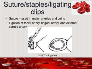
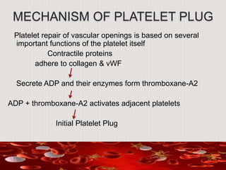
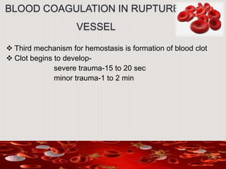
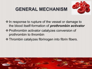
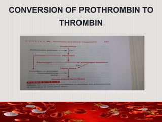
Dr.Niti Sarawgi
II MDS
HAEMOSTASIS
Contents
• Introduction
• Events in haemostasis
• Intrinsic pathway & extrinsic pathway
• Mechanical haemostasis
• Chemical haemostasis
• Thermal haemostasis
• Conclusion
INTRODUCTION
Hemostasis is the process of forming clots in
the walls of damaged blood vessels and
preventing blood loss while maintaining blood in
the fluid state within the vascular system.
EVENTS IN
HAEMOSTASIS
Haemostasis means prevention of ‘Blood
Loss’. Haemostasis is achieved by several
mechanism:-
Vascular constriction
Formation of platelet plug
Formation of blood clot
Growth of fibrous tissue into the clot.
VASCULAR CONSTRICTION
The contraction results from:-
 Local myogenic spasms
 Local autacoid factors
 Nervous reflexes
Platelets release, thromboxane-A2 which is responsible
for vasoconstriction of smaller vessels.
The more severely a vessel is traumatized, the greater the
degree of vascular spasm.
Mechanism
Vasoconstriction is as a result of increased Ca ion
conc. in smooth muscles.
Hormonal components :
Circulating epinephrine & activation of sympathetic nervous
system.
Interact with cell surface adrenergic receptors
Signal transduction
Increased intercellular Ca from sarcoplasmic reticulum
Ca-calmodulin complex
Activates myosin light- chain kinase
stimulate cross bridge cycle
• Factors :
• -Exogenous – temperature (cold)
• -Endogenous-Autonomic nervous system, hormones and
mechanisms inherent to vasculature (myogenic
response)
-Medications: antihistamines , amphetamines , cocaine
FORMATION OF
PLATELET PLUG
 PHYSICAL AND CHEMICAL CHARACTERISTICS OF
PLATELETS
 MECHANISM OF PLATELET PLUG
• Platelet adhesion
• Platelet activation
• Platelet aggregation
• Formation of temporary haemostatic plug
NORMAL ARCHITECTURE OF
A BLOOD VESSEL
PLATELETS
 Platelets are enucleate cells
 1-4 micro meters in size
 Normal blood concentration of
platelets is 1.5L-3L/micro liters
 Formed in bone marrow from
megakaryocytes
 T-1/2 is 8-12 days
 Eliminated from the circulation
by tissue macrophages.
CONTD…
Platelet cytoplasm contains active factors such
as:-
 Actin and myosin molecules
Thrombosthenin is another contractile protein which can
cause platelets to contract.
 ER and the Golgi apparatus
 Mitochindria and enzyme system.
PLATELET-CELL MEMBRANE
 Surface coat of glycoproteins
a) repulse adherence to normal endothelium
b) but adherence to injured vessel wall i.e., endothelial
cells, and even more to the exposed collagen from deep
with in the vessel wall
 Large amounts of phospholipids – activates multiple
stages in blood-clotting process.
PLATELET ADHESON
PLATELET ACTIVATION
PLATELET
AGGREGATION
• The activated sticky platelets stick to each other form
Platelet aggregation
• TXA2 powerful vasoconsrictor &mobilisation Ca from
dense tubular system –activates myosin -actin cause
release reaction –Platelet aggregation
• This involves a series of self sustaining events
• Leads to formation of platelet plug
MECHANISM OF PLATELET PLUG
Platelet repair of vascular openings is based on several
important functions of the platelet itself
Contractile proteins
adhere to collagen & vWF
Secrete ADP and their enzymes form thromboxane-A2
ADP + thromboxane-A2 activates adjacent platelets
Initial Platelet Plug
BLOOD COAGULATION IN RUPTURED
VESSEL
 Third mechanism for hemostasis is formation of blood clot
 Clot begins to develop-
severe trauma-15 to 20 sec
minor trauma-1 to 2 min
MECHANISM OF BLOOD COAGULATION
BASIC
THEORY
GENERAL
MECHANISM
GENERAL MECHANISM
In response to rupture of the vessel or damage to
the blood itself-formation of prothrombin activator
Prothrombin activator catalyzes conversion of
prothrombin to thrombin
Thrombin catalyzes fibrinogen into fibrin fibers.
CONVERSION OF PROTHROMBIN TO
THROMBIN
Contd…
 Then Fibrin Stabilizing Factor is released from platelets
entrapped in the clot
 Same thrombin that causes fibrin formation activates the Fibrin
Stabilizing Factor, before FSF can have effect on fibrin fibers
 Activated FSF forms strong covalent bond b/w monomer of the
fibrin and multiple cross linkage b/w adjacent fibrin fibers.
BLOOD CLOT
 The clot is a meshwork
 Fibrin fibers also adhere to damaged surfaces of blood
vessels.
CLOT RETRACTION-SERUM
 Contraction causes expression of fluid from clot-serum
 Platelet contractile proteins contributes greatly to the clot
retraction by activating Platelet Thrombosthenin
 They also helps compress fibrin mesh work into smaller mass.
 As the clot contracts, the edges are further pulled together,
contributing ultimate state of Hemostasis.
VISCIOUS CIRCLE OF CLOT
FORMATION
 Positive feed back for clot formation
 Most important cause of this is the the proteolytic action
of thrombin
 For instance, thrombin has a direct proteolytic effect on
prothrombin itself, hence more of thrombin formation
 Critical amount of thrombin causes more blood clotting
and hence further more production of thrombin and
hence called a vicious circle of clotting.
INITIATION OF COAGUALTION
FORMATION OF PROTHROMBIN ACTIVATOR
These mechanisms are set into play by:-
 Trauma to the vascular wall and the adjacent tissues
 Contact of the blood with damaged endothelial cells
 Prothrombin activator is generally considered to be formed in
these ways-
a) Extrinsic pathway for initiating blood clotting
b) Intrinsic pathway for initiating blood clotting
CLOTTING FACTORS
• Factor I (fibrinogen)-
Fibrinogen is a soluble plasma protein (MW 330000) which
is acted upon by thrombin to form insoluble fibrin clot.
• Factor II (prothrombin)-
Inactive precursor of thrombin is formed in liver.
• Factor III (tissue factor, tissue extract, thromboplastin)-
This converts prothrombin in the presence of factors V, VII,
and Xa, Calcium, and phospholipid.
.
 Factor IV (calcium)-
 Factor V (labile factor, thrombogene or proaccelerin)-
This factor is reqd. for the conversion of prothrombin to
thrombin by tissue extract and plasma factors.
 Factor VII (stable factor, autoprothrombin I)-
Factor VII is reqd. for the formation of prothrombin activator
by tissue extract.
 Factor VIII (antihemophilic globulin [AHG],
antihemophilic factor)-
Factor VIII is reqd. for the formation of prothrombin activator
from blood constituents; it’s consumed during clotting and
hence
absent from serum. In vivo the half life of factor VIII is 10-20h.
 Factor IX (Christmas factor, plasma thromboplastin
component, autoprothrombin II)-
It’s needed for the formation of prothrombin activator
from blood constituents..
 Factor X (stuart-prower factor)-
Converted to factor Xa either by factors IXa & VIII or
factor VII & tissue factor. Factor X can be activated by
other proteases such as trypsin or Russel’s viper venom.
 Factor XI (plasma thromboplastin acntecedent-PTA)-
Also a beta2 globulin present both in serum & alumina
treated plasma. It’s thermolabile. Unlike factor XII, its
activity increases when stored frozen.
CONTD..
 Factor XII ( Hageman factor or contact factor)-
It takes part in the formation of prothrombin activator from
blood constituents. It’s present in both serum & plasma.
 Factor XIII (fibrin stabilizing factor)-
This is plasma protein which causes polymerization of soluble
fibrin to produce insoluble fibrin.
 Fletcher factor-
Described by Hathway (1965). Deficiency resembles factor XII
deficiency. It’s a prekallikerin. Evidence indicating that
prekallikerin is activated by limited proteolysis.
 Fitzgerald factor-
Its heat stable. This appears to act after the activation of
Hageman factor & Fletcher factor but before the activation of
factor XI. It’s necessary for conversion of factor XI by Kaolin
activated factor XII. It’s reqd. for normal fibrinolysis & kinin
formation.
CONTD..
EXTRINSIC PATHWAY FOR INITIATING
BLOOD CLOTTING
INTRINSIC PATHWAY FOR INITIATING
BLOOD CLOTTING
INTRINSIC
PATHWAY
COMMON PATHWAY
ROLE OF CALCIUM IONS IN BOTH
PATHWAYS
• Except for first two steps in the intrinsic pathway Ca2+ ions are
required for the promotion/acceleration of all the blood-clotting
reactions
• In the living body Ca2+ ion concentration seldom falls low
enough to significantly affect the kinetics
INTERACTIONS B/W THE EXTRINSIC &
INTRINSIC PATHWAYS-SUMMARY OF
INITIATION
LYSIS OF BLOOD CLOTS
• Plasminogen or profibrinolysin when activated forms
plasmin (fibrinolysin)
• Plasmin is a proteolytic enzyme that digests fibrin fibers
and protein coagulants such as fibrinogen, Factor V, Factor
VIII, prothrombin & Factor XII.
Streptokinase is an exogenous activator derived from beta-haemolytic streptococci
FIBRINOLYTIC SYSTEM
ACTIVATION OF PLASMINOGEN TO FORM
PLASMIN: THEN LYSIS OF CLOT
 Injured tissues and vascular endothelium gradually, after the
clot has stopped the bleeding, release powerful activator, t-PA
(tissue plasminogen activator)
 t-PA converts plasminogen to plasmin, which in turn removes
the remaining unnecessary blood clot.
 Infact, many small blood vessels in which blood flow has been
blocked are reopened by this mechanism.
Methods of haemostasis
Mechanical
Chemical
Thermal
Mechanical haemostasis
Direct pressure
Gauze pack
Suture and ligation
Staples
Direct pressure
• First choice to control bleeding
• Fast and simplest
• Small Arterial bleeding
• Venous bleeding
• 15-20 sec
• Not recommended in major artery and veins.
Fabric
pads/gauze/sponge
• Used with direct pressure
• It is used in
- only pressure is not an option
-systemic bleeding due to infection, trauma,
massive blood loss, and platelet dysfunction.
Suture/staples/ligating
clips
• Suture – used in major arteries and veins
• Ligation of facial artery, lingual artery, and external
carotid artery
Stick Tie Ligation
Types of Ligation
• Stick Tie:
• Also called as transfixation.
Used for High Blood pressure
Proximal part of the vessels
• Regular Tie
Used for Distal part of the vessels
Also used for tubectomy .
Regular Tie
• Staples- sterile and disposable
titanium staples
• Ligating clips-
quick and easy
decrease foreign body reaction
various size
Use of Hemostats
• Hemostats (Mosquito and Artery) are designed to catch
bleeders.
• Can be straight or curved.
• Various sizes –Micro mosquito,Hartman artery, Halstead
mosquito, Crile ,Crile Ranklin, Kelly Ranklin,Long , Loop
or sponge locking
Bone wax
• Is a mixture of Beeswax (70%) and Vaseline (30%).
• It is a non-absorbable material , becoming soft and
malleable in the hand when warmed
• Its Hemostatic effect is based on physical rather than
biochemical properties.
• It has been used in bone surgeries
• COMPLICATIONS:ALLERGIC, GRANULOMA, INFECTION,
INTERFERES WITH BONE HEALING
Trans Catheter arterial
embolization
-Restricts tumors blood supply .
-Arterial embolization preferentially interrupts tumors blood
supply and stalls growth until neovascularization
- Used to control bleeding in Hemangiomas
Thermal Energy
Method
• Heat (Cautery)
• Electro cautery: it is the use of high frequency alternating
current for cutting, coagulating, dessication or fulgurating
tissue in both open and laparoscopic procedure
monopolar electro surgery
bipolar electro surgery
bipolar electrosurgery vessel sealing technology
argon enhanced coagulation technology
• Ultrasonic device
• Lasers
Monopolar electro surgery
• Most frequently used
• Two electodes- active (the pencil)
- dispersive
• Modes - coagulation mode
- cutting mode
- blend mode
• Current flows through the patient from electrode (active)
to electrode (dispersive)
Bipolar electro surgery
• Current does not flow through the patient’s body
• Lower voltage
• Indicated in limited thermal spread
• Delicate tissue, small anatomical tissue
• Safe for implanted medical devices such as pacemaker,
internal cardioconverter fibrillator etc.
Bipolar electrosurgery
sealing technology
• Advances electrosurgery modality in which the intimal
layers of the vessel are fused and permanent seal is
formed.
• Heat with compression
• Capable of simultaneously sealing and transecting
vessel upto 7mm in diameter, large tissue pedicle and
vascular bundles
Argon enhanced
coagulation technology
• Used a stream of inert non combustible argon gas.
• Argon gas makes more conductive in electrosurgery
• Acts as a bridges between patientt and electrode
• In this monopolar current is transmitted to a tissue
through the flow of argon gas .The tip of the coagulator
is held 1 cm from the tissue . A flow of argon gas clears
the surgical site of fluids to allow current to be focused
directly on the tissue .
Ultrasonic device
• Converts electrical energy into mechanical energy
• Oscillate longitudinally at the point of contact, vibrating at
55,500/sec.
• Simultaneously cuts and coagulates
• Seal vessel upto 5 mm diameter
• Limits thermal damage to surrounding tissue
• No current through the body
• Laser : Light amplification by stimulated emission of
radiations.
• results in bloodless surgery. As they coagulate the
small blood vessel during cutting of tissues.
• Cryosurgery :- Extreme cooling has been used for
hemostasis .temperature ranging from -20 to -180
are used. Tissue capillaries ,small arterioles undergo
cryogenic necrosis . This is caused by dehydration
and denaturation of lipid molecules
Chemical methods
• Pharmacological agents
• Topical haemostatic agent
Passive
active
Pharmacological
agents
• Sterile haemocoagulase solution
• Epinephrine
• Vitamin k
• Protamine
• Desmopressin
• Lysin analogs
• Etamsylate
Sterile
haemocoagulase
• Eg. Botro clot, Reptilase inj.
• Contents –
• Haemocoagulaes- isolated from bothrox atrox
• Chlorhexidine solution
• Water
• Topical application of 5-10 drops , 1ml IM
Styptics
• Monsels solution – ferric subsulphate :
Acts by precipitating protein.
Used for capillary bleeding and post-extraction bleeding
• Tannic acid :
Acts by precipitating proteins
Home remedy for an emergency
• Mann Hemostatic – tannic acid , alum and chlorobutanol
• Others:
-Silver nitrate
-Ferric chloride
Epinephrine
• Causes direct vasoconstriction
• Can be applied topically and can be injected with LA
• Prolong analgesic effect
• Reduces bleeding during surgery
• Topical - The drug is applied with the help of gauze
pack in concentration of 1:1000 over a oozing
• It is also injected along with local anesthetics in
concentration of 1:80,000 and 1:2,00,000.
Vitamin K
• Plays important role in coagulation process
• Helps in production of fibrinogen and prothrombin in
liver
• Route- orally and IV(slow)
• IM and subcutaneous is not recommended because
irratic absorption
• Dose- Males: 120 mcg/day PO
• Females: 90 mcg/day PO
• 5-10 mg IV (dilute in 50 mL IV fluid and infuse over 20
min
Protamine
• Reverse heparin anticoagulation activity
• Adverse effect- anaphylaxis, acute pulmonary
vasoconstriction, right ventricular failure
• Contraindication
-diabetic
-pt undergone vasectomy
-drug allergy
-previous protamine exposure
• Dose -1.0 -to- 1.5 mg protamine sulfate IV for every 100 IU
of active heparin
Desmopressin
• Synthetic vasopressin analogues
• Stimulates the release of von willibrand factor and factor
Viii from the endothelial cells.
• Enhances primary haemostasis
• Slow iv infusion
• Dose- 0.3 µg/kg diluted in 50ml saline and infused i.v
over 30 min.
• Reduces perioperative bleeding
Lysine Analogues
• Eg. Epsilon Aminocaproic acid, tranexamic acid
• Inhibits the activation of plasminogen
• Reduces the conversion of plasminogen into plasmin
• Dose iv- Epsilon aminocaproic acid- 1-15gm(loading
dose) followed by maintenance dose of 1-2gm hourly
• Total dose of 10-30gm
• Oral- 500mg ,
• Inj – 5g/20 ml
• Tranexamic acid- loading dose 2-7gm
• Follwed by 20-250 mg hourly
• Total dose of 3-10gm
• Oral dose; 500 mg 6-8 hrly
• Children; 1.25g/5 ml of syrup
• Inj- 0.5-1g slow i.v infusion TID
Ethamsylate
• Reduces capillary bleeding
• Increase the ability of platelets to stick together
• Dose; 250-500 mg 8 hourly
• Contraindication- hypersensitivity and blood porphyria.
Adenochrome
Monosemicarbazone
-Doubtful efficacy
1mg/ml inj is given as 2ml/6 hourly before surgery
-Acts by correcting abnormal platelet adhesion
Contraindications
-Allergy to ingredients
-Pregnancy & Lactation
Topical Haemostatic
Agents
• Passive- collagen based product
- oxidised regenerated cellulose
- gelatine
• Active haemostat
- thrombin product
- pooled human plasma thrombin
- recombinant thrombin
Collagen base
products
• Derived from bovine tendon or bovine dermal collagen
• Further divides into –
 microfibrillar collagen haemostat
eg. Avitene, ultrafoam, Instat.
 Absorable collagen haemstat sponge
eg. Helistat
Oxidised regenerated
cellulose
• Eg. Surgicel, surgicel NU KIT
• Absorbable white knitted fabric sheet with high or low
density.
• It should be use dry.
Gelatins
• Eg. Gelfoam , gelfoam plus, surgiform
• Absorbable porcine gelatin haemostatic agent
• Sponge or powder
• Frequently used with saline with thrombin or epinephrine
Polysaccharides
hemospheres
• Eg. Arista, hemostase, vitasure
• Topical hemostatic agent derived from vegetables starch
• Should not be used in closed spaces
Bovine thrombine
• Powder in vial form
• Spray kit or gelatin sponge
• Eg. Thrombin JMI
Pooled human
thrombin
• Frozen liquid in vial form
• Applied via saturated kneaded absorable gelatin sponge
• Eg. evithrom
Recombinant thrombin
• Lyophilized powder in vial form
• Used with sterile saline
• Should be used within 24hr after reconstitution
• Applied with pump or spray or via saturated kneaded
absorbable gelatin sponge
• Eg recothrom
Flowable haemostat
agent
• Combination of active and passive
• Blocking the blood flow as well as converting
fibrinogen into fibrin at the site of bleeding
• Combination of
• buvine gelatine and pooled human thrombin
• Absorbable porcine gelatin + either of the 3 thrombin
types
Sealant
Fibrin sealant
Polyethylene glycol polymers
Albumin and glutaraldehyde
cyanoacrylates
Fibrin Sealant
• Concentrated fibrinogen and thrombin
• Three types
• Pooled human plasma
• Individual human plasma with bovine and bovine
thrombine
• Pooled human plasma and equine sealant
Polyethylene glycol
polymers
• Three types
• Coseal- PEG polymer
• Duraseal- PEG with trilysine amine and blue dye
• Progel- PEG human serumm albumin
Albumin
glutaraldehyde
• Eg bioglue
• 10% glutaraldehayde Crossed linked 45% bovine
serum albumin sealant
Cyanoacrylate
• 2-Octyl and Butyle lactoyl cyanoacrylate
• Mainly used on the skin
• Suture less closure of the wound
Conclusion
References
• Human Physiology, vol.1; Dr. C.C.Chaterjee.
• Review of Medical Physiology; W.F.Ganong
• Essentials of Medical Physiology – A. Sembulingum
• Physiological Basis of Medical Practice; John B. West.
• Haematology; Martin R. Howard, Peter J. Hamilton.
• Internet sources

Hemostasis

  • 1.
  • 2.
    Contents • Introduction • Eventsin haemostasis • Intrinsic pathway & extrinsic pathway • Mechanical haemostasis • Chemical haemostasis • Thermal haemostasis • Conclusion
  • 3.
    INTRODUCTION Hemostasis is theprocess of forming clots in the walls of damaged blood vessels and preventing blood loss while maintaining blood in the fluid state within the vascular system.
  • 4.
    EVENTS IN HAEMOSTASIS Haemostasis meansprevention of ‘Blood Loss’. Haemostasis is achieved by several mechanism:- Vascular constriction Formation of platelet plug Formation of blood clot Growth of fibrous tissue into the clot.
  • 5.
    VASCULAR CONSTRICTION The contractionresults from:-  Local myogenic spasms  Local autacoid factors  Nervous reflexes Platelets release, thromboxane-A2 which is responsible for vasoconstriction of smaller vessels. The more severely a vessel is traumatized, the greater the degree of vascular spasm.
  • 6.
    Mechanism Vasoconstriction is asa result of increased Ca ion conc. in smooth muscles. Hormonal components : Circulating epinephrine & activation of sympathetic nervous system. Interact with cell surface adrenergic receptors Signal transduction Increased intercellular Ca from sarcoplasmic reticulum Ca-calmodulin complex Activates myosin light- chain kinase stimulate cross bridge cycle
  • 7.
    • Factors : •-Exogenous – temperature (cold) • -Endogenous-Autonomic nervous system, hormones and mechanisms inherent to vasculature (myogenic response) -Medications: antihistamines , amphetamines , cocaine
  • 8.
    FORMATION OF PLATELET PLUG PHYSICAL AND CHEMICAL CHARACTERISTICS OF PLATELETS  MECHANISM OF PLATELET PLUG • Platelet adhesion • Platelet activation • Platelet aggregation • Formation of temporary haemostatic plug
  • 9.
  • 10.
    PLATELETS  Platelets areenucleate cells  1-4 micro meters in size  Normal blood concentration of platelets is 1.5L-3L/micro liters  Formed in bone marrow from megakaryocytes  T-1/2 is 8-12 days  Eliminated from the circulation by tissue macrophages.
  • 11.
    CONTD… Platelet cytoplasm containsactive factors such as:-  Actin and myosin molecules Thrombosthenin is another contractile protein which can cause platelets to contract.  ER and the Golgi apparatus  Mitochindria and enzyme system.
  • 12.
    PLATELET-CELL MEMBRANE  Surfacecoat of glycoproteins a) repulse adherence to normal endothelium b) but adherence to injured vessel wall i.e., endothelial cells, and even more to the exposed collagen from deep with in the vessel wall  Large amounts of phospholipids – activates multiple stages in blood-clotting process.
  • 13.
  • 14.
  • 15.
    PLATELET AGGREGATION • The activatedsticky platelets stick to each other form Platelet aggregation • TXA2 powerful vasoconsrictor &mobilisation Ca from dense tubular system –activates myosin -actin cause release reaction –Platelet aggregation • This involves a series of self sustaining events • Leads to formation of platelet plug
  • 16.
    MECHANISM OF PLATELETPLUG Platelet repair of vascular openings is based on several important functions of the platelet itself Contractile proteins adhere to collagen & vWF Secrete ADP and their enzymes form thromboxane-A2 ADP + thromboxane-A2 activates adjacent platelets Initial Platelet Plug
  • 17.
    BLOOD COAGULATION INRUPTURED VESSEL  Third mechanism for hemostasis is formation of blood clot  Clot begins to develop- severe trauma-15 to 20 sec minor trauma-1 to 2 min
  • 18.
    MECHANISM OF BLOODCOAGULATION BASIC THEORY GENERAL MECHANISM
  • 19.
    GENERAL MECHANISM In responseto rupture of the vessel or damage to the blood itself-formation of prothrombin activator Prothrombin activator catalyzes conversion of prothrombin to thrombin Thrombin catalyzes fibrinogen into fibrin fibers.
  • 20.
  • 21.
    Contd…  Then FibrinStabilizing Factor is released from platelets entrapped in the clot  Same thrombin that causes fibrin formation activates the Fibrin Stabilizing Factor, before FSF can have effect on fibrin fibers  Activated FSF forms strong covalent bond b/w monomer of the fibrin and multiple cross linkage b/w adjacent fibrin fibers.
  • 22.
    BLOOD CLOT  Theclot is a meshwork  Fibrin fibers also adhere to damaged surfaces of blood vessels.
  • 23.
    CLOT RETRACTION-SERUM  Contractioncauses expression of fluid from clot-serum  Platelet contractile proteins contributes greatly to the clot retraction by activating Platelet Thrombosthenin  They also helps compress fibrin mesh work into smaller mass.  As the clot contracts, the edges are further pulled together, contributing ultimate state of Hemostasis.
  • 24.
    VISCIOUS CIRCLE OFCLOT FORMATION  Positive feed back for clot formation  Most important cause of this is the the proteolytic action of thrombin  For instance, thrombin has a direct proteolytic effect on prothrombin itself, hence more of thrombin formation  Critical amount of thrombin causes more blood clotting and hence further more production of thrombin and hence called a vicious circle of clotting.
  • 25.
    INITIATION OF COAGUALTION FORMATIONOF PROTHROMBIN ACTIVATOR These mechanisms are set into play by:-  Trauma to the vascular wall and the adjacent tissues  Contact of the blood with damaged endothelial cells  Prothrombin activator is generally considered to be formed in these ways- a) Extrinsic pathway for initiating blood clotting b) Intrinsic pathway for initiating blood clotting
  • 26.
    CLOTTING FACTORS • FactorI (fibrinogen)- Fibrinogen is a soluble plasma protein (MW 330000) which is acted upon by thrombin to form insoluble fibrin clot. • Factor II (prothrombin)- Inactive precursor of thrombin is formed in liver. • Factor III (tissue factor, tissue extract, thromboplastin)- This converts prothrombin in the presence of factors V, VII, and Xa, Calcium, and phospholipid.
  • 27.
    .  Factor IV(calcium)-  Factor V (labile factor, thrombogene or proaccelerin)- This factor is reqd. for the conversion of prothrombin to thrombin by tissue extract and plasma factors.  Factor VII (stable factor, autoprothrombin I)- Factor VII is reqd. for the formation of prothrombin activator by tissue extract.  Factor VIII (antihemophilic globulin [AHG], antihemophilic factor)- Factor VIII is reqd. for the formation of prothrombin activator from blood constituents; it’s consumed during clotting and hence absent from serum. In vivo the half life of factor VIII is 10-20h.
  • 28.
     Factor IX(Christmas factor, plasma thromboplastin component, autoprothrombin II)- It’s needed for the formation of prothrombin activator from blood constituents..  Factor X (stuart-prower factor)- Converted to factor Xa either by factors IXa & VIII or factor VII & tissue factor. Factor X can be activated by other proteases such as trypsin or Russel’s viper venom.  Factor XI (plasma thromboplastin acntecedent-PTA)- Also a beta2 globulin present both in serum & alumina treated plasma. It’s thermolabile. Unlike factor XII, its activity increases when stored frozen. CONTD..
  • 29.
     Factor XII( Hageman factor or contact factor)- It takes part in the formation of prothrombin activator from blood constituents. It’s present in both serum & plasma.  Factor XIII (fibrin stabilizing factor)- This is plasma protein which causes polymerization of soluble fibrin to produce insoluble fibrin.  Fletcher factor- Described by Hathway (1965). Deficiency resembles factor XII deficiency. It’s a prekallikerin. Evidence indicating that prekallikerin is activated by limited proteolysis.  Fitzgerald factor- Its heat stable. This appears to act after the activation of Hageman factor & Fletcher factor but before the activation of factor XI. It’s necessary for conversion of factor XI by Kaolin activated factor XII. It’s reqd. for normal fibrinolysis & kinin formation. CONTD..
  • 30.
    EXTRINSIC PATHWAY FORINITIATING BLOOD CLOTTING
  • 31.
    INTRINSIC PATHWAY FORINITIATING BLOOD CLOTTING INTRINSIC PATHWAY
  • 32.
  • 33.
    ROLE OF CALCIUMIONS IN BOTH PATHWAYS • Except for first two steps in the intrinsic pathway Ca2+ ions are required for the promotion/acceleration of all the blood-clotting reactions • In the living body Ca2+ ion concentration seldom falls low enough to significantly affect the kinetics
  • 34.
    INTERACTIONS B/W THEEXTRINSIC & INTRINSIC PATHWAYS-SUMMARY OF INITIATION
  • 35.
    LYSIS OF BLOODCLOTS • Plasminogen or profibrinolysin when activated forms plasmin (fibrinolysin) • Plasmin is a proteolytic enzyme that digests fibrin fibers and protein coagulants such as fibrinogen, Factor V, Factor VIII, prothrombin & Factor XII.
  • 36.
    Streptokinase is anexogenous activator derived from beta-haemolytic streptococci FIBRINOLYTIC SYSTEM
  • 37.
    ACTIVATION OF PLASMINOGENTO FORM PLASMIN: THEN LYSIS OF CLOT  Injured tissues and vascular endothelium gradually, after the clot has stopped the bleeding, release powerful activator, t-PA (tissue plasminogen activator)  t-PA converts plasminogen to plasmin, which in turn removes the remaining unnecessary blood clot.  Infact, many small blood vessels in which blood flow has been blocked are reopened by this mechanism.
  • 38.
  • 39.
    Mechanical haemostasis Direct pressure Gauzepack Suture and ligation Staples
  • 40.
    Direct pressure • Firstchoice to control bleeding • Fast and simplest • Small Arterial bleeding • Venous bleeding • 15-20 sec • Not recommended in major artery and veins.
  • 41.
    Fabric pads/gauze/sponge • Used withdirect pressure • It is used in - only pressure is not an option -systemic bleeding due to infection, trauma, massive blood loss, and platelet dysfunction.
  • 42.
    Suture/staples/ligating clips • Suture –used in major arteries and veins • Ligation of facial artery, lingual artery, and external carotid artery Stick Tie Ligation
  • 43.
    Types of Ligation •Stick Tie: • Also called as transfixation. Used for High Blood pressure Proximal part of the vessels • Regular Tie Used for Distal part of the vessels Also used for tubectomy . Regular Tie
  • 44.
    • Staples- sterileand disposable titanium staples • Ligating clips- quick and easy decrease foreign body reaction various size
  • 45.
    Use of Hemostats •Hemostats (Mosquito and Artery) are designed to catch bleeders. • Can be straight or curved. • Various sizes –Micro mosquito,Hartman artery, Halstead mosquito, Crile ,Crile Ranklin, Kelly Ranklin,Long , Loop or sponge locking
  • 46.
    Bone wax • Isa mixture of Beeswax (70%) and Vaseline (30%). • It is a non-absorbable material , becoming soft and malleable in the hand when warmed • Its Hemostatic effect is based on physical rather than biochemical properties. • It has been used in bone surgeries • COMPLICATIONS:ALLERGIC, GRANULOMA, INFECTION, INTERFERES WITH BONE HEALING
  • 47.
    Trans Catheter arterial embolization -Restrictstumors blood supply . -Arterial embolization preferentially interrupts tumors blood supply and stalls growth until neovascularization - Used to control bleeding in Hemangiomas
  • 48.
    Thermal Energy Method • Heat(Cautery) • Electro cautery: it is the use of high frequency alternating current for cutting, coagulating, dessication or fulgurating tissue in both open and laparoscopic procedure monopolar electro surgery bipolar electro surgery bipolar electrosurgery vessel sealing technology argon enhanced coagulation technology • Ultrasonic device • Lasers
  • 49.
    Monopolar electro surgery •Most frequently used • Two electodes- active (the pencil) - dispersive • Modes - coagulation mode - cutting mode - blend mode • Current flows through the patient from electrode (active) to electrode (dispersive)
  • 50.
    Bipolar electro surgery •Current does not flow through the patient’s body • Lower voltage • Indicated in limited thermal spread • Delicate tissue, small anatomical tissue • Safe for implanted medical devices such as pacemaker, internal cardioconverter fibrillator etc.
  • 51.
    Bipolar electrosurgery sealing technology •Advances electrosurgery modality in which the intimal layers of the vessel are fused and permanent seal is formed. • Heat with compression • Capable of simultaneously sealing and transecting vessel upto 7mm in diameter, large tissue pedicle and vascular bundles
  • 52.
    Argon enhanced coagulation technology •Used a stream of inert non combustible argon gas. • Argon gas makes more conductive in electrosurgery • Acts as a bridges between patientt and electrode • In this monopolar current is transmitted to a tissue through the flow of argon gas .The tip of the coagulator is held 1 cm from the tissue . A flow of argon gas clears the surgical site of fluids to allow current to be focused directly on the tissue .
  • 53.
    Ultrasonic device • Convertselectrical energy into mechanical energy • Oscillate longitudinally at the point of contact, vibrating at 55,500/sec. • Simultaneously cuts and coagulates • Seal vessel upto 5 mm diameter • Limits thermal damage to surrounding tissue • No current through the body
  • 54.
    • Laser :Light amplification by stimulated emission of radiations. • results in bloodless surgery. As they coagulate the small blood vessel during cutting of tissues. • Cryosurgery :- Extreme cooling has been used for hemostasis .temperature ranging from -20 to -180 are used. Tissue capillaries ,small arterioles undergo cryogenic necrosis . This is caused by dehydration and denaturation of lipid molecules
  • 55.
    Chemical methods • Pharmacologicalagents • Topical haemostatic agent Passive active
  • 56.
    Pharmacological agents • Sterile haemocoagulasesolution • Epinephrine • Vitamin k • Protamine • Desmopressin • Lysin analogs • Etamsylate
  • 57.
    Sterile haemocoagulase • Eg. Botroclot, Reptilase inj. • Contents – • Haemocoagulaes- isolated from bothrox atrox • Chlorhexidine solution • Water • Topical application of 5-10 drops , 1ml IM
  • 58.
    Styptics • Monsels solution– ferric subsulphate : Acts by precipitating protein. Used for capillary bleeding and post-extraction bleeding • Tannic acid : Acts by precipitating proteins Home remedy for an emergency • Mann Hemostatic – tannic acid , alum and chlorobutanol • Others: -Silver nitrate -Ferric chloride
  • 59.
    Epinephrine • Causes directvasoconstriction • Can be applied topically and can be injected with LA • Prolong analgesic effect • Reduces bleeding during surgery • Topical - The drug is applied with the help of gauze pack in concentration of 1:1000 over a oozing • It is also injected along with local anesthetics in concentration of 1:80,000 and 1:2,00,000.
  • 60.
    Vitamin K • Playsimportant role in coagulation process • Helps in production of fibrinogen and prothrombin in liver • Route- orally and IV(slow) • IM and subcutaneous is not recommended because irratic absorption • Dose- Males: 120 mcg/day PO • Females: 90 mcg/day PO • 5-10 mg IV (dilute in 50 mL IV fluid and infuse over 20 min
  • 61.
    Protamine • Reverse heparinanticoagulation activity • Adverse effect- anaphylaxis, acute pulmonary vasoconstriction, right ventricular failure • Contraindication -diabetic -pt undergone vasectomy -drug allergy -previous protamine exposure • Dose -1.0 -to- 1.5 mg protamine sulfate IV for every 100 IU of active heparin
  • 62.
    Desmopressin • Synthetic vasopressinanalogues • Stimulates the release of von willibrand factor and factor Viii from the endothelial cells. • Enhances primary haemostasis • Slow iv infusion • Dose- 0.3 µg/kg diluted in 50ml saline and infused i.v over 30 min. • Reduces perioperative bleeding
  • 63.
    Lysine Analogues • Eg.Epsilon Aminocaproic acid, tranexamic acid • Inhibits the activation of plasminogen • Reduces the conversion of plasminogen into plasmin • Dose iv- Epsilon aminocaproic acid- 1-15gm(loading dose) followed by maintenance dose of 1-2gm hourly • Total dose of 10-30gm • Oral- 500mg , • Inj – 5g/20 ml
  • 64.
    • Tranexamic acid-loading dose 2-7gm • Follwed by 20-250 mg hourly • Total dose of 3-10gm • Oral dose; 500 mg 6-8 hrly • Children; 1.25g/5 ml of syrup • Inj- 0.5-1g slow i.v infusion TID
  • 65.
    Ethamsylate • Reduces capillarybleeding • Increase the ability of platelets to stick together • Dose; 250-500 mg 8 hourly • Contraindication- hypersensitivity and blood porphyria.
  • 66.
    Adenochrome Monosemicarbazone -Doubtful efficacy 1mg/ml injis given as 2ml/6 hourly before surgery -Acts by correcting abnormal platelet adhesion Contraindications -Allergy to ingredients -Pregnancy & Lactation
  • 67.
    Topical Haemostatic Agents • Passive-collagen based product - oxidised regenerated cellulose - gelatine • Active haemostat - thrombin product - pooled human plasma thrombin - recombinant thrombin
  • 68.
    Collagen base products • Derivedfrom bovine tendon or bovine dermal collagen • Further divides into –  microfibrillar collagen haemostat eg. Avitene, ultrafoam, Instat.  Absorable collagen haemstat sponge eg. Helistat
  • 69.
    Oxidised regenerated cellulose • Eg.Surgicel, surgicel NU KIT • Absorbable white knitted fabric sheet with high or low density. • It should be use dry.
  • 70.
    Gelatins • Eg. Gelfoam, gelfoam plus, surgiform • Absorbable porcine gelatin haemostatic agent • Sponge or powder • Frequently used with saline with thrombin or epinephrine
  • 71.
    Polysaccharides hemospheres • Eg. Arista,hemostase, vitasure • Topical hemostatic agent derived from vegetables starch • Should not be used in closed spaces
  • 72.
    Bovine thrombine • Powderin vial form • Spray kit or gelatin sponge • Eg. Thrombin JMI
  • 73.
    Pooled human thrombin • Frozenliquid in vial form • Applied via saturated kneaded absorable gelatin sponge • Eg. evithrom
  • 74.
    Recombinant thrombin • Lyophilizedpowder in vial form • Used with sterile saline • Should be used within 24hr after reconstitution • Applied with pump or spray or via saturated kneaded absorbable gelatin sponge • Eg recothrom
  • 75.
    Flowable haemostat agent • Combinationof active and passive • Blocking the blood flow as well as converting fibrinogen into fibrin at the site of bleeding • Combination of • buvine gelatine and pooled human thrombin • Absorbable porcine gelatin + either of the 3 thrombin types
  • 76.
    Sealant Fibrin sealant Polyethylene glycolpolymers Albumin and glutaraldehyde cyanoacrylates
  • 77.
    Fibrin Sealant • Concentratedfibrinogen and thrombin • Three types • Pooled human plasma • Individual human plasma with bovine and bovine thrombine • Pooled human plasma and equine sealant
  • 78.
    Polyethylene glycol polymers • Threetypes • Coseal- PEG polymer • Duraseal- PEG with trilysine amine and blue dye • Progel- PEG human serumm albumin
  • 79.
    Albumin glutaraldehyde • Eg bioglue •10% glutaraldehayde Crossed linked 45% bovine serum albumin sealant
  • 80.
    Cyanoacrylate • 2-Octyl andButyle lactoyl cyanoacrylate • Mainly used on the skin • Suture less closure of the wound
  • 81.
  • 82.
    References • Human Physiology,vol.1; Dr. C.C.Chaterjee. • Review of Medical Physiology; W.F.Ganong • Essentials of Medical Physiology – A. Sembulingum • Physiological Basis of Medical Practice; John B. West. • Haematology; Martin R. Howard, Peter J. Hamilton. • Internet sources