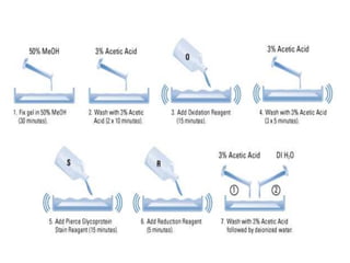
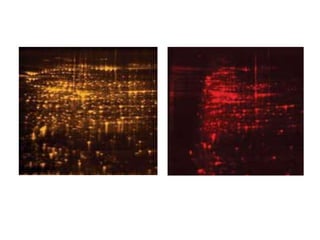
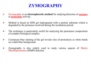
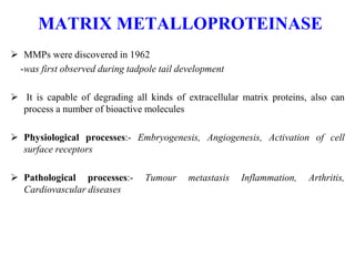
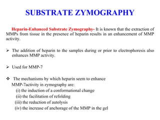
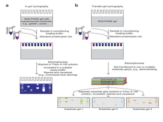
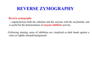
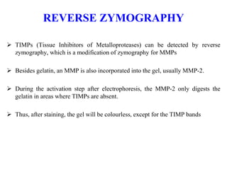
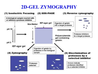
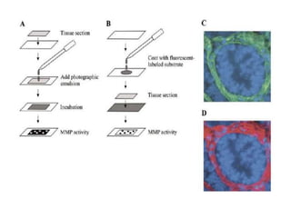
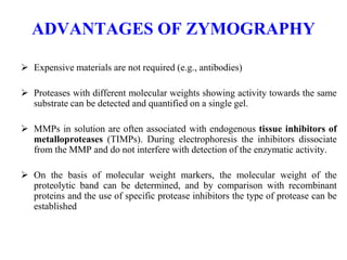
Zymography is an electrophoretic technique for the detection of hydrolytic enzymes, based on the substrate repertoire of the enzyme. ... Zymography also refers to a collection of related, fermented products, considered as a body of work.
2. PERIODIC ACID-SCHIFF
Periodic acid–Schiff (PAS) is a staining method used to detect polysaccharides
such as glycogen, and muco-substances- glycoproteins, glycolipids
The reaction of periodic acid oxidizes the vicinal diols in these sugars, usually
breaking up the bond between two adjacent carbons not involved in the
glycosidic linkage or in the ring of the monosaccharide units that are parts of the
long polysaccharides, and creating a pair of aldehydes at the two free tips of
each broken monosaccharide ring.
The oxidation condition has to be sufficiently regulated so as to not oxidize the
aldehydes further. These aldehydes then react with the Schiff reagent to give a
purple-magenta color. A suitable basic (Schiff) stain is often used as a
counterstain.
6. FLUORESCENT STAINING
Oriole Gel Stain
Easy-to-use, fast (90 min) and sensitive
No destain or fixation required
Compactable with downstream process and mass spectrometric analysis
Nano gram sensitivity, no background
Flamingo Gel Stain
Two steps, time consuming (5hr)
Compactable with mass spectrometric analysis and Edman-based sequencing
To be used with Laser based scanners
SYPRO Ruby Gel Stain
Detection of glycoprotein, lipoproteins and metalloproteins
No detection of nucleic acids in the sample
9. ZYMOGRAPHY
Zymography is an electrophoretic method for studying/detection of enzymes
or proteolytic activity.
Method is based on SDS gel impregnated with a protein substrate which is
degraded by the proteases resolved during the incubation period.
The technique is particularly useful for analyzing the proteinase composition
of complex biological samples.
Coomassie blue staining of the gel reveals sites of proteolysis as white bands
on a dark blue background.
Zymography is also widely used to study various aspects of Matrix
Metalloproteinase (MMP) function.
10. MATRIX METALLOPROTEINASE
MMPs were discovered in 1962
-was first observed during tadpole tail development
It is capable of degrading all kinds of extracellular matrix proteins, also can
process a number of bioactive molecules
Physiological processes:- Embryogenesis, Angiogenesis, Activation of cell
surface receptors
Pathological processes:- Tumour metastasis Inflammation, Arthritis,
Cardiovascular diseases
11. TYPES OF ZYMOGRAPHY
Types of Zymography
– Substrate Zymography :
- Gelatin Zymography
- Casein Zymography
- Collagen Zymography
- Heparin-Enhanced Zymography
– Reverse Zymography
– In Situ Zymography
12. SUBSTRATE ZYMOGRAPHY
When specific substrate is co-polymerized with the acrylamide
In zymography, the proteins are separated by electrophoresis under denaturing
conditions- SDS (sodium dodecyl sulfate) with non-reducing sample buffer
The separation occurs in a polyacrylamide gel containing a specific substrate
that is co-polymerized with the acrylamide
13. SUBSTRATE ZYMOGRAPHY
Gelatin Zymography– Gelatin zymography is mainly used for the detection of the
Gelatinases, MMP-2 and MMP-9
Casein Zymography– suitable for the detection of MMP-1, MMP-7, MMP-12, and MMP-
13
Collagen Zymography– used for the detection of MMP-1 and MMP-13, but MMP-2 and
MMP-9 can also be detected
-The incorporation of native collagen fibers in polyacrylamide gels appears unsuitable
for zymography because of their complicated structure, but SDS disrupts most of the
fibrillar organization of the collagen, allowing proteins to run into the gel.
14. SUBSTRATE ZYMOGRAPHY
Heparin-Enhanced Substrate Zymography- It is known that the extraction of
MMPs from tissue in the presence of heparin results in an enhancement of MMP
activity.
The addition of heparin to the samples during or prior to electrophoresis also
enhances MMP activity.
Used for MMP-7
The mechanisms by which heparin seem to enhance
MMP-7activity in zymography are:
(i) the induction of a conformational change
(ii) the facilitation of refolding
(iii) the reduction of autolysis
(iv) the increase of anchorage of the MMP in the gel
15.
16. REVERSE ZYMOGRAPHY
Reverse zymography
- copolymerizes both the substrate and the enzyme with the acrylamide, and
is useful for the demonstration of enzyme inhibitor activity.
-Following staining, areas of inhibition are visualized as dark bands against a
clear (or lightly stained) background.
17. REVERSE ZYMOGRAPHY
TIMPs (Tissue Inhibitors of Metalloproteases) can be detected by reverse
zymography, which is a modification of zymography for MMPs
Besides gelatin, an MMP is also incorporated into the gel, usually MMP-2.
During the activation step after electrophoresis, the MMP-2 only digests the
gelatin in areas where TIMPs are absent.
Thus, after staining, the gel will be colourless, except for the TIMP bands
20. In situ ZYMOGRAPHY
It is a unique laboratory technique that enables the localisation of matrix-
degrading metalloproteinase (MMP) activity in histological sections.
Frozen sections are placed on glass slides coated with fluorescently labelled
matrix proteins
After incubation MMP activity can be observed as black holes in the
fluorescent background due to proteolysis of the matrix protein.
This technique can be combined with immunohistochemistry to enable co-
location of proteins such as cell type markers or other proteins of interest.
Additionally, this technique can be adapted for use with cell cultures,
permitting precise location of MMP activity within cells,
21.
22. ADVANTAGES OF ZYMOGRAPHY
Expensive materials are not required (e.g., antibodies)
Proteases with different molecular weights showing activity towards the same
substrate can be detected and quantified on a single gel.
MMPs in solution are often associated with endogenous tissue inhibitors of
metalloproteases (TIMPs). During electrophoresis the inhibitors dissociate
from the MMP and do not interfere with detection of the enzymatic activity.
On the basis of molecular weight markers, the molecular weight of the
proteolytic band can be determined, and by comparison with recombinant
proteins and the use of specific protease inhibitors the type of protease can be
established
23. If you would like to donate us?
Scan below and donate us 0.013$ (US dollar) (5Rs Indian rupee)
Contact: If you want PPT/PDF files, please contact below.
Email: gnccmysore@gmail.com
Telegram:+919738137533(only for Chat)