An illustrative presentation on Biotin (Vitamin B7), clinical indications and technological applications for Medical, Dental, Pharmacology & Biotechnology students to facilitate easy- learning.
this presentation is about Vitamin B6 which include structure , biochemical function , biochemical reaction, effect of deficiency of vitamin B6, Toxicity and function of Vitamin B6.
this presentation is about Vitamin B6 which include structure , biochemical function , biochemical reaction, effect of deficiency of vitamin B6, Toxicity and function of Vitamin B6.
This is a presentation on probiotic foods, where I have described what probiotic food is, their mechanism of action, adequacy, and some popular forms of probiotic foods present in the market.
At the start of the 20th century, Russian noble prize winner and father of modern immunology, Elie Metchnikoff, a scientist at the Pasteur institute, was the first conceptualize “probiotics”.
In 1907 Metchnokoff proposed that the acid producing bacteria in fermented milk products could prevent “fouling” in the large intestine and if consumed regularly, lead to a longer, healthier life.
In early 1930’s, in Japan, Minoru shirota developed a fermented milk product called Yakult (probiotic yogurt like product made by fermenting a mixture of skimmed milk with a special strain of Lactobacillus casei shirota).
Probiotic term coined in 1965 by Lilly and StillwellThe human gastrointestinal (GI) tract is a highly specialised ecosystem that has evolved over
time, both physiologically and microbiologically. At least in part, this is a consequence of the
host and environmental pressures that it must counteract in order to maintain eubiosis. The
GI tract is one of the most diverse and metabolically active organs in the human body. The
human gut and its microbiota cannot be realistically considered as separate entities as they
represent a dynamic biological system that has co-evolved from birth. The human GI tract
is composed of highly adapted regions for mediation of its diverse functions, many of which
impact markedly upon host health and welfare. Physiological considerations in each unique
region infl uence the degree and type of colonisation and initial colonisers also modify the
physiological conditions therein. This results in the development of distinct microhabitats
along the length of the GI tract, which infl uence metabolism, protection and immune stimulation.
Such effects are both local and systemic as the GI tract is connected to the vascular,
lymphatic and nervous systems. The ability of the gut to sustain its benefi cial microbiota,
against harmful or opportunistic microbiota, in a desirable community structure, is critical
for host health and reduction of disease risk. The focus of this chapter is to discuss how the
complex interplays between the human GI tract and its indigenous microbiota affect host
health and how certain benefi cial microbial species, with their potential for manipulation,
are crucial to this processThe human gastrointestinal tract is sterile up until birth, when microbial colonisation begins
during the delivery process. The inoculum may be largely derived either from the mother’s
vaginal or faecal fl ora (in a conventional birth) or from the environment (in a caesarean
delivery).Hence, the microbiota that colonise the newborn tract are acquired post-natally.
This is of extreme importance in the choice of delivery, as newborns delivered by caesarean
section are exposed to a different microbiota than that of a vaginal delivery. Bacterial
populations develop progressively during the fi rst few days of life; facultative anaerobes
predominate initially and create a reduced environment that allows for the growth of strict
An illustrative and lucid presentation on Scurvy (deficiency of vitamin C) for Medical, Dental, Pharmacology & Biotechnology students to facilitate easy- learning.
An illustrative presentation on Vitamin C (Ascorbic acid) and Scurvy for Medical, Dental, Pharmacology & Biotechnology students to facilitate easy- learning.
An illustrative presentation on Microscopic examination of Urine for Medical, Dental, Pharmacology and Biotechnology students to facilitate easy- learning and self-study..
Urinalysis for detection of abnormal constituentsrohini sane
An illustrative presentation on Urinalysis for detection of abnormal constituents for medical ,dental , pharmacology and biotechnology students to facilitate easy-learning.
Urinalysis for detection of normal inorganic and organic constituentsrohini sane
An illustrative presentation on urinalysis for detection of normal inorganic and organic constituents for medical, dental , pharmacology and biotechnology students to facilitate easy-learning.
Biochemical kidney function tests with their clinical applicationsrohini sane
An illustrative presentation on Biochemical kidney function tests with their clinical applications for medical ,dental, pharmacology and biotechnology student to facilitate easy-learning.
A comprehensive presentation on Total parenteral nutrition(TPN) to facilitate easy -learning for medical , dental , pharmacology and biotechnology students.
Nutritional management of clinical disordersrohini sane
A lucid presentation Nutritional management of clinical disorders to facilitate easy-learning for medical , dental , pharmacology and biotechnology students.
Nutritional importance of vitamins and mineralsrohini sane
A lucid presentation on Nutritional importance of vitamins and minerals for medical , dental , pharmacology and biotechnology students to facilitate easy-learning.
A lucid presentation on Basal metabolic rate ( BMR) and nutrition for medical ,dental ,pharmacology and biotechnology students to facilitate easy-learning.
Physical activity of the human body and nutritionrohini sane
A lucid presentation on Physical activity of the human body and Nutrition for medical ,dental ,pharmacology and biotechnology students for easy learning.
Pulmonary Thromboembolism - etilogy, types, medical- Surgical and nursing man...VarunMahajani
Disruption of blood supply to lung alveoli due to blockage of one or more pulmonary blood vessels is called as Pulmonary thromboembolism. In this presentation we will discuss its causes, types and its management in depth.
Report Back from SGO 2024: What’s the Latest in Cervical Cancer?bkling
Are you curious about what’s new in cervical cancer research or unsure what the findings mean? Join Dr. Emily Ko, a gynecologic oncologist at Penn Medicine, to learn about the latest updates from the Society of Gynecologic Oncology (SGO) 2024 Annual Meeting on Women’s Cancer. Dr. Ko will discuss what the research presented at the conference means for you and answer your questions about the new developments.
These simplified slides by Dr. Sidra Arshad present an overview of the non-respiratory functions of the respiratory tract.
Learning objectives:
1. Enlist the non-respiratory functions of the respiratory tract
2. Briefly explain how these functions are carried out
3. Discuss the significance of dead space
4. Differentiate between minute ventilation and alveolar ventilation
5. Describe the cough and sneeze reflexes
Study Resources:
1. Chapter 39, Guyton and Hall Textbook of Medical Physiology, 14th edition
2. Chapter 34, Ganong’s Review of Medical Physiology, 26th edition
3. Chapter 17, Human Physiology by Lauralee Sherwood, 9th edition
4. Non-respiratory functions of the lungs https://academic.oup.com/bjaed/article/13/3/98/278874
Knee anatomy and clinical tests 2024.pdfvimalpl1234
This includes all relevant anatomy and clinical tests compiled from standard textbooks, Campbell,netter etc..It is comprehensive and best suited for orthopaedicians and orthopaedic residents.
NVBDCP.pptx Nation vector borne disease control programSapna Thakur
NVBDCP was launched in 2003-2004 . Vector-Borne Disease: Disease that results from an infection transmitted to humans and other animals by blood-feeding arthropods, such as mosquitoes, ticks, and fleas. Examples of vector-borne diseases include Dengue fever, West Nile Virus, Lyme disease, and malaria.
Lung Cancer: Artificial Intelligence, Synergetics, Complex System Analysis, S...Oleg Kshivets
RESULTS: Overall life span (LS) was 2252.1±1742.5 days and cumulative 5-year survival (5YS) reached 73.2%, 10 years – 64.8%, 20 years – 42.5%. 513 LCP lived more than 5 years (LS=3124.6±1525.6 days), 148 LCP – more than 10 years (LS=5054.4±1504.1 days).199 LCP died because of LC (LS=562.7±374.5 days). 5YS of LCP after bi/lobectomies was significantly superior in comparison with LCP after pneumonectomies (78.1% vs.63.7%, P=0.00001 by log-rank test). AT significantly improved 5YS (66.3% vs. 34.8%) (P=0.00000 by log-rank test) only for LCP with N1-2. Cox modeling displayed that 5YS of LCP significantly depended on: phase transition (PT) early-invasive LC in terms of synergetics, PT N0—N12, cell ratio factors (ratio between cancer cells- CC and blood cells subpopulations), G1-3, histology, glucose, AT, blood cell circuit, prothrombin index, heparin tolerance, recalcification time (P=0.000-0.038). Neural networks, genetic algorithm selection and bootstrap simulation revealed relationships between 5YS and PT early-invasive LC (rank=1), PT N0—N12 (rank=2), thrombocytes/CC (3), erythrocytes/CC (4), eosinophils/CC (5), healthy cells/CC (6), lymphocytes/CC (7), segmented neutrophils/CC (8), stick neutrophils/CC (9), monocytes/CC (10); leucocytes/CC (11). Correct prediction of 5YS was 100% by neural networks computing (area under ROC curve=1.0; error=0.0).
CONCLUSIONS: 5YS of LCP after radical procedures significantly depended on: 1) PT early-invasive cancer; 2) PT N0--N12; 3) cell ratio factors; 4) blood cell circuit; 5) biochemical factors; 6) hemostasis system; 7) AT; 8) LC characteristics; 9) LC cell dynamics; 10) surgery type: lobectomy/pneumonectomy; 11) anthropometric data. Optimal diagnosis and treatment strategies for LC are: 1) screening and early detection of LC; 2) availability of experienced thoracic surgeons because of complexity of radical procedures; 3) aggressive en block surgery and adequate lymph node dissection for completeness; 4) precise prediction; 5) adjuvant chemoimmunoradiotherapy for LCP with unfavorable prognosis.
Ozempic: Preoperative Management of Patients on GLP-1 Receptor Agonists Saeid Safari
Preoperative Management of Patients on GLP-1 Receptor Agonists like Ozempic and Semiglutide
ASA GUIDELINE
NYSORA Guideline
2 Case Reports of Gastric Ultrasound
Flu Vaccine Alert in Bangalore Karnatakaaddon Scans
As flu season approaches, health officials in Bangalore, Karnataka, are urging residents to get their flu vaccinations. The seasonal flu, while common, can lead to severe health complications, particularly for vulnerable populations such as young children, the elderly, and those with underlying health conditions.
Dr. Vidisha Kumari, a leading epidemiologist in Bangalore, emphasizes the importance of getting vaccinated. "The flu vaccine is our best defense against the influenza virus. It not only protects individuals but also helps prevent the spread of the virus in our communities," he says.
This year, the flu season is expected to coincide with a potential increase in other respiratory illnesses. The Karnataka Health Department has launched an awareness campaign highlighting the significance of flu vaccinations. They have set up multiple vaccination centers across Bangalore, making it convenient for residents to receive their shots.
To encourage widespread vaccination, the government is also collaborating with local schools, workplaces, and community centers to facilitate vaccination drives. Special attention is being given to ensuring that the vaccine is accessible to all, including marginalized communities who may have limited access to healthcare.
Residents are reminded that the flu vaccine is safe and effective. Common side effects are mild and may include soreness at the injection site, mild fever, or muscle aches. These side effects are generally short-lived and far less severe than the flu itself.
Healthcare providers are also stressing the importance of continuing COVID-19 precautions. Wearing masks, practicing good hand hygiene, and maintaining social distancing are still crucial, especially in crowded places.
Protect yourself and your loved ones by getting vaccinated. Together, we can help keep Bangalore healthy and safe this flu season. For more information on vaccination centers and schedules, residents can visit the Karnataka Health Department’s official website or follow their social media pages.
Stay informed, stay safe, and get your flu shot today!
Title: Sense of Taste
Presenter: Dr. Faiza, Assistant Professor of Physiology
Qualifications:
MBBS (Best Graduate, AIMC Lahore)
FCPS Physiology
ICMT, CHPE, DHPE (STMU)
MPH (GC University, Faisalabad)
MBA (Virtual University of Pakistan)
Learning Objectives:
Describe the structure and function of taste buds.
Describe the relationship between the taste threshold and taste index of common substances.
Explain the chemical basis and signal transduction of taste perception for each type of primary taste sensation.
Recognize different abnormalities of taste perception and their causes.
Key Topics:
Significance of Taste Sensation:
Differentiation between pleasant and harmful food
Influence on behavior
Selection of food based on metabolic needs
Receptors of Taste:
Taste buds on the tongue
Influence of sense of smell, texture of food, and pain stimulation (e.g., by pepper)
Primary and Secondary Taste Sensations:
Primary taste sensations: Sweet, Sour, Salty, Bitter, Umami
Chemical basis and signal transduction mechanisms for each taste
Taste Threshold and Index:
Taste threshold values for Sweet (sucrose), Salty (NaCl), Sour (HCl), and Bitter (Quinine)
Taste index relationship: Inversely proportional to taste threshold
Taste Blindness:
Inability to taste certain substances, particularly thiourea compounds
Example: Phenylthiocarbamide
Structure and Function of Taste Buds:
Composition: Epithelial cells, Sustentacular/Supporting cells, Taste cells, Basal cells
Features: Taste pores, Taste hairs/microvilli, and Taste nerve fibers
Location of Taste Buds:
Found in papillae of the tongue (Fungiform, Circumvallate, Foliate)
Also present on the palate, tonsillar pillars, epiglottis, and proximal esophagus
Mechanism of Taste Stimulation:
Interaction of taste substances with receptors on microvilli
Signal transduction pathways for Umami, Sweet, Bitter, Sour, and Salty tastes
Taste Sensitivity and Adaptation:
Decrease in sensitivity with age
Rapid adaptation of taste sensation
Role of Saliva in Taste:
Dissolution of tastants to reach receptors
Washing away the stimulus
Taste Preferences and Aversions:
Mechanisms behind taste preference and aversion
Influence of receptors and neural pathways
Impact of Sensory Nerve Damage:
Degeneration of taste buds if the sensory nerve fiber is cut
Abnormalities of Taste Detection:
Conditions: Ageusia, Hypogeusia, Dysgeusia (parageusia)
Causes: Nerve damage, neurological disorders, infections, poor oral hygiene, adverse drug effects, deficiencies, aging, tobacco use, altered neurotransmitter levels
Neurotransmitters and Taste Threshold:
Effects of serotonin (5-HT) and norepinephrine (NE) on taste sensitivity
Supertasters:
25% of the population with heightened sensitivity to taste, especially bitterness
Increased number of fungiform papillae
Couples presenting to the infertility clinic- Do they really have infertility...Sujoy Dasgupta
Dr Sujoy Dasgupta presented the study on "Couples presenting to the infertility clinic- Do they really have infertility? – The unexplored stories of non-consummation" in the 13th Congress of the Asia Pacific Initiative on Reproduction (ASPIRE 2024) at Manila on 24 May, 2024.
263778731218 Abortion Clinic /Pills In Harare ,sisternakatoto
263778731218 Abortion Clinic /Pills In Harare ,ABORTION WOMEN’S CLINIC +27730423979 IN women clinic we believe that every woman should be able to make choices in her pregnancy. Our job is to provide compassionate care, safety,affordable and confidential services. That’s why we have won the trust from all generations of women all over the world. we use non surgical method(Abortion pills) to terminate…Dr.LISA +27730423979women Clinic is committed to providing the highest quality of obstetrical and gynecological care to women of all ages. Our dedicated staff aim to treat each patient and her health concerns with compassion and respect.Our dedicated group ABORTION WOMEN’S CLINIC +27730423979 IN women clinic we believe that every woman should be able to make choices in her pregnancy. Our job is to provide compassionate care, safety,affordable and confidential services. That’s why we have won the trust from all generations of women all over the world. we use non surgical method(Abortion pills) to terminate…Dr.LISA +27730423979women Clinic is committed to providing the highest quality of obstetrical and gynecological care to women of all ages. Our dedicated staff aim to treat each patient and her health concerns with compassion and respect.Our dedicated group of receptionists, nurses, and physicians have worked together as a teamof receptionists, nurses, and physicians have worked together as a team wwww.lisywomensclinic.co.za/
HOT NEW PRODUCT! BIG SALES FAST SHIPPING NOW FROM CHINA!! EU KU DB BK substit...GL Anaacs
Contact us if you are interested:
Email / Skype : kefaya1771@gmail.com
Threema: PXHY5PDH
New BATCH Ku !!! MUCH IN DEMAND FAST SALE EVERY BATCH HAPPY GOOD EFFECT BIG BATCH !
Contact me on Threema or skype to start big business!!
Hot-sale products:
NEW HOT EUTYLONE WHITE CRYSTAL!!
5cl-adba precursor (semi finished )
5cl-adba raw materials
ADBB precursor (semi finished )
ADBB raw materials
APVP powder
5fadb/4f-adb
Jwh018 / Jwh210
Eutylone crystal
Protonitazene (hydrochloride) CAS: 119276-01-6
Flubrotizolam CAS: 57801-95-3
Metonitazene CAS: 14680-51-4
Payment terms: Western Union,MoneyGram,Bitcoin or USDT.
Deliver Time: Usually 7-15days
Shipping method: FedEx, TNT, DHL,UPS etc.Our deliveries are 100% safe, fast, reliable and discreet.
Samples will be sent for your evaluation!If you are interested in, please contact me, let's talk details.
We specializes in exporting high quality Research chemical, medical intermediate, Pharmaceutical chemicals and so on. Products are exported to USA, Canada, France, Korea, Japan,Russia, Southeast Asia and other countries.
Prix Galien International 2024 Forum ProgramLevi Shapiro
June 20, 2024, Prix Galien International and Jerusalem Ethics Forum in ROME. Detailed agenda including panels:
- ADVANCES IN CARDIOLOGY: A NEW PARADIGM IS COMING
- WOMEN’S HEALTH: FERTILITY PRESERVATION
- WHAT’S NEW IN THE TREATMENT OF INFECTIOUS,
ONCOLOGICAL AND INFLAMMATORY SKIN DISEASES?
- ARTIFICIAL INTELLIGENCE AND ETHICS
- GENE THERAPY
- BEYOND BORDERS: GLOBAL INITIATIVES FOR DEMOCRATIZING LIFE SCIENCE TECHNOLOGIES AND PROMOTING ACCESS TO HEALTHCARE
- ETHICAL CHALLENGES IN LIFE SCIENCES
- Prix Galien International Awards Ceremony
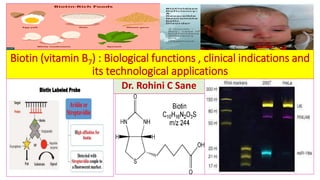
Biotin (vitamin b7) biological functions, clinical indications and its technological applications
1. Biotin (vitamin B7) : Biological functions , clinical indications and
its technological applications
Dr. Rohini C Sane
2. Historical background of Biotin
• Biotin = vitamin B7 = vitamin H = anti-egg white injury factor(intake of raw
→not boiled egg may cause Biotin deficiency).
• Boas(1927) fed rats with huge quantity of raw eggs → rats developed
dermatitis, nervous manifestations and retardation in growth.
• Vincent du Vigneaud (Noble prize 1955): isolated Biotin.
3. Structure of Biotin (Vitamin B7 / Vitamin H)
❑Structure of Biotin (Vitamin H) :
❖Sulphur-containing water soluble
heterocyclic monocarboxylic acid
(Vitamin B7) .
❖Imidazole ring fused with
tetrahydrothiophene ring with a
Valeric acid side chain.
• The carboxyl group of biotin forms an
amide linkage with the epsilon(ε)
nitrogen of Lysine residue in the
apoenzyme forming a biotinyl
enzyme.
• Biocytin : a coenzyme form (Active
form)→ (Biotin is covalently bound to
ε- amino group of Lysine (Lysine -
linked by amide bond)in the enzyme.
O
II
C
N H
C H
CH (CH2)4 COOH
H N
H C
H2C
S
ǀ
ǀ
ǀ
ǀ
ǀ
ǀ
CO2 Binding site
Site for binding with Lysine
Biotin = cis-tetrahydro-2-oxo-
1-thienol-(3,4-d)-imidazoline -
4-valeric acid .
Imidazole ring
Tetrahydrothiophene ring
1
2
3
4
5
1
2
3
C10H16 O3N2 S
Molecular weight : 244
5. Sources of Biotin in human
❖Human beings cannot synthesize Biotin and hence Biotin has to be supplied in diet.
❖Sources of Biotin in human : synthesis by intestinal bacterial flora and dietary
sources .
❖Normal bacterial flora of the intestine provides adequate quantities of
Biotin(biosynthesis of Biotin by E.coli). Moreover, it is distributed ubiquitously(widely)
in animal tissue, fruits and vegetables.
❖Rich food sources of Biotin:
• Milk, cheese , yogurt , yeast , cauliflower , berries , soyabean , tomatoes , sweet
potatoes , spinach , Avocado , lentils , banana , carrots , white mushrooms , Molasses,
wheat germs , oats , sunflower seeds , almonds, walnuts , hazelnuts, peanuts and
grains. (water soluble form in most plants material except cereals and nuts).
• Egg yolk , liver, kidney , pancreas, Sardine , Yellow Tuna, pork , beef , Turkey , chicken ,
lamb and Calf silver. (water insoluble form in animal tissues).
7. Co-enzyme-R(Biotin)from Rhizobium
• Co-enzyme R is growth essential for
Rhizobium (nitrogen-fixing
organisms) in the root nodules of
Leguminous plant .
• Co-enzyme R is proved to be Biotin.
• Pimelic acid is possible precursor.
• Desthiobiotin is a probable
intermediate.
8. Daily dietary requirement of Biotin( RDA)
• Daily dietary requirement of Biotin( RDA) = 200-300 μg
• Estimated Average requirement : Adequate intake(AI)based on urinary excretion of Biotin
and the metabolite 3-hydroxy isovaleric acid.
• Tolerable upper intake level (UL)→ applies to chronic daily use of fortified foods/dietary
supplements → no value established for Biotin .
➢Daily dietary requirement of Biotin(RDA) increases in pregnancy and lactation
(additional 5 μg /kg of body weight) .
➢Intravenous supply of Biotin for adults during TPN = 60 μg /day.
➢Patients receiving hemodialysis or peritoneal dialysis or with biotinidase deficiency require
more .
Age in years Adequate intake(AI)of Biotin (μg/day)
≥ 19 30
14-18(aldolescents) 25
9-13 20
4-8 12
1-3 8
<1 year(Infants) 0.7 μg /kg of body weight
9. Metabolism of Biotin in the human body
• Sources of Biotin in human : dietary sources(largely protein bound)and free form .It is
biosynthesized biotin by bacterial flora.
• Occurrence /existence in dietary food source : widely distributed in many food sources as
Biocytin (ε- amino-biotinyl lysine)→ Biotin is released after proteolysis.
• Biosynthesis : by intestinal bacteria (human body cannot synthesize Biotin).
• Digestion: proteolysis by gastrointestinal enzyme to produce biotinylate peptides →release of free
Biotin further hydrolysis by intestinal biotinidase.
• Absorption : readily absorbed by intestinal epithelial cells using a biotin carrier (the sodium
dependent multi-vitamin transporter →SMVT). Avidin (present in raw egg white)prevents its
absorption.
• Transport : by circulating blood
• Secretion : in milk
• Uptake of absorbed Biotin : by liver , kidney and muscles (localized in cytosolic and mitochondrial
carboxylases).
• Storage :in Liver and kidneys (limited extent→ 14% of administered dose). Total body content of
Biotin = 1mg
• Excretion : Urinary excretion(10-180 μg/day) > dietary intake (28-100 μg/day) .
Fecal excretion (15-200μg/day) > 3-6 times of dietary intake . Fecal excretion represents unabsorbed
Biotin synthesized by intestinal bacteria.
10. Absorption and transport of Biotin
Biotin containing food/enzymes
Covalently linked to proteins
Proteolytic enzymes
Biocytin
Pancreatic Biotinidase
Biotin + Lysine
sodium
dependent
multi-vitamin
transporter
→SMVT)
Intestinal Brush border
Biotin
GPR109A
Hydroxycarboxylic acid
(HCA-2) receptor mutation causes biotin –
responsive basal ganglia
disease(encephalopathy )
SLC19A3 Low concentration of
Biotin : active
transport
High concentration
of Biotin : Passive
transport
Basal lateral
membrane
11. The sodium-dependent multivitamin transporter (SMVT) to facilitate intestinal
absorption of Biotin
• Is located in the intestinal brush border membrane.
• Na+ dependent carrier-mediated process .
• Transports biotin against sodium ion concentration gradient.
• Not specific for vitamin (Biotin) transport.
• Functions in cellular uptake of Biotin ,Pantothenic acid and Lipoic acid with
similar affinities .
• Biotin uptake by intestinal epithelial cells is inhibited by activation of protein
kinase C apparently through phosphorylation of SMVT.
12. Facilitated transport of Biotin during intestinal absorption
Intestinal Absorption of Biotin at its Low
concentration
Intestinal Absorption of Biotin at its High
concentration
Saturable ,Active and facilitated mechanism
dependent on Na+ .
Passive transport
Facilitated transport inhibited by certain anti-
convulsant drugs and chronic exposure to
ethanol.
13. Catabolism of Biotin
• The enzyme biocytinase (biotin amidohydrolase) in plasma and erythrocytes
catalyze the hydrolysis biocytin to yield free biotin.
• Free biotin is taken up by tissues (such as liver, muscle and kidney) and
localized in cytosolic and mitochondrial carboxylase.
• Small fraction of Biotin is oxidized to D- and L- sulfoxides ( ureido ring intact
not otherwise degraded).
• Side chain of larger portion of Biotin is degraded via mitochondrial β-oxidation
to yield bis-nor biotin and its degradation products.
• Biotin catabolism in smokers > Biotin catabolism in non-smokers.
14. Excretion of Biotin
• Source for excretion of Biotin : Half of the absorbed biotin.
• Mean urinary excretion is a reflective of dietary intake (28-100 μg/day for
adult).
• Metabolite Forms for excretion of Biotin:
1. Bis-norbiotin (occurring from β-oxidation of the Valeric acid side chain).
2. Biotin sulfoxide (occurring from oxidation of the sulfur in the heterocyclic
ring).
• Ratio of Biotin : Bis-norbiotin : Biotin sulfoxide = 3:2:1(in circulating plasma
and urine).
• Minor metabolites of biotin for excretion : Bis-norbiotin methyl ketone and
Biotin sulfone.
15. Laboratory Assessment of Biotin status
❖Bioassay methods
❖Microbiological assay : growth stimulation of yeast cells (Saccharomyces cerevisiae or
Lactobacillus plantarum) is measured . Whole Blood is first digested with papain or acid
hydrolysis to release free biotin . This sample is then added to a Biotin-deficient medium
inoculated with a test organism , such as Lactobacillus plantarum .
❖Measurement of unfound Biotin include Avidin-binding assays : a competitive protein
binding radio-assay with 3H-labelled Biotin or a nonradioactive enzyme-linked sorbent
using Streptavidin as a binding agent .
❖ Measurement of Urinary Biotin and 3-hydroisovaleric acid : by HPLC
Urinary excretion of Biotin and 3-hydroisovaleric acid appear to be better indicator of
biotin status than whole blood concentrations.
❖Urinary Biotin and 3-hydroisovaleric acid : gas chromatography-mass spectrometry.
❖Lymphocyte Propionyl-CoA carboxylase using H14 CO3 - : early indicator and sensitive
indicator of biotin deficiency in patients on prolonged TPN without biotin supplementation
and in children with protein-energy malnutrition.
16. Reference intervals of Biotin
• Whole blood Biotin levels (physiological) by microbiological method :
200 - 500 pg /ml (0.5- 2.2 nmol/ L with mean 1.31nmol/L)
• Biotin deficiency : Whole blood biotin < 0.5 nmols/L
• Lowered circulating blood levels and urinary excretion are observed in
1. alcoholics
2. Patients with achlorhydria
3. Elderly
4. Athletes
• Biotin content of red cells is similar to that of plasma for a given method.
17. Co-enzyme and non-coenzyme roles of Biotin
Coenzyme role of Biotin Non-coenzyme role of Biotin
Pyruvate carboxylase Cell proliferation
Acetyl-CoA carboxylase Gene silencing
Propionyl-CoA carboxylase DNA repair
β-methyl crotonyl-CoA Carboxylase Gene expression and cell signaling
Biotin is a coenzyme of carboxylase
reactions . Biotin is a carrier of activated
carbon dioxide (CO2) for the mechanism
of Biotin-dependent carboxylations .
Biotin is the prosthetic of certain enzymes (carboxylases and decarboxylases) that catalyze
CO2 transfer reaction (CO2 fixation reaction/ carboxylation) in human tissue.
18. Biotin-dependent carboxylases
Biotin
VitaminB7
Vitamin H
Pyruvate Carboxylase
(key enzyme of
Gluconeogenesis, TCA and
Transamination)
Acetyl-CoA Carboxylase
(First committed step in
biosynthesis and
elongation of fatty acids)
Propionyl-CoA
Carboxylase (oxidation of odd chain
fatty acids and synthesis of Succinyl
CoA)
β-methyl crotonyl-CoA
Carboxylase(catabolism of
Leucine)
Biotin is covalently
bound to the ε- amino
groups of Lysine
residues in Biotin –
dependent enzymes.
19. Functions of Biotin as a prosthetic group of ATP-dependent carboxylases
Enzyme Substrate Product Importance enzyme
Pyruvate Carboxylase Pyruvate Oxaloacetate Gluconeogenesis(synthesisof
Glucosefromnon-carbohydrate
substance),providesoxaloacetate
forTCACycle,Transamination
Acetyl-CoA
Carboxylase
Acetyl-CoA Malonyl-CoA Limiting reaction in Fatty Acid
biosynthesis
Propionyl-CoA
Carboxylase
Propionyl-
CoA
D-Methyl Malonyl-CoA
→Succinyl-CoA
Succinyl-CoA→HemeSynthesis,
Succinyl-CoAoxidizedInTCAcycle
β-methyl crotonyl-
CoA Carboxylase
β-methyl
Crotonyl-CoA
β-Methyl glutaconyl-
CoA
Leucine metabolism (Branched
ChainAminoAcids)
Biotinfunctionsasaprostheticgroupforcarboxylasesandisattachedtotheenzymebyanamidebond
betweenthecarboxylgroupofBiotinandtheterminalε-aminogroupofLysineresidueoftheenzyme,
formingaBiotin-enzyme.
20. Mechanism of Biotin during carboxylation reactions in the human body
• The peptide biocytin (ε-N-biotinyl lysine) is resistant to hydrolysis by proteolytic
enzymes in intestinal tract but together with biotin is readily absorbed .
• A biotin carrier , sodium-dependent multivitamin transporter (SMVT) for which
Pantothenic acid and lipoate compete.
• (SMVT) is located in brush borders membrane and transports biotin against a
sodium ion concentration gradient.
• The enzyme biocytinase (Biotin amidohydrolase) is located in plasma and
erythrocytes catalyzes hydrolysis of biocytin to yield free biotin.
• Covalent attachment of Biotin to apoenzyme involves ATP-dependent conversion of
the vitamin to biotinyl-5’-adenylate followed by condensation of the biotinyl moiety
with ε-amino groups of specific Lysyl residues in apoenzyme preformed from
subunits.
• Enzyme responsible for formation of ε- N-biotinyl-l-Lysyl ( biocytinyl) moiety of
proteins is holocarboxylase synthetases (HCS).
21. Biotin recycling
Holocarboxylase synthetase (HCS) uses ATP to catalyze the covalent bonding of different
apocarboxylases with Biotin to form different biotin-carboxylase complexes called
holocarboxylase . In holocarboxylase –amide bond binds the carboxyl terminal of valeric acid
side chain of Biotin with ε–NH2 group at the end of the side chain of lysine residue of
apocarboxylases.
22. Mechanism of carboxylation reactions facilitated by Biotin
• In biological system, Biotin functions as the co-enzyme for the enzyme called
carboxylase which catalyze the carbon fixation(carboxylation).
• These enzymes operate via a common mechanism ,which involves
phosphorylation of bicarbonate by ATP to form carbonyl phosphate , followed
by transfer of the carbonyl group to the sterically less hindered nitrogen of the
biotin moiety.
• In this process , Biotin is first gets converted to carboxy-biotin complex by
reaction with ATP and HCO3
- .
• The resulting N(1)-carboxybiotinyl enzyme can then exchange the carboxylate
function with a reactive center in a substrate i.e. CO2 -Biotin complex is the
source of active CO2 which is transferred to the substrate .
• CO2 becomes attached to the biotin coenzyme as above.
23. Mechanism of carboxylation reactions facilitated by Biotin
Biotin
enzyme
Carbonic
phosphoric
anhydride
Carboxy-
biotin-
enzyme
CO2
ATP
ADP
Pi
Carboxylated substrate
substrate
Biotin acts as
co-enzyme for
carboxylation
reactions. It
captures a
molecule of
CO2 which is
attached to
nitrogen of the
Biotin
molecule.
The energy required for this reaction is provided by ATP . Then the activated carboxyl group is
transferred to the substrate.
Substrate + CO2 + ATP → Product
(Carboxylated substrate) + ADP+ Pi
24. Biochemical functions of Biotin in carboxylation of Pyruvate to Oxaloacetate
❖ Carboxylation of Pyruvate to Oxaloacetate :
Substrate : Pyruvate
Enzyme : Pyruvate carboxylase
Coenzyme (Carrier of CO2) : Biotin
Energy source : ATP
Product : Oxaloacetate
Mechanism of reaction : Pyruvate carboxylase has Biotin which is bound to the
apoenzyme linked to the ε-amino group of Lysine , forming the active enzyme
(holoenzyme).
Biotin-enzyme reacts with CO2 in presence of ATP to form a carboxy-biotin-
enzyme complex (high energy complex). This high energy complex then hands
over the CO2 to Pyruvate (carboxylation reaction) to produce Oxaloacetate ..
25. Role of Biotin in conversion of Pyruvate to Oxaloacetic acid by
Pyruvate carboxylase
COOH
CH2
CO
COOH
Pyruvic acid Oxaloacetic acid
CH3
CO
COOH
Pyruvate carboxylase
CO2
Biotin
ATP ADP + Pi
I
I
I
I
I
Acetyl-CoA
Mn2+
❖ Carboxylation of Pyruvate to Oxaloacetate by Pyruvate carboxylase is Biotin-dependent
reaction. Hydrolysis of ATP drives the formation of enzyme-biotin-CO2 intermediate (high
energy complex). This complex subsequently carboxylates Pyruvate to OAA.
❖ Mitochondrial Pyruvate carboxylase(liver and kidney) catalyzes formation of Oxaloacetate,
which together with Acetyl-CoA forms Citrate .
Two important aspects of this reaction:
1. Provides oxaloacetate , that replenishes TCA cycle intermediates that may be depleted,
depending on the synthetic needs of the cell.
2. Is important enzyme in the gluconeogenesis pathway .
26. Mechanism of carboxylation reaction catalyzed by Biotinylated enzyme
(mitochondrial Pyruvate carboxylase) in formation of Oxaloacetate
Biotin-enzyme + CO2
Biotin-enzyme
ATP
ADP +Pi
S
Lys
NH
(CH 2)4-CO
N NH
H H
O
Enzyme
S
O
O- C-
II
II
- ←Caroxy-biotin-enzyme complex
I
H
O
CH3- C- COO
II -
←Pyruvate
←Oxaloacetate
O
OOC-CH2- C- COO
- II
Pyruvate carboxylase with
covalently attached Biotin
-
Protein portion of enzyme : Acetyl-CoA
carboxylase , Propionyl-CoA carboxylase, Pyruvate
carboxylase, Methyl crotonyl-CoA carboxylase to
catalyze carboxylation of substrate into
corresponding carboxylated product.
Biotin covalently
bound to Lysyl
residue
of a biotin-
dependent enzyme .
27. Glucose
Glucose -6-phosphate
Glyceraldehyde-3-phosphate
1,3 –Bi phosphoglycerate
3-Phosphoglycerate
2-Phosphoglycerate
Phosphoenolpyruvate
Pyruvate
Oxaloacetate Pyruvate Acetyl-CoA
Oxaloacetate
Malate Malate Citrate
Fumarate α-Ketoglutarate
Succinyl-CoA
TCA cycle
Glucogenic amino acids
Lactate
Glucogenic amino acids
Glucogenic amino acids
Propionyl CoA
Glucogenic amino acids
Pyruvate carboxylase
ATP , Mg 2+ , Biotin
ATP
ADP + Pi
Glucokinase /Hexokinase
Pi
H2O
Glucose-6-phosphatase
Pi
H2O
Fructose1,6-
biphosphatase
Fructose -6-phosphate
Fructose -1,6-phosphate
GDP +Pi + CO2
GTP
Phosphoenolpyruvate Carboxykinase
ATP
ADP+Pi
Phosphofructokinase
Pathway of Gluconeogenesis(red)
and Glycolysis(blue)
Mitochondrion
28. Importance of Carboxylation of Pyruvate to Oxaloacetate by Biotin-dependent
Pyruvate carboxylase in Gluconeogenesis and TCA cycle
❖Importance of Carboxylation of
Pyruvate to Oxaloacetate by Biotin-
dependent Pyruvate carboxylase :
• Activated by Acetyl-CoA.
• ATP-dependent.
• Biotin-dependent reaction.
• Replenishes Oxaloacetate which is
an intermediate of TCA cycle
(ensures continuous operation of
Citric acid cycle) in liver and kidney .
• Provides non-carbohydrate
substrates for Gluconeogenesisinliver
andkidneycells .
• An irreversible reaction.
• Pyruvate carboxylase from muscle
cells use OAA produced for
replenishingTCAanddonotsynthesize
glucose.
Pyruvate carboxylase
Biotin
ATP
ADP + Pi
CO2
Acetyl-CoA
+
NADH + H+
NAD
Mitochondrial matrix
Cytosol
Malate
NAD +
NADH + H+
Oxaloacetate Phosphoenol Pyruvate
CO2
Glucose
Gluconeogenesis
*PEPcarboxykinase
+GTP
*
Malate
Oxaloacetate
Malate
dehydrogenase
Pyruvate
29. Importance of Biotin- dependent carboxylation of Acetyl-CoA to Malonyl-CoA
❖Carboxylation of Acetyl-CoA to Malonyl-CoA(cytosolic reaction) is
• initial (first) and the rate limiting reaction in fatty acid biosynthesis.
• irreversible reaction catalyzed by an enzyme complex , Acetyl-CoA carboxylase, that
requires Biotin as a prosthetic group and utilizes bicarbonate (as a source of CO2) in
presence of ATP.
Acetyl-CoA+ CO2 + ATP Malonyl-CoA +ADP+ Pi
❖Biotin dependent Acetyl-CoA carboxylase is:
▪ an allosteric enzyme activated by Citrate. It is a storage vehicle for biotin.
▪ Inhibited by its end product Palmitoyl-CoA.
➢In addition to high allosteric control , high carbohydrate and low fat diet stimulates
the synthesis of enzyme.
❖Malonyl-CoA is a substrate for fatty acid synthase complex. Fatty acid synthase
subsequently adds 2-carbon units from Malonyl-CoA to growing fatty acid acyl
chain to form Palmitate.
Acetyl-CoA carboxylase
Biotin
30. Role of Biotin formation of Malonyl-CoA by carboxylation of Acetyl-CoA
O
CH3-C-SCoA
Acetyl-CoA
O
-OOC-CH2-C-SCoA
Malonyl-CoA
Acetyl-CoA carboxylase
CO2
ATP
ADP + Pi
Biotin
II
II
Malonyl-CoA is used for
fatty acid biosynthesis .
Biotin-dependent Acetyl-
CoA carboxylase is
regulatory enzyme in fatty
acid synthesis.
1 Acetyl-CoA
8 Acetyl-CoA Palmitic acid
7 Acetyl-CoA 7 Malonyl-CoA
Site de novo synthesis of fatty
acids : Cytoplasm of liver,
adipose tissue , kidney , brain
and mammary glands
Acetyl-CoA is the starting
material for the biosynthesis
of fatty acids.
The energy for the carbon –
carbon condensations in
fatty acid synthesis is
supplied by the process of
carboxylation and then
decarboxylation of acetyl
groups in cytosol.
31. Role of Malonyl-CoA in biosynthesis of Palmitic acid
Acetyl-CoA Malonyl-CoA
16 15 14 13 12 11 10 9 8 7 6 5 4 3 2 1
CH3- CH2- CH2- CH2- CH2- CH2- CH2- CH2-CH2- CH2-CH2-CH2-CH2- CH2- CH2- COOH
Cycle I II III IV V VI VII
Carbon atoms 15 and 16 are from Acetyl-CoA and Carbon 1-14 from Malonyl-CoA .
8 Acetyl-CoA are required to make one molecule of Palmitic acid . But in the reaction
mechanism which is explained above only one Acetyl-CoA takes part and other 7 Acetyl-CoA
take part after they are converted into 7 Malonyl-CoA.
Palmitic acid
32. Role of Biotin in fatty acid biosynthesis
The cytosolic pathway (extra-mitochondrial pathway, De Novo synthesis of fatty acids) is a
major pathway for synthesis of fatty acids. Synthesis of fatty acid from Acetyl-CoA takes place
outside mitochondria . Acetyl-CoA forms Citrate which comes out of the mitochondria can
cleave to give Acetyl-CoA (as such cannot come out of the mitochondria).
Tissue involved in cytosolic fatty
acid biosynthesis: Liver, adipose
tissue, mammary gland ,brain,
kidney
33. Role of Biotin in fatty acid biosynthesis
Other carboxylases are involved in the
metabolism of odd chain fatty acids and
branched-chain fatty acids.
34. Carboxylation of Acetyl-CoA to Malonyl-CoA by Biotin-dependent Acetyl-CoA
carboxylase
Energy for the carbon-to-carbon condensation in fatty acid synthesis is supplied by process of
carboxylation and then decarboxylation of acetyl groups in cytosol . The carboxylation of
Acetyl-CoA to Malonyl-CoA is catalyzed by Acetyl-CoA carboxylase which needs CO2 and ATP.
The coenzyme is the vitamin Biotin which is covalently bound to Lysyl residue of carboxylase.
Acetyl-CoA carboxylase (inactive dimer)
Acetyl-CoA carboxylase
(active polymer)
Acetyl-CoA Malonyl-CoA
CO 2
ATP ADP +Pi
Biotin
Allosteric regulation of
Malonyl CoA synthesis by
Acetyl CoA carboxylase. The
carboxyl group is contributed
by dissolved CO 2.
O
CH3-C-S-CoA
O
C-CH2-C-S-CoA
O
O
II II
-
+ H +
2 carbon
compound
3 carbon
compound
→ →
Citrate
+
Long-chain fatty acyl-CoA -
35. Role of Biotin in Propionyl-CoA metabolism
Propionyl-CoA D-Methyl malonyl-CoA
L-Methyl malonyl-CoA
Biotin
Methyl malonyl-CoA
racemase
Methyl malonyl-CoA
mutase
Deoxy adenosyl cobalamin( vitamin B12)
ATP ADP+ Pi
Heme synthesis TCA
Produced in
metabolism of
Valine ,
Isoleucine,
Threonine
Synthesis of D-Methyl malonyl-CoA : Propionyl CoA is carboxylated forming D-Methyl malonyl-
CoA. The enzyme Propionyl-CoA carboxylase has an absolute requirement for the coenzyme Biotin.
The D-form is isomerized to L-Methyl malonyl-CoA by enzyme Methyl malonyl-CoA racemase.
CO2
Propionyl-CoA carboxylase
Succinyl-CoA
Gluconeogenesis (only example of glucogenic
precursor from fatty acid oxidation)
β-oxidation of odd chain fatty acids
Mn2+
36. Gluconeogenesis (only
example of glucogenic
precursor from fatty
acid oxidation)
Metabolism of Propionyl-CoA to Succinyl-CoA
β-Oxidation
odd chain fatty acid
CH3
CH2
CO-S-CoA
Propionyl-CoA
Propionyl-CoA carboxylase
CH3
H C COO
-
CO-S-CoA
ATP
AMP +PPi
CO2
D-Methyl malonyl-CoA
CH3
- OOC C H
CO S–CoA
Methyl malonyl-CoA racemase /
epimerase
L-Methyl malonyl-CoA
COO
-
CH2
CH2
CO S CoA
Methyl malonyl-CoA mutase
Deoxy adenosyl
cobalamin( vitamin B12)
Succinyl-CoA
Heme synthesis
TCA
I
I
I
I
I
I
I
I
I
ǀ
ǀ
ǀ
ǀ
ǀ
ǀ
ǀ
Produced in metabolism of Valine ,
Isoleucine, Threonine
Propionyl-CoA carboxylase has an absolute
requirement for the coenzyme Biotin
Biotin
37. Importance of Biotin dependent metabolism of Leucine
❖In the metabolism of Leucine following reaction is dependent on Biotin.
β-Methyl crotonyl-CoA β-MethylGlutaconyl-CoA
❖Importance of Hydroxymethyl glutaryl-CoA (HMG) which is produced during
catabolism of Leucine:
• Precursor for of Cholesterol biosynthesis.
• Precursor for ketone body formation.
Biotin
β-methyl crotonyl-CoA Carboxylase
Hydroxymethyl glutaryl-CoA (HMG)
38. Biotin-dependent reactions in catabolism of branched chain amino acids
Valine Isoleucine* Leucine*
α-keto isovalerate α-keto-β-methyl valerate α-keto isocaproate
Iso butyryl-CoA 2-methylbutyryl-CoA Iso valeryl-CoA
Methacrylyl-CoA Tiglyl-CoA β-Methyl crotonyl-CoA
HMG-CoA
Propionyl-CoA
Succinyl-CoA
Transamination
Oxidative
decarboxylation
α-keto acid dehydrogenase
TPP TPP TPP
CO2 CO2 CO2
CO2 , ATP , Biotin
β-methyl crotonyl-CoA Carboxylase
Acetoacetate
Lyase
Acetyl-CoA
CO2 , ATP , Biotin
Methyl malonyl-CoA
Deoxy adenosyl cobalamin( vitamin B12)
α-keto acid dehydrogenase
Maple syrup
urine disease
Valine and Isoleucine that generate
Propionyl-CoA ,which is converted to
Succinyl-CoA by Biotin and Vitamin B12
requiring reactions.
* Ketogenic
Glucogenic
39. Biotin Dependent Enzymes from amino acid metabolism
❑Biotin Dependent Enzymes is : Threonine Deaminase
40. Role of Biotin in catabolism of Threonine(Threonine Deaminase activity)
• Threonine dehydratase produces
α-ketobutyrate .
• Subsequently , α-ketobutyrate is
oxidatively decarboxylated by
Threonine Deaminase to yield
Propionyl-CoA .
• Propionyl-CoA is then carboxylated
to Methyl malonyl-CoA .
• Methyl malonyl-CoA is isomerized to
Succinyl-CoA.
• Succinyl-CoA enters the Kreb’s
cycle(TCA) and give rise to Pyruvate.
It is a precursor for heme synthesis .
• Threonine is glucogenic amino acid.
Threonine Deaminase activity
Threonine
α-ketobutyrate
Propionyl-CoA
Methyl malonyl-CoA
Threonine dehydratase PLP
NH4
+
Threonine Deaminase Oxidative decarboxylation
Biotin
Glucose
Pyruvate Heme
Methyl malonyl-CoA
mutase
Succinyl-CoA
Deoxyadenosyl
cobalamin(vitaminB12)
TCA
Propionyl-CoAcarboxylase
42. Biotin independent carboxylation reactions
1. Carbamoyl phosphate synthetase , which is stepping stone for urea and
pyrimidine synthesis .
2. Malic enzyme , converting Pyruvate to Malate during gluconeogenesis .
3. Carboxylation of Glutamate to form γ-carboxyglutamate (Gla) in activation of
blood clotting factors.
43. Role of Biotin in purine biosynthesis
• Biotin is required for fixation of CO2 in carbon 6 of the purine nucleus during
the de novo purine biosynthesis.
Biotin→
Incorporation of CO2 in
Purine synthesis do not
require Biotin .
44. Role of Biotin in biosynthesis of Carbamoyl phosphate of Urea cycle
Formation of Carbamoyl phosphate in urea cycle do not require Biotin.
45. Vitamin K cycle in carboxylation reaction
• Vitamin K is required in the hepatic
synthesis of prothrombin and blood clotting
factors II, VII, IX and X . These proteins are
synthesized as inactive precursor molecules
.
• Formation of the clotting factors requires
the vitamin K-dependent carboxylation of
Glutamic acid residue to γ-
carboxyglutamate (Gla) .
• This forms a mature clotting factor that
contains γ-carboxyglutamate (Gla) and
capable of subsequent activation.
• The reaction requires O2 , CO2 and
Hydroquinone form of vitamin K .
• The formation of (Gla) is sensitive to
inhibition by Dicumarol (an anticoagulant
occurring in naturally in spoiled sweet
clover)and by Warfarin (synthetic analog of
vitamin K) .
46. Biotin-independent Carboxylation of Glutamate to form γ-
carboxyglutamate (Gla) in activation of blood clotting factors
H
N CH C
CH2 O
CH2
COO-
H
N CH C
CH2 O
CH
II
I
I
I
I
-
COO- COO-
I
- - II
I
I
γ-carboxyglutamate (Gla) residue
Precursors of clotting factors II , VII ,IX, X
Mature of clotting factors II , VII ,IX, X
Glutamyl residue
Polypeptide
of clotting
factor
CO 2
O 2
Hydroquinone(active Vitamin K)
⃝
⃝ +
Warfarin,
Dicumarol
-
vvvvvvvv-
-vvvvvvvv
-vvvvvvvv vvvvvvvv-
-
Liver : site of synthesis
and activation of clotting
factors II , VII ,IX, X
50. Regulation of gene expression by Biotin
Biotin
Biotinyl-AMP
soluble Guanylate cyclase
Guanosine triphosphate c-GMP
Protein kinase G
Phosphorylated proteins
Transcriptional activation of gene
Holocarboxylase synthetase
Proteins
⃝
⃝
⃝
+
+
+
Altered gene expression
during Biotin deficiency and
new enzymatic activities of
the enzyme biotinidase is
confirming suggestions of a
role for Biotin in the
regulation of gene expression.
54. Deficiency of Biotin
• The addition of raw egg white to the diet as a source of protein induces
symptoms of biotin deficiency.
• Raw egg contains a glycoprotein Avidin ,which tightly binds Biotin and
prevents its absorption from the intestine.
• With normal diet ,however it has been estimated that 20 eggs / day would be
required to induce deficiency syndrome of Biotin.
• Thus ,inclusion of an occasional raw egg in the diet does not lead to Biotin
deficiency.
• Although, eating of raw eggs is generally not recommended due to possibility
of salmonella infection.
• Multiple carboxylase deficiency: results from a defect in ability to link Biotin
to carboxylases or to remove it from carboxylases during their degradation.
Treatment is Biotin supplementation.
Intake of perfect boiled eggs → No danger of Biotin
deficiency
55. Causes of Deficiency of Biotin
❖Deficiency of Biotin is uncommon and does not occur naturally . Since it is widely
distributed in food and also supplied by the intestinal bacteria.
❖Causes of deficiency manifestations include:
1. Prolonged use of intestinal-sterilizing antibiotics e.g. Sulphonamides
supplementation, broad spectrum oral antibiotics → causes destruction of
intestinal flora→ biosynthesis of Biotin is affected/decreased .
2. Prolonged / High consumption of raw (uncooked) egg whites which contain
Avidin (a glycoprotein). Avidin has high affinity for Biotin → binds with the
imidazole ring of Biotin → blocks its absorption of Biotin by intestinal epithelial
cells . (intake of 20 more eggs/day causes Biotin deficiency) . Therefore, Avidin
from egg white serves as an antagonist/ anti-vitamin.
3. Genetic Deficiency of Holo-carboxylase synthetase (HCS is required for
attachment of Biotin to apoenzyme) or genetic deficiency of Biotinidase.
4. Leiner’s Disease : In breast fed infants with persistent diarrhea.
5. Patients who are on long term Total parental nutrition (TPN) with inadequate
Biotin supplementation .
56. Symptoms in deficiency manifestations of Biotin
1. Nausea ,vomiting
2. Anorexia (Loss of appetite) → weakness
3. Anemia(pallor)
4. Seborrheic /dry scaly dermatitis
5. Hair : loss (Alopecia) , graying and thinning
6. Spectacle eyed –due to circum-ocular alopecia
7. Brittle nails
8. Glossitis
9. Muscle pain
10. Depression/ Hallucination/sleepiness/ insomnia
11. neurological manifestations
Injection of Biotin 100-300mg
will bring about rapid cure of
these symptoms .
Symptoms develop after
5-7 weeks.
57. Leiner’sDisease(Acquireddeficiencyof Biotin)
• Leiner’s Disease (Acquired deficiency of Biotin): Erythroderma
desquamative or exfoliative dermatitis in young infants.
• Occurrence of Leiner’s disease : in breast-fed infants frequently in
association with persistent diarrhea.
• Cause of deficiency of biotin in Leiner’s Disease : low Biotin content
of human milk and poor absorption of biotin due to diarrhea.
• Management of Leiner’s Disease : administration of Biotin.
58. Congenital deficiencyof Biotin
❖Congenital deficiency of Biotin :
• A rare genetic deficiency of holocarboxylase(holoenzyme synthetase →
reflected in inadequate conversion of apocarboxylases to holocarboxylases ) .
• Genetic enzyme defect in Propionyl-CoA carboxylase(reflected in a
distinguishing acidemia).
• Affected child cannot utilize biotin in metabolic role.
• Clinical manifestation of Congenital deficiency of Biotin :
1. Dry-scaly dermatitis
2. Greying of hair
3. Alopecia (loss of hair)
4. Incoordination of movement
5. Urine : high content lactate, β-hydroxy propionate and β-methyl crotonate
(due to the failure of corresponding enzyme activities).
60. Pregnancy (more utilization of Biotin)
Deficiency of Biotin
ReducedactivityBiotindependentenzymes(Acetyl-CoAcarboxylaseandPropionyl-CoAcarboxylase)
Alteration of lipid metabolism
Alteration in metabolism of Polyunsaturated fatty acids (PUFA) and Prostaglandins
Deranged(defective) muscle development
Fetal birth defects like cleft palate , Micrognathia and Micromyelia
Molecular basis of fetal birth defects in Biotin deficiency
61. Pathophysiology of Biotinidase deficiency
❖Biotinidase deficiency ( OMIM number :253260):
• Enzyme /transport defect : Biotinidase deficiency (<10% of normal serum activity)
(disorder of organic acid Metabolism) .
• Incidence in USA → > 1: 75000 population , carrier frequency for heterozygous for
biotinidase gene mutation → 1:120 in general population.
• Symptoms observed :
1. Alopecia
2. Periorificial skin rash
3. Conjunctivitis
4. Developmental delay
5. Hypotonia , seizures
• Modality of acute episodes : ketoacidosis in early life
• Major biochemical markers in newborns : Biotinidase (serum) , 3-hydroxy isovaleric
acid (urine)
• Prenatal diagnosis : enzyme assay in amniocytes
• Sudden unexpected death : Anecdotal reports
62. Formation of 3-hydroxyisovaleric acid under conditions of Biotin deficiency
β-Methylcrotonyl-CoA carboxylase
Biotin
β-Methylcrotonyl-CoA
Leucine
Biotin deficiency
3-hydroxyisovaleric acid →
excreted in urine
β-Methylglutaconyl-CoA
H3C SCoA
CH3 O
H3C OH OH
CH3 O
HO SCoA
O CH3 O
Biotinidase deficiency
63. Management of Biotinidase deficiency
• Treatable genetic condition with supplementation of Biotin (not dietary) .
• Life long supplementation of Biotin(5-20mg/day) in preventing or relieving
most symptoms .
• Seizures resolves within hours to days .
• Cutaneous symptoms resolve within weeks.
• Resolution of Neurological defects variable.
64. Multiple carboxylase deficiency (Juvenile/late form)
Active
Inactive
Multiple carboxylase deficiency : results from a defect in ability to link Biotin to
carboxylases or to remove it from carboxylases during their degradation. Treatment is
Biotin supplementation.
65. Dermatitis in Biotin deficiency :1
Seborrheic dermatitis
Biotin responsive dermatoses
Dermatitis red rash around genital area
66. Spectacle eyed appearance due to circum –
ocular alopecia .
Dermatitis red rash around eyes ,nose and
mouth.
Dermatitis in Biotin deficiency : 2
69. Toxicity of Biotin
• No adverse effects of biotin in doses up to 300 times normal dietary intake
(as in patients with biotinidase deficiency).
70. Summary of Biotin
Name of
water
soluble
vitamin
Co-
enzyme
form
RDA
(μg/day)
Main reaction
using
co-enzyme
Direct and indirect
assay
Deficiency disease
Biotin Biocytin 200-300 Carboxylation
of
Pyruvate,
Acetyl CoA ,
Propionyl CoA,
β-methyl
crotonyl CoA
Microbiological,
Competitive
protein binding
(CPB),carboxylases,
avidin binding
No specific disease.
Consumption of
large amount of
raw egg whites
(which contains
protein Avidin that
binds Biotin) can
induce a biotin
deficiency.
72. Signal amplification by Biotin-Avidin Complex in ELISA
• Biotin and other affinity labels do not generate detectable signals on
their own.
• They can initiate signal amplification mediated by high-affinity binding
antibodies or in case of biotin , with Avidin or streptavidin.
• Biotin will tightly bind with Avidin .
• Instead of enzyme directly fixed over antibody ,Biotin is labelled on the
first antibody. The avidin-conjugated enzyme is added and color
reaction is done as before using horse radish peroxidase or alkaline
phosphatase.
• The advantage here is that for each Biotin fixed ,4 Avidin molecules
therefore so 4 enzyme molecules are fixed .
• The intensity of color the assay is thus increased many times .
73. Signal amplification by Biotin-Avidin Complex in ELISA
Biotin can be used to introduce amplification into an immunoassay . The
binding constant of Biotin-Avidin complex is extremely high (10 15 L/mol ) ;
capitalizing on this system allows immunoassay systems to be devised even
more sensitive than the simple antibody systems.
74. Signal amplification by Biotin –Avidin Complex in ELISA
Biotin-Avidin System : is Used in Elisa reaction for detection of pathogens in Elisa test(great
affinity of avidin amplifies the signal) .
It uses Biotin-labeled antibody . Biotin can be attached to the antibody without loss of
immunoreactivity by the antibody . When Avidin-conjugated label is added , a complex of
Ag: Ab: Ab-Biotin: Avidin label is formed . Further amplification is achieved by a Biotin : Avidin
linkage because the binding ratio of Biotin : Avidin is 4:1.
76. Non-isotopic probes using Biotin
• The first practical example of non-isotopic probe labelling used a
biotin label analog of dUTP.
• Despite of altered steric configuration ,this nucleotide is incorporated
by DNA polymerase and terminal transferase.
• Alternately ,oligonucleotide probes can be label during synthesis
with biotin for subsequent attachment to indicator molecules.
• Labels at either the 5’ or 3’ end of the molecule are preferred
(because central modifications may interfere with hybridization).
• If label is biotinylated enzyme ,then large number of enzyme
molecules in the complete complex provide a large increase of
enzyme activity coupled with small amount of antigen to be
determined and the antigen assay is correspondingly more sensitive.
77. In situ hybridization using Biotinylated probes for detection of target DNA using
gel electrophoresis
• Technique used to detect DNA or m-RNA/
miRNA by in situ hybridization using
Biotinylated probes : Gel electrophoresis
.
• Procedure : DNA fragment that
hybridizes with the Biotinylated probe in
gel and can be made visible by immersing
gel in a solution of fluorescent dye-
coupled Avidin.
• Results : After washing away the excess
Avidin , the DNA fragment that binds the
probe is fluorescent and is displayed.
• Application : DNA/mRNA/ miRNA can be
viewed in the context of the tissue
morphology.
When a probe is with fluorescein ,it
can be seen under UV microscope ,
then it is called FISH( fluorescent in situ
hybridization) .
78. Biotinylated hybridization probes for detection of target DNA or m-RNA
Alkaline phosphatase and Horse- radish
peroxidase can act on luminescent
substrate to emit light.
79. Principle and technique of in situ hybridization using biotinylated probes
• Avidin or Streptavidin are linked to enzymes
(e.g. Horse-radish peroxidase or Alkaline
phosphatase) connecting single target to a
single enzyme.
• Enzyme activity is monitored according to
enzyme substrate used (colorimetric,
fluorescent or chemiluminescent).
• Affinity labels can be used to capture and
localize targets to an area of solid support.
• Biotinylated probes can affixed to a
streptavidin-coated surface.
• After incubation with target nucleic acid , a
second probe is added ,which is either
directly labeled with fluorescence or
conjugated with affinity label to an enzyme.
• Multiple separation and washing steps to
decrease the background OR non specific
localization of reagents (results in
amplification of undesired signals along
with desired signals) .
80. Analytical detection of limitsof immunoassays can be increased using enzyme labels
❖If the label is an biotinylated enzyme , then large numbers of enzyme
molecules in complete complex provide a large increase of enzyme activity
coupled with small amount of antigen being determined and the antigen
assays is correspondingly more sensitive.
❖Enzymes predominate in Elisa :
1. Alkaline phosphatase
2. Horseradish peroxidase
3. Glucose -6-phosphate dehydrogenase
4. β-galactosidase
❖Alkaline phosphatase and Horse-raddish
peroxidase can act on luminescent
substrate to emit light .
81. In situ hybridization
• Allows to examine the tissue first by microscope.
• Is a modified version of DNA –DNA hybridization.
• If a metaphase spread chromosome preparation is probed with a gene , location of the
gene on a specific chromosome can be identified e.g. Philadelphia chromosome
abnormality.
• Principle may be applied to histology slide also. In tissue preparation ,DNA is denatured ,the
specific probe is tagged with fluorescent labels ,incubated , washed and seen under
fluorescent microscope.
• The process is known as in situ hybridization(FISH).
• Histology section, single cells may be treated with specific antibodies tagged with
biotinylated fluorescent tag and seen under microscope .Histology section may be treated
with antibody linked with peroxidase and color developed.
Philadelphia
82. In situ hybridization
cells with specific antibody showing surface
immunofluorescence.
Human breast cancer cells with specific
antibody showing Immuno-peroxidase
technique.
84. DNA-DNA hybridization
Double stranded DNA
DNA denatured and
strands separated by
heat or alkali.
Add biotinylated probes
Parent DNA and probe
hybridized if
complementary
sequences available.
Fluorescent in situ (FISH) is a molecular technique that uses
fluorescent probes that bind to only those parts of
chromosomes with complementary sequences.
86. Application biotinylated
DNA probe in southern
blotting technique
Genomic DNA
DNA fragments
DNA cut by Restriction endonuclease
AgaroseGelelectrophoresis
Agarose Gel
Denature by mild alkali NaOH
Blot transfer on Nitrocellulose (or nylon
membrane) followed by fixation of DNA
on the membrane by baking at 80 ⁰C.
Biotinylated DNA probes
_ _
_ _
- -
- -
_ _
_ _
- -
- -
Long DNA fragments
Small DNA fragments
Southern blotting refers
to the transfer of DNA
from Agarose gel to
nitrocellulose or nylon
for DNA hybridization.
detection of complementary
nucleotide sequence in the host DNA
Band visualization by Autoradiography
87. Applications of Southern blotting technique
• An invaluable method in gene analysis( i.e. to detect a specific
segment of DNA in the whole genome).
• Important for conformation of DNA cloning.
• Forensically applied to detect minute quantities of DNA (to identify
parenthood , thieves and rapists etc.)
• Highly useful for the determination of restriction fragment length
polymorphism (RFLP) associated with pathological conditions.
• Mutant genes such as HbS , Cystic fibrosis, Phenylketonuria ,DMD as
well as presence of viral DNA (Hepatitis B and C) can be identified by
this method.
88. Principle and technique of in situ hybridization using biotinylated probes
Northern blotting is a procedure by which RNA molecules are
transferred from agarose gel to diazo benzyloxymethyl(DBM)
paper or nylon membrane by capillary action for hybridization.
Denaturation of RNA by
formaldehyde .
RNA immobilized on the
membrane is hybridized
to single-stranded c-DNA
probes . Determination
of the size of a transcript.
89. Applications of Northern blotting technique
❖Applications of Northern
blotting:
• is theoretically a good
technique for determining the
number of genes (through
specific mRNA).
• Determinationofhybridization
patternsinmRNAsamples
(RNA-DNAhybridization).
• Analysis of gene expression in a
tissue.
• Determination of the size of a
transcript.
RNA extract
Agarose Gel electrophoresis
rRNA brands
Diazobenzyloxymethyl paper(DMB)
Biotinylated c-DNA probes
Hybridization of Biotinylated c-DNA
probes with rRNA
90. Results of Multiplex miRNA northern blots via hybridization
Near infrared fluorescent
Northern blot
Advances in oligonucleotide synthesis and fluorescence detection systems have made
fluorescently labeled biotinylated affinity probes , the preferred reporter for nucleic acid
analysis.
91. Application of biotinylated probes in western blotting:1
❖Western blotting is a technique by which a protein is transferred from a
polyacrylamide gel to nitrocellulose or nylon membrane after electrophoresis.
• The proteins are isolated from the tissue and electrophoresis is done.
• The separated proteins are the transferred on to a nitrocellulose membrane.
• After fixation, it is probed with radioactive antibody and autoradiographed.
• Alternately ,the specific antibody is poured over, washed and a second
antibody carrying biotinylated horse-radish peroxidase is added .
• Hydrogen peroxide and a chromogen are layered .
❖Application of western blotting: very useful to identify the specific protein in
tissue , thereby showing the expression of a particular gene.
93. Blot transfer techniques
Southern (for DNA) Northern blot(for RNA) Western blot(for proteins)
DNA* C-DNA* Antibody*
DNA/RNA fragments or proteins
placed in the well and then
electrophoresed.
Transfer to nitrocellulose or
DMB or nylon membrane .
Biotinylated probes added.
Autoradiograph/
colorimetric, fluorescent or
chemiluminescent analysis
_ _ _ _
_ _
_ _
- -
- -
_ _
_ _
_ _
- -
- -
_ _
_ _
_ _
_ _
- -
- -
- -
94. Examples of diagnostic applications of ISH and FISH
1. Assessment of gene rearrangement in leukemia.
2. Diagnosis of B-cell lymphoma by demonstration of reduced light-chain
mRNA.
3. Determination of amplification of HER2 in breast cancer.
4. Diagnosis of various types of lymphomas.
5. Chromogenic in situ hybridization (CISH) for diagnosis of melanoma and
lymphomas using mRNA probes for kappa and lambda chains.
95. Acute promyelocytic leukemia with cryptic insertion with
RARA into PML Diagnosis of B-cell lymphoma
HER2 receptors and HER2 positive breast cancer Melanoma cells
96. Use of Biotin-labeled probes in Elisa and Polymerase chain reaction(PCR)
DuringPCRamplification,thePCRproductstypicallyislabeledwithnucleotidesthateitherradioactiveor
fluorescentorhaveattachedaffinitylabels(e.g.biotinylatedprobes) .Ifdesired,singlestrandedprobescanbe
obtainedbyusingBiotinlabeledprimerfollowedbysolidphaseseparationwithstreptavidin.
98. Role of Biotin azide in purification of DNA –protein complex using iPOND methaodology
The iPOND methodology enables purification of proteins bound directly or indirectly to the nascent DNA at replication forks.
The method relies on labeling short fragments of nascent DNA with EdU, a nucleoside analog of thymidine. EdU contains an
alkyne functional group that permits copper-catalyzed cycloaddition to a biotin azide to yield a stable covalent linkage This
facilitates a single-step purification of DNA-protein complexes based on the high affinity biotin-streptavidin interaction.
PMCID: PMC3671908
NIHMSID: NIHMS362
809
PMID: 22383038
99. Role of Biotin in isolation of proteins on Nascent DNA in normoxia,
hypoxia and reoxygenation conditions