Downloaded 318 times
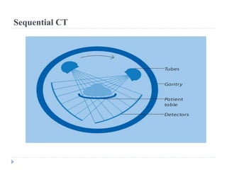


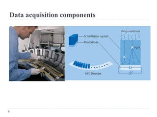


CT scans use X-rays and computers to create detailed images of the inside of the body. CT scanning was independently developed in the 1970s by Godfrey Hounsfield and Allan Cormack. A CT scan works by using an X-ray device and detector that rotate around the body, detecting differences in radiation absorption as they pass through tissues. The data is then used to construct a series of cross-sectional images of the bones, muscles, fat, and organs inside the body. CT scans allow doctors to see internal structures in great detail and find diseases that may not be visible on regular X-rays.


![Human Reproduction [ Reproductive System ] Notes @irfanullah_mehar Irfanullah...](https://cdn.slidesharecdn.com/ss_thumbnails/humanreproductionreproductivesystemnotesirfanullahmeharirfanullahmeharjanantantra-260111172350-56e85778-thumbnail.jpg?width=640&height=640&fit=bounds)


