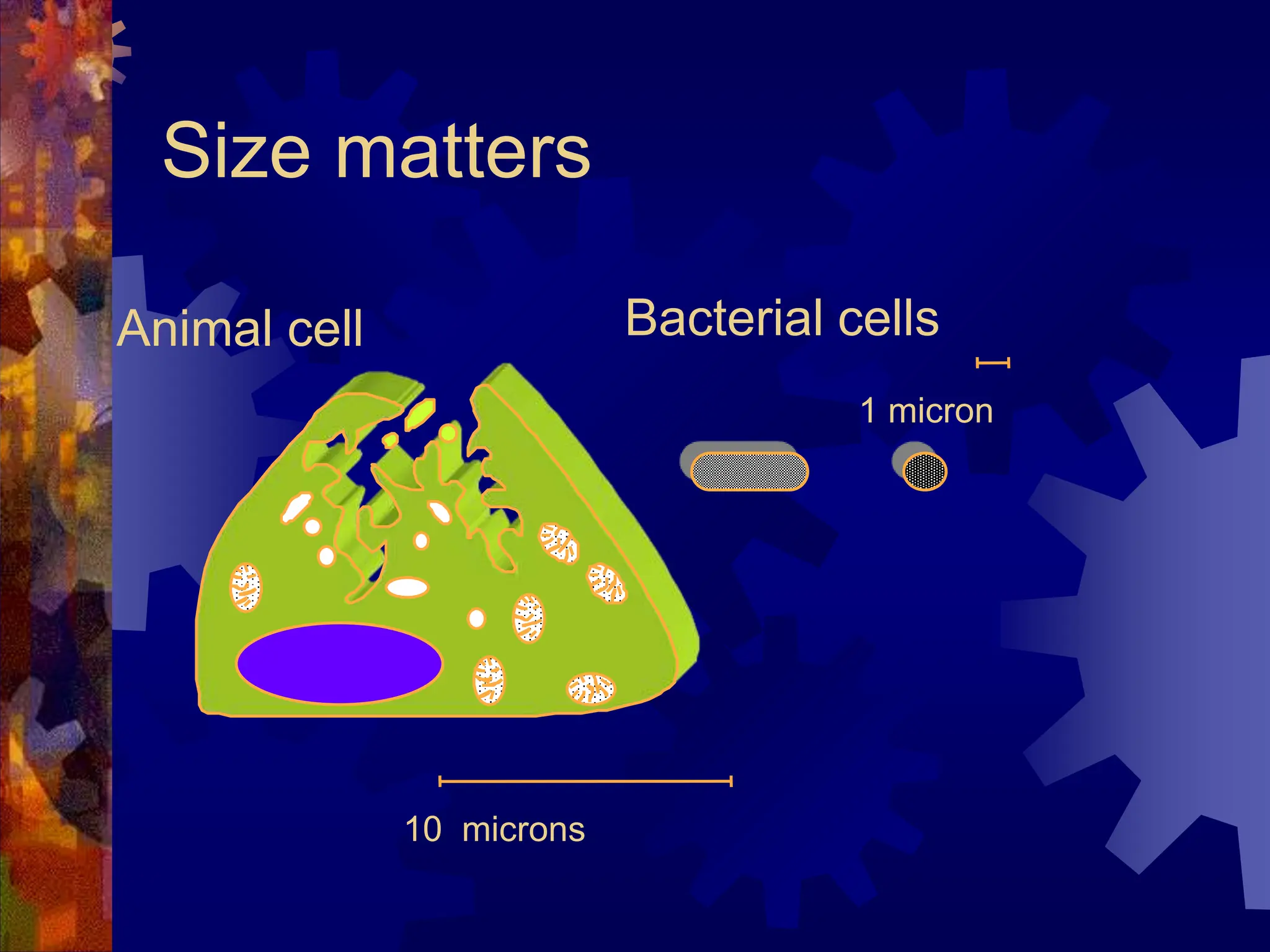
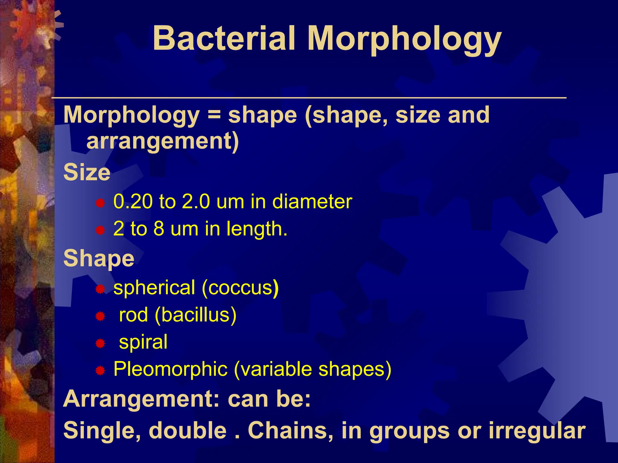
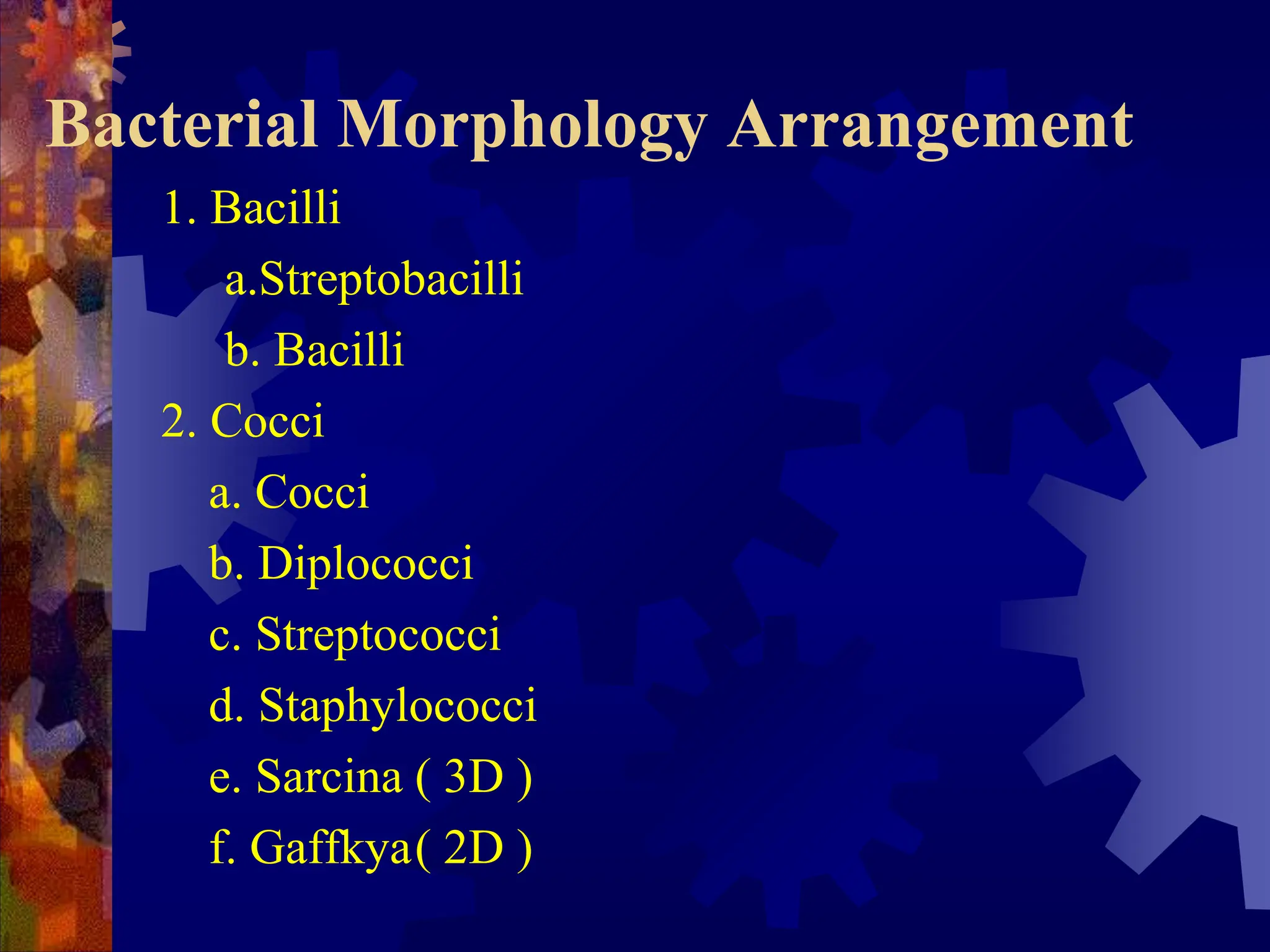
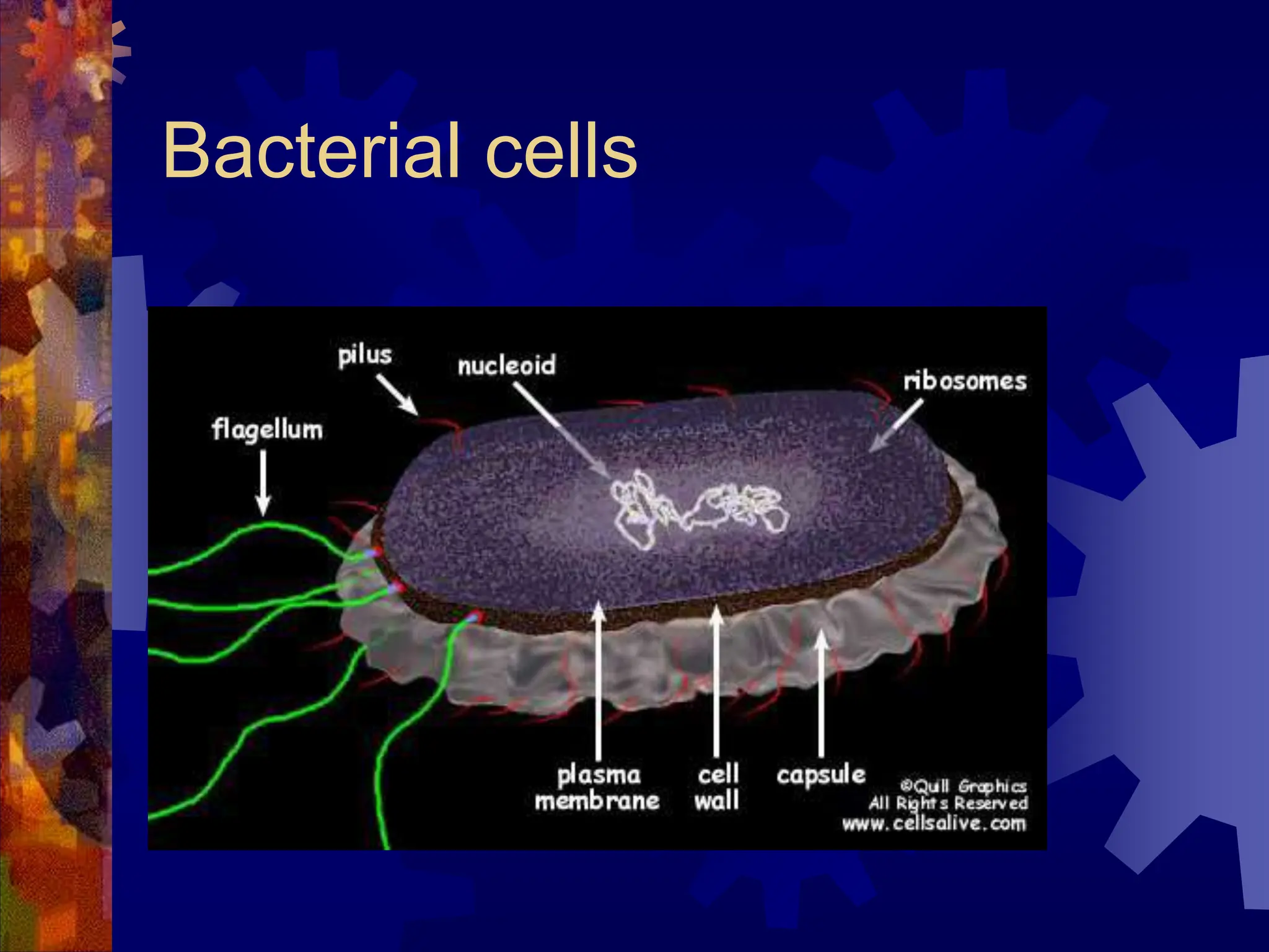
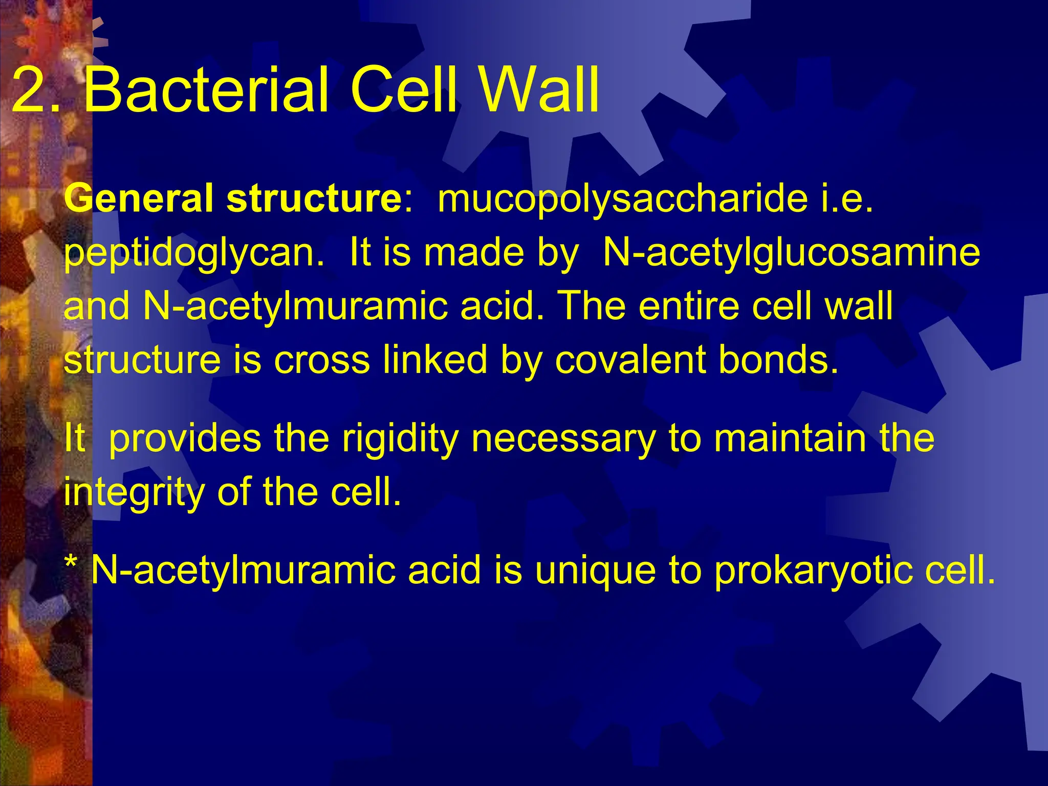
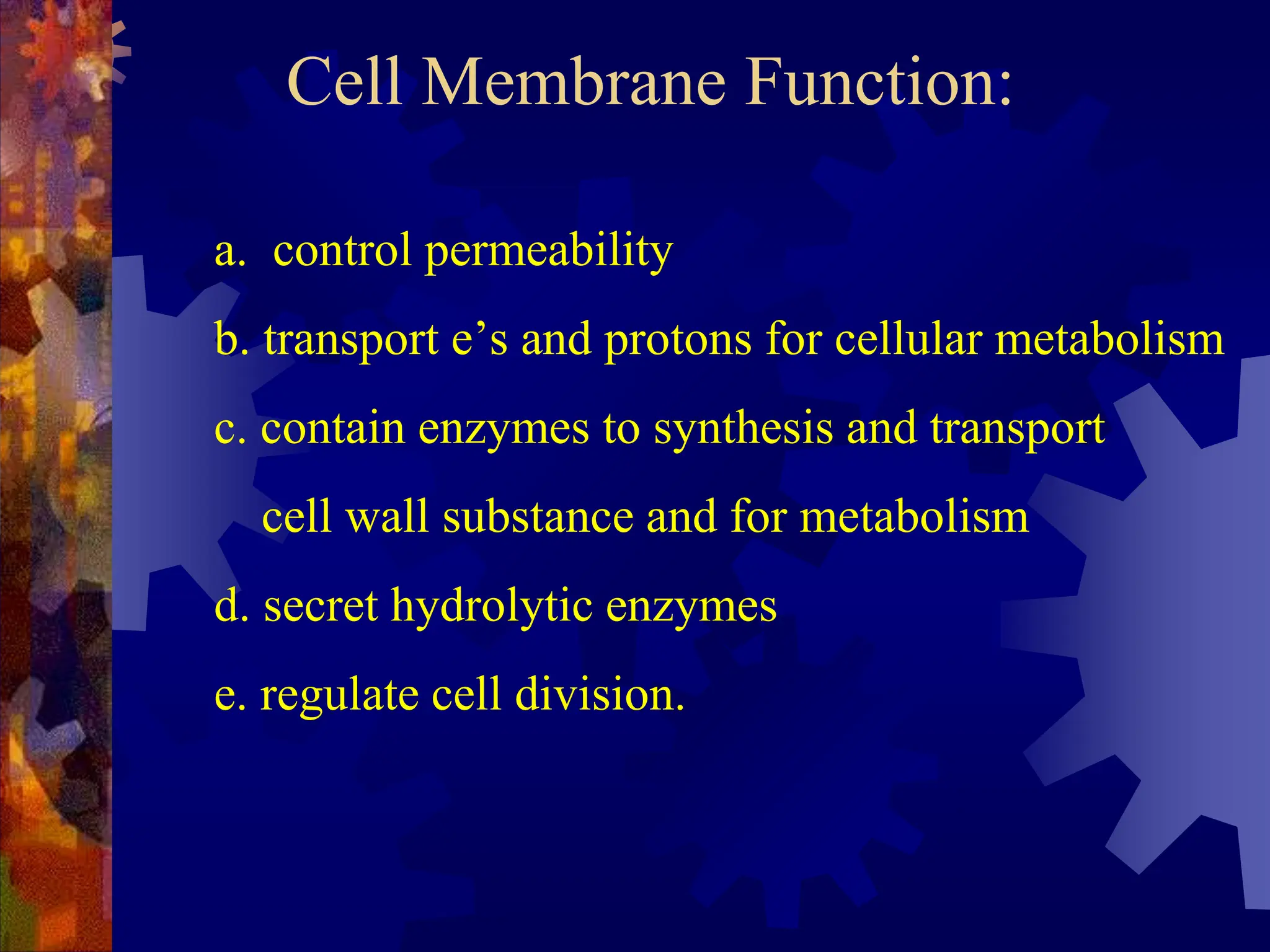
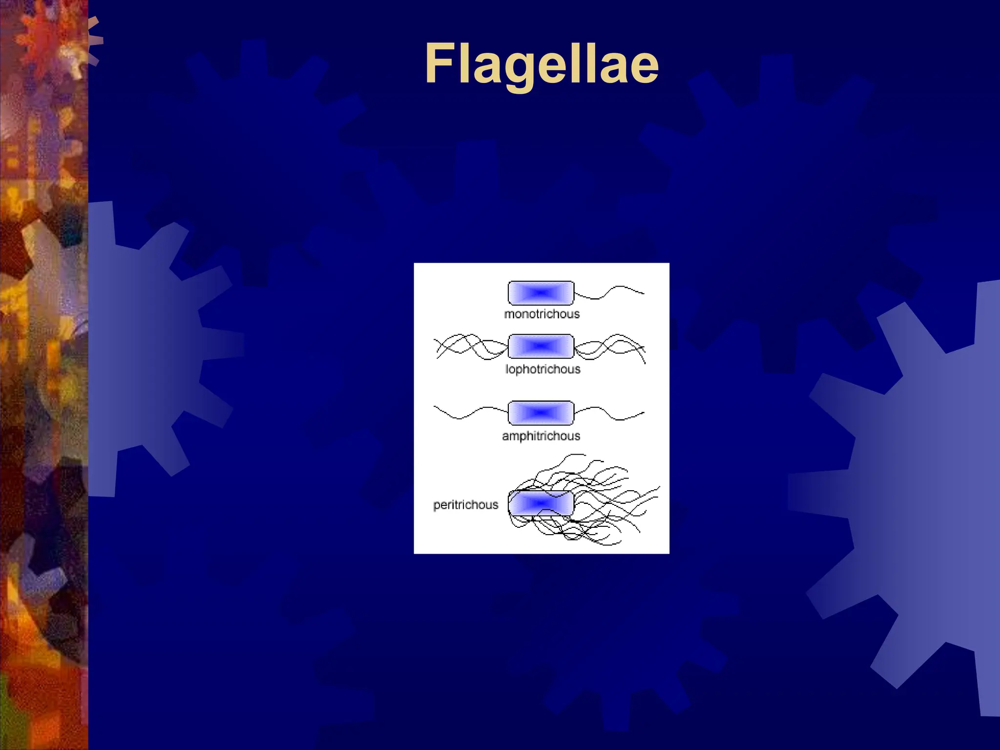
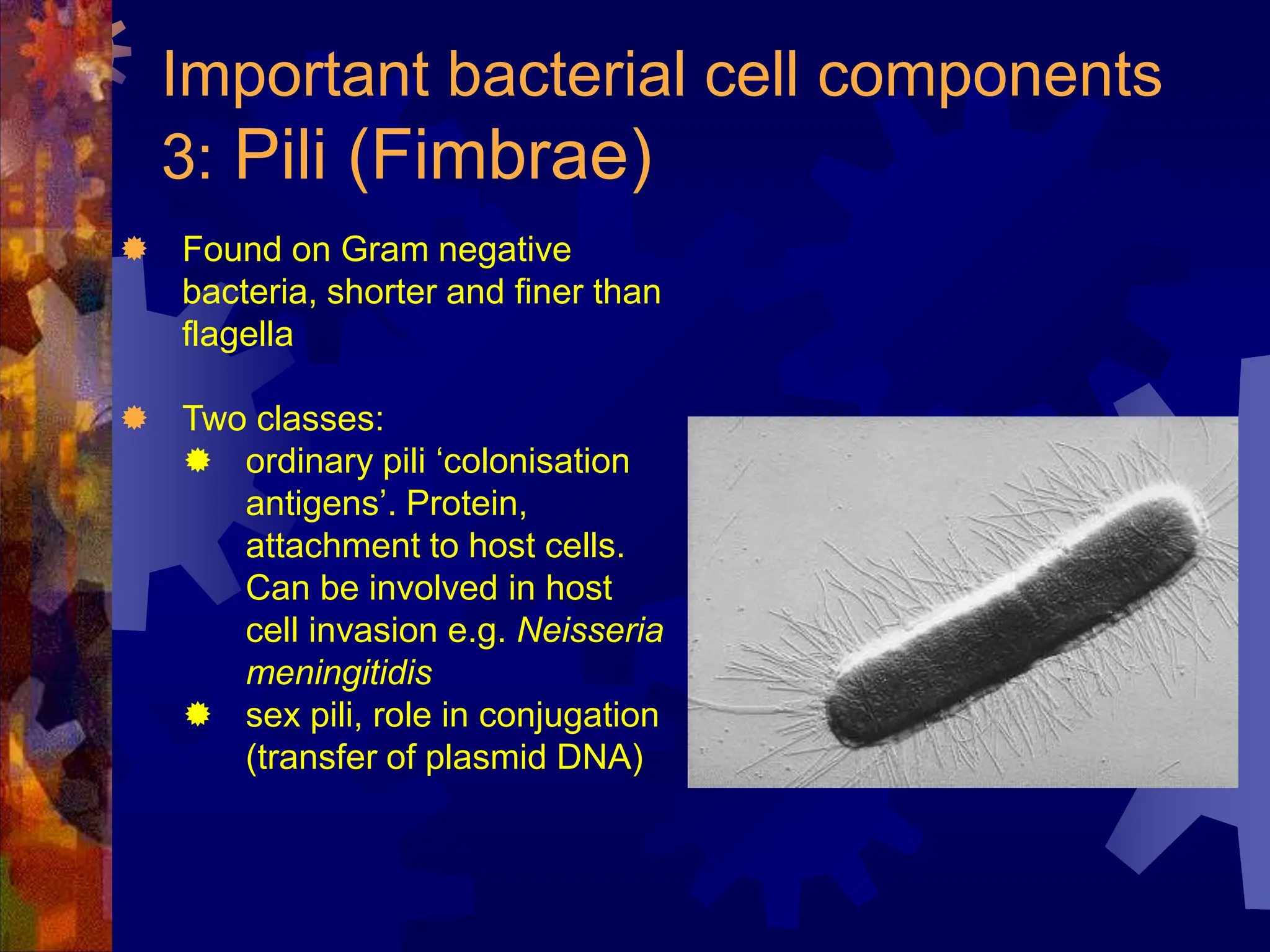
MICROOBIOLOGY
Dr.S.Sundaresan
Tagore Medical College and Hospital
Bacterial Cell Structures &
Morphology
Bacteria are:
 Unicellular, prokaryotes
 Have both DNA and RNA
 Divide by Binary fission
 No mitochondria
 Rigid cell wall containing
peptidoglycan
Size matters
Animal cell
1 micron
10 microns
Bacterial cells
Bacterial Morphology and Structure
Bacterial Morphology
Morphology = shape (shape, size and
arrangement)
Size
 0.20 to 2.0 um in diameter
 2 to 8 um in length.
Shape
 spherical (coccus)
 rod (bacillus)
 spiral
 Pleomorphic (variable shapes)
Arrangement: can be:
Single, double . Chains, in groups or irregular
Common Shapes & Arrangement
Bacterial Morphology Arrangement
1. Bacilli
a.Streptobacilli
b. Bacilli
2. Cocci
a. Cocci
b. Diplococci
c. Streptococci
d. Staphylococci
e. Sarcina ( 3D )
f. Gaffkya( 2D )
Bacterial Cell Structure
 Surface layers - capsule, cell wall, cell
membrane
 Cytoplasm - nuclear material, ribosome,
mesosome, inclusions etc.
 Appendages - flagella, pili or fimbriae
 Special structure - Spore
Bacterial cells
Bacterial Cell Structure
General structure: mucopolysaccharide i.e.
peptidoglycan. It is made by N-acetylglucosamine
and N-acetylmuramic acid. The entire cell wall
structure is cross linked by covalent bonds.
It provides the rigidity necessary to maintain the
integrity of the cell.
* N-acetylmuramic acid is unique to prokaryotic cell.
2. Bacterial Cell Wall
(a). Gram positive bacterial cell wall
Thick peptidoglycan layer
pentaglycin cross linkage.
All have lipoteichoic acid.
(b) Gram -negative
 Thin peptidoglycan
 Tetrapeptide cross linkage
 A second membrane structure: protein and
lipopolysaccharide
 Toxicity : endotoxin on lipid A of lipopolysaccharide.
glucosamine- glucosamine-long
 polysaccharide- repeated sequences of a few .
Cell walls of bacteria(1)
Cell Membrane Function:
a. control permeability
b. transport e’s and protons for cellular metabolism
c. contain enzymes to synthesis and transport
cell wall substance and for metabolism
d. secret hydrolytic enzymes
e. regulate cell division.
III. Cytoplasm
80% water, nucleic acids, proteins, carbohydrates, lipid
and inorganic ions etc.
1. Bacterial chromosomes
a single large circular double stranded DNA no histone
proteins. The only proteins associated with the
bacterial chromosomes are the ones for DNA
replication, transcription etc.
2. Ribosome
protein synthesis
The bacterial chromosome and
supercoiling
3. Mesosomes
A large invaginations of the plasma membrane,
irregular in shape.
a. increase in membrane surface, which may be
useful as a site for enzyme activity in respiration
and transport.
b. may participate in cell replication by serving as a
place of attachment for the bacterial chromosome.
4. Inclusions
Not separate by a membrane but distinct.
Granules of various kinds:
* glycogen,
*polyhydroxybutyric acid droplets (PHB)
i.e. fat droplets
* inorganic metaphosphate (metachromatic granules) - in
general, starvation of cell for almost any nutrients
leads to the formation of this to serve as an
intracellular phosphate reservoir.
II. CELL SURFACE LAYER
1. Capsule or slime layer
Many bacteria are able to secrete material
that adheres to the bacterial cell but is
actually external to the cell.
It consists of polypeptide and
polysaccharide on bacilli. Most of them
have only polysaccharide. It is a protective
layer that resists host phagocytosis.
Medically important.
Appendages
1. flagella
Some rods and spiral form have this.
a). function: motility
b). origin : cell membrane flagella attach to the cell by hook
and basal body which consists of set(s) of rings and rods
Gram - : 2 sets of ring and rods, L, P, S, M rings and
rods
e.g. E. coli
Gram + : S, M rings and rods
e.g. B. megaterium
Organ of bacterial locomotion
Structure of the flagellum
Flagella movement(1)
Flagella movement(2)
Flagella movement(3)
b).Origin (continued)
– The structure of the bacterial flagella allows it to spin
like a propeller and thereby propel the bacterial cell;
clockwise or counter clockwise ( Eucaryotic , wave
like motion.
– Bacterial flagella provides the bacterium with
mechanism for swimming toward or away from
chemical stimuli, a behavior is knows as
CHEMOTAXIX, chemosenors in the cell envelope
can detect certain chemicals and signal the flagella to
respond.
c). position
monotrichous
lophotrichous
peritrichous
d). structure
protein in nature: subunit flagellin
Flagellae
Flagellae
2. Pili or Fimbriae
Shorter than flagella and straighter , smaller.
Only on some gram- bacteria.
a). function: adhere. One of the invasive
mechanism on bacteria. Some pathogens
cause diseases due to this. If mutant
(fimbriae) not virulent. Prevent
phagocytosis.
Important bacterial cell components
3: Pili (Fimbrae)
 Found on Gram negative
bacteria, shorter and finer than
flagella
 Two classes:
 ordinary pili ‘colonisation
antigens’. Protein,
attachment to host cells.
Can be involved in host
cell invasion e.g. Neisseria
meningitidis
 sex pili, role in conjugation
(transfer of plasmid DNA)
pili - sex factor. If they make pili, they are + or
donors of F factor.
It is necessary for bacterial conjugation
resulting in the transfer of DNA from one cell to
another.
It have been implicated in the ability of
bacteria to recognize specific receptor sites on
the host cell membrane. In addition, number of
bacteria virus infect only those bacteria have F
pilus.
b). Origin: Cell membrane
c). Position: common pili , numerous over the cell,
usually called fimbriae sex pile, 1-4/cell
d). Structure: composed of proteins which can be
dissociated into smaller unit
Pilin . It belongs to a class of protein Lectin which
bond to cell surface polysaccharide.
IV. Special Structure
* Endospores
Spore former: sporobactobacilli and sporosarcinae - no
medical importance. bacillus and clostridium have
medical importance.
* Position: median, sub-terminal and terminal have
small water, high calcium content and dipicolinic acid
(calcium dipicolinate)
extremely resistant to heat, UV, chemicals etc. may be
due to many S containing A.A for disulfide groups.
Gram positive, bacillus,
anaerobic, spores
Clostridium spp
• After the active growth period approaching
the stationary growth phase, a structure
called forespore develops within the cells.
• It consists of coat, cortex and nuclear
structure.
The process of endospore
formation
Vegetative/spore-containing
cells(1)
Vegetative/spore-containing
cells(2)

Bacteria Structure, Cell wall, Cell Membrane, Cytoplasm, Ribosomes.pptx

  • 1.
  • 2.
  • 3.
    Bacteria are:  Unicellular,prokaryotes  Have both DNA and RNA  Divide by Binary fission  No mitochondria  Rigid cell wall containing peptidoglycan
  • 4.
    Size matters Animal cell 1micron 10 microns Bacterial cells
  • 5.
  • 6.
    Bacterial Morphology Morphology =shape (shape, size and arrangement) Size  0.20 to 2.0 um in diameter  2 to 8 um in length. Shape  spherical (coccus)  rod (bacillus)  spiral  Pleomorphic (variable shapes) Arrangement: can be: Single, double . Chains, in groups or irregular
  • 7.
    Common Shapes &Arrangement
  • 8.
    Bacterial Morphology Arrangement 1.Bacilli a.Streptobacilli b. Bacilli 2. Cocci a. Cocci b. Diplococci c. Streptococci d. Staphylococci e. Sarcina ( 3D ) f. Gaffkya( 2D )
  • 9.
    Bacterial Cell Structure Surface layers - capsule, cell wall, cell membrane  Cytoplasm - nuclear material, ribosome, mesosome, inclusions etc.  Appendages - flagella, pili or fimbriae  Special structure - Spore
  • 10.
  • 11.
  • 12.
    General structure: mucopolysaccharidei.e. peptidoglycan. It is made by N-acetylglucosamine and N-acetylmuramic acid. The entire cell wall structure is cross linked by covalent bonds. It provides the rigidity necessary to maintain the integrity of the cell. * N-acetylmuramic acid is unique to prokaryotic cell. 2. Bacterial Cell Wall
  • 13.
    (a). Gram positivebacterial cell wall Thick peptidoglycan layer pentaglycin cross linkage. All have lipoteichoic acid.
  • 14.
    (b) Gram -negative Thin peptidoglycan  Tetrapeptide cross linkage  A second membrane structure: protein and lipopolysaccharide  Toxicity : endotoxin on lipid A of lipopolysaccharide. glucosamine- glucosamine-long  polysaccharide- repeated sequences of a few .
  • 15.
    Cell walls ofbacteria(1)
  • 16.
    Cell Membrane Function: a.control permeability b. transport e’s and protons for cellular metabolism c. contain enzymes to synthesis and transport cell wall substance and for metabolism d. secret hydrolytic enzymes e. regulate cell division.
  • 17.
    III. Cytoplasm 80% water,nucleic acids, proteins, carbohydrates, lipid and inorganic ions etc. 1. Bacterial chromosomes a single large circular double stranded DNA no histone proteins. The only proteins associated with the bacterial chromosomes are the ones for DNA replication, transcription etc. 2. Ribosome protein synthesis
  • 18.
    The bacterial chromosomeand supercoiling
  • 19.
    3. Mesosomes A largeinvaginations of the plasma membrane, irregular in shape. a. increase in membrane surface, which may be useful as a site for enzyme activity in respiration and transport. b. may participate in cell replication by serving as a place of attachment for the bacterial chromosome.
  • 20.
    4. Inclusions Not separateby a membrane but distinct. Granules of various kinds: * glycogen, *polyhydroxybutyric acid droplets (PHB) i.e. fat droplets * inorganic metaphosphate (metachromatic granules) - in general, starvation of cell for almost any nutrients leads to the formation of this to serve as an intracellular phosphate reservoir.
  • 21.
    II. CELL SURFACELAYER 1. Capsule or slime layer Many bacteria are able to secrete material that adheres to the bacterial cell but is actually external to the cell. It consists of polypeptide and polysaccharide on bacilli. Most of them have only polysaccharide. It is a protective layer that resists host phagocytosis. Medically important.
  • 22.
    Appendages 1. flagella Some rodsand spiral form have this. a). function: motility b). origin : cell membrane flagella attach to the cell by hook and basal body which consists of set(s) of rings and rods Gram - : 2 sets of ring and rods, L, P, S, M rings and rods e.g. E. coli Gram + : S, M rings and rods e.g. B. megaterium
  • 23.
  • 24.
  • 25.
  • 26.
  • 27.
  • 28.
    b).Origin (continued) – Thestructure of the bacterial flagella allows it to spin like a propeller and thereby propel the bacterial cell; clockwise or counter clockwise ( Eucaryotic , wave like motion. – Bacterial flagella provides the bacterium with mechanism for swimming toward or away from chemical stimuli, a behavior is knows as CHEMOTAXIX, chemosenors in the cell envelope can detect certain chemicals and signal the flagella to respond.
  • 29.
  • 30.
  • 31.
  • 32.
    2. Pili orFimbriae Shorter than flagella and straighter , smaller. Only on some gram- bacteria. a). function: adhere. One of the invasive mechanism on bacteria. Some pathogens cause diseases due to this. If mutant (fimbriae) not virulent. Prevent phagocytosis.
  • 33.
    Important bacterial cellcomponents 3: Pili (Fimbrae)  Found on Gram negative bacteria, shorter and finer than flagella  Two classes:  ordinary pili ‘colonisation antigens’. Protein, attachment to host cells. Can be involved in host cell invasion e.g. Neisseria meningitidis  sex pili, role in conjugation (transfer of plasmid DNA)
  • 34.
    pili - sexfactor. If they make pili, they are + or donors of F factor. It is necessary for bacterial conjugation resulting in the transfer of DNA from one cell to another. It have been implicated in the ability of bacteria to recognize specific receptor sites on the host cell membrane. In addition, number of bacteria virus infect only those bacteria have F pilus.
  • 35.
    b). Origin: Cellmembrane c). Position: common pili , numerous over the cell, usually called fimbriae sex pile, 1-4/cell d). Structure: composed of proteins which can be dissociated into smaller unit Pilin . It belongs to a class of protein Lectin which bond to cell surface polysaccharide.
  • 36.
    IV. Special Structure *Endospores Spore former: sporobactobacilli and sporosarcinae - no medical importance. bacillus and clostridium have medical importance. * Position: median, sub-terminal and terminal have small water, high calcium content and dipicolinic acid (calcium dipicolinate) extremely resistant to heat, UV, chemicals etc. may be due to many S containing A.A for disulfide groups.
  • 37.
  • 38.
    • After theactive growth period approaching the stationary growth phase, a structure called forespore develops within the cells. • It consists of coat, cortex and nuclear structure. The process of endospore formation
  • 39.
  • 40.