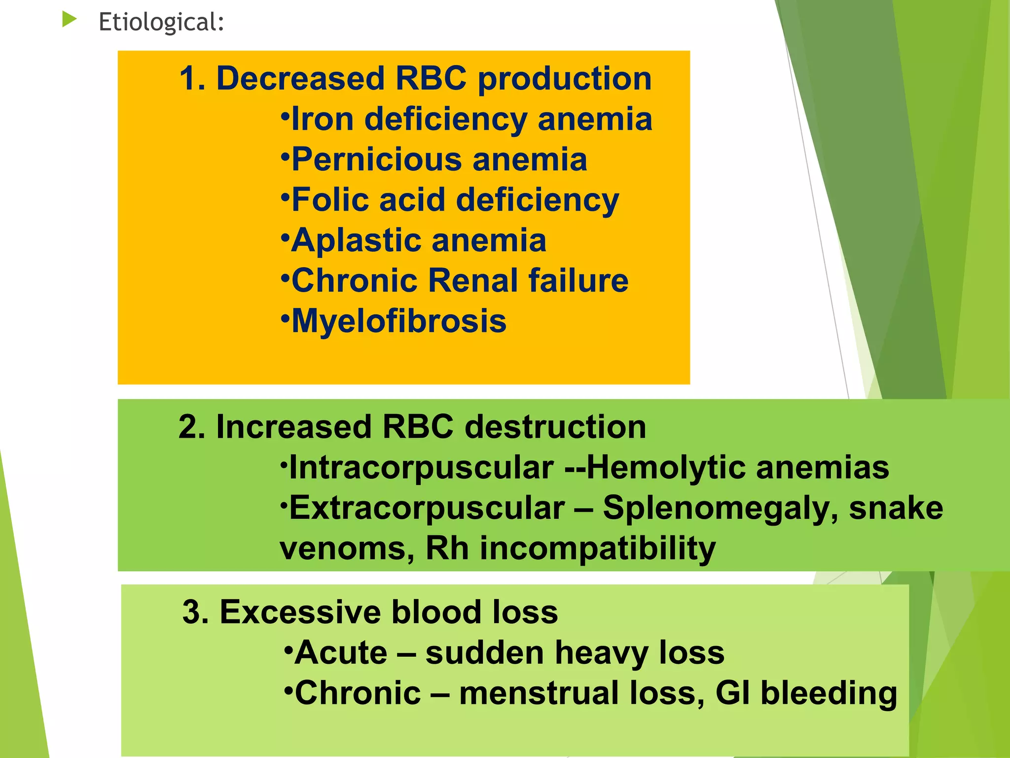
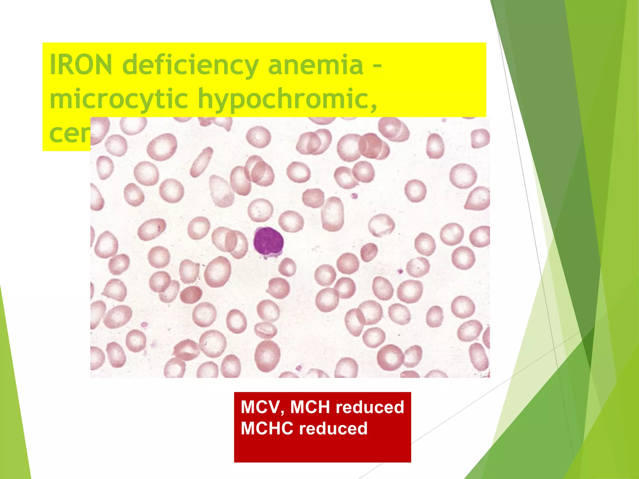
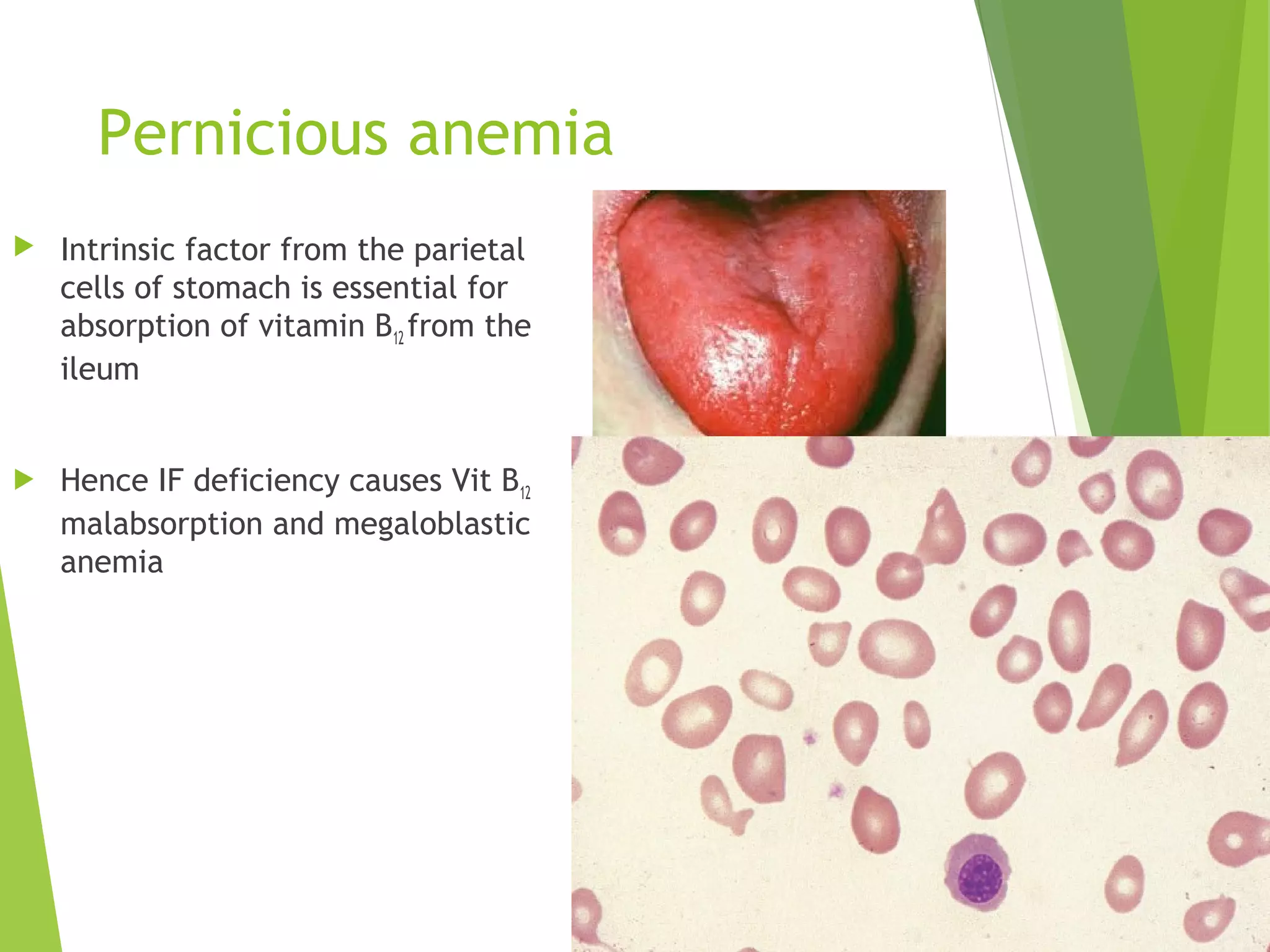
ANEMIA
Dr.Sundarsan
Pathology
Loyola college
ANEMIA
Specific Learning Objectives
At the end of the class, the student should be able to
 Define anemia
 Discuss the classification of anemia
 Enumerate the blood cell indices
 Discuss the cause, morphological features and treatment of various forms of
anemia
 Explain the effects of anemia
 Explain the basis of physiological anemia of pregnancy
Definition
Qualitative or quantitative reduction in RBC count or
Hemoglobin levels or both resulting in decrease in
oxygen carrying capacity of blood
Normal – Males  14 to 16 g/dL
Females  12 to 14 g/dL
Clinical features
 Fatigue, Tiredeness
 Breathlessness
 Lethargy,headache
 Pallor of skin and
mucous membranes
 Palpitations
 Menstrual disturbances
 Reduction in
BMR
Investigations
 Hb
 RBC count
 Blood indices
 Peripheral Blood smear
 Serum Iron, Ferritin, Vitamin B12 and folate levels
 Plasma Electrophoresis
Classification of anemia
 Morphological - according to the shape and amount of
Hb
 Etiological – according to the cause of anemia
 Morphological:
1. Normocytic normochromic
•Hemolytic anemias
•After blood loss
•Aplastic anemia
2. Microcytic hypochromic
•Iron deficiency anemia
•Thalassemia
3. Macrocytic normochromic
•Megaloblastic anemia
•Folic acid deficiency
 Etiological:
1. Decreased RBC production
•Iron deficiency anemia
•Pernicious anemia
•Folic acid deficiency
•Aplastic anemia
•Chronic Renal failure
•Myelofibrosis
2. Increased RBC destruction
•Intracorpuscular --Hemolytic anemias
•Extracorpuscular – Splenomegaly, snake
venoms, Rh incompatibility
3. Excessive blood loss
•Acute – sudden heavy loss
•Chronic – menstrual loss, GI bleeding
Blood indices
Indices Formula Normal
vaule
Mean Corpuscular
Volume – average
volume of a single
RBC
80 to 100
fL
Mean
Corpuscular
Hemoglobin-
amount of Hb in a
single RBC
26 to 32
pg
Mean
Corpuscular
Hemoglobin
Concentration –
concentration of Hb
in a packed volume
of RBCs
32 to 36
%
IRON deficiency anemia
• Inadequate iron intake
• Hookworm infestation (children)
• Heavy menstrual loss
• Bleeding Hemorrhoids (piles)
• Peptic ulcer bleed
IRON deficiency anemia –
microcytic hypochromic,
central pallor
MCV, MCH reduced
MCHC reduced
Treatment
 Oral FeSO4 tablets
 Parenteral iron therapy
 Blood transfusion
Response to treatment
 Reticulocytes – 0.5 to 1.5 %
Megaloblastic anemia
• Def of B12, FA, IF
• nutritional
• Gastric atrophy, gastrectomy
• tropical sprue  malsbsortption
• Sources of B12 –milk, meat, liver of animals
• Sources of FA – leafy veg, pulses, liver
 Methylene Tetrahydrofolate is essential for formation of DNA
 Formation of this requires vitamin B12 as a cofactor
 Hence deficiency of B12 or Folic acid both causes defect in formation and
maturation of DNA
• Slow reproduction of RBCs
• Red cells too large with odd shapes with friable membranes
 Formation of megaloblasts
MCV, MCH increased
MCHC normal
Megaloblasts +
hypersegmented neutrophils
Pernicious anemia
 Intrinsic factor from the parietal
cells of stomach is essential for
absorption of vitamin B12 from the
ileum
 Hence IF deficiency causes Vit B12
malabsorption and megaloblastic
anemia
Megaloblastic anemia
 Clinical features:
 Fatigue,
breathlessness
 Soreness of tongue
 Glossitis, angular
stomatitis
 Tingling of hands and
feet due to
peripheral neuropathy
 Severe  Subacute
Combined
Degeneration of Spinal
cord (SACD)
• Treatment –
• FA tabs
• Vit B12
injections
Please note…
 Folate deficiency during pregnancy is associated with
Neural tube defects in babies
 Hence Folate supplementation before and during
pregnancy is mandatory along with iron tablets
Aplastic anemia
• X-rays
• drugs
• Gamma radiations from nuclear
bombs
• chemicals from industries
Treatment:
•Antithymocyte globulin
• BM transplant
Microcytic hypochromic
Sickle cell anemia
 Valine is present instead of glutamic acid in the 6th
position of beta chain – HbS
• HbS – heterozygous – only half of Hb is abnormal
-- homozygous – all are abnormal hbs
• In deoxygenated state, HbS tends to polymerize,
forms sickle shaped cells, hemolyze and blocks
blood vessels
• Treatment
• HbF – decreases polymerization
• Hydroxyurea – increases HbF synthesis
• Severe – bone marrow transplantation
Thalassemia
 Defect in the regulatory portion of globin chain
• α Thalassemia – rare
 β Thalassemia
 Major – complete absence of β chain
- minor – reduced synthesis of β chain
 Compensatory increase in HbA2
and HbF
 Microcytic hypochromic anemia
 Failure to thrive
• Treatment:
• blood transfusion
• Splenectomy
• BM transplantation
Hereditary spherocytosis
•Mutation in spectrin, the
transmembrane protein
which maintains the shape
of the RBC cell membrane
•And the linker protein,
ankyrin
•Spherocytes, trapped and
hemolyze in spleen
•Treatment : splenectomy
G6PDH deficiency
• G6PDH is required for generation of NADPH which maintains RBC fragility
• NADPH maintains Glutathione in reduced state
• Def of G6PD leads to def of NADPH
• RBC becomes more friable
• Hemolysis
• Especially when exposed to drugs and toxins, infections like malaria
Anemia of chronic disease
 Chronic renal failure
 Normocytic normochromic anemia
 Treat the cause
 Erythropoeitin therapy
Severe anemia leads to
cardiac failure
• Normal viscosity of
blood -- 3 times that
of waterr
• In anemia - falls to
1.5 times that of
water
• Decrease in
peripheral resistance
 increase in cardiac
output
• And also ,hypoxia
induced peripheral
vasodilation increases
cardiac output
• Increase in cardiac output
maintains enough oxygenation
of tissues
• However it also increases
cardiac workload
• During exercise, the
compensation cannot be
done , hence leads to heart
failure

Anemia

  • 1.
  • 2.
  • 3.
    Specific Learning Objectives Atthe end of the class, the student should be able to  Define anemia  Discuss the classification of anemia  Enumerate the blood cell indices  Discuss the cause, morphological features and treatment of various forms of anemia  Explain the effects of anemia  Explain the basis of physiological anemia of pregnancy
  • 4.
    Definition Qualitative or quantitativereduction in RBC count or Hemoglobin levels or both resulting in decrease in oxygen carrying capacity of blood Normal – Males  14 to 16 g/dL Females  12 to 14 g/dL
  • 5.
    Clinical features  Fatigue,Tiredeness  Breathlessness  Lethargy,headache  Pallor of skin and mucous membranes  Palpitations  Menstrual disturbances  Reduction in BMR
  • 6.
    Investigations  Hb  RBCcount  Blood indices  Peripheral Blood smear  Serum Iron, Ferritin, Vitamin B12 and folate levels  Plasma Electrophoresis
  • 7.
    Classification of anemia Morphological - according to the shape and amount of Hb  Etiological – according to the cause of anemia
  • 8.
     Morphological: 1. Normocyticnormochromic •Hemolytic anemias •After blood loss •Aplastic anemia 2. Microcytic hypochromic •Iron deficiency anemia •Thalassemia 3. Macrocytic normochromic •Megaloblastic anemia •Folic acid deficiency
  • 9.
     Etiological: 1. DecreasedRBC production •Iron deficiency anemia •Pernicious anemia •Folic acid deficiency •Aplastic anemia •Chronic Renal failure •Myelofibrosis 2. Increased RBC destruction •Intracorpuscular --Hemolytic anemias •Extracorpuscular – Splenomegaly, snake venoms, Rh incompatibility 3. Excessive blood loss •Acute – sudden heavy loss •Chronic – menstrual loss, GI bleeding
  • 10.
    Blood indices Indices FormulaNormal vaule Mean Corpuscular Volume – average volume of a single RBC 80 to 100 fL Mean Corpuscular Hemoglobin- amount of Hb in a single RBC 26 to 32 pg Mean Corpuscular Hemoglobin Concentration – concentration of Hb in a packed volume of RBCs 32 to 36 %
  • 11.
    IRON deficiency anemia •Inadequate iron intake • Hookworm infestation (children) • Heavy menstrual loss • Bleeding Hemorrhoids (piles) • Peptic ulcer bleed
  • 12.
    IRON deficiency anemia– microcytic hypochromic, central pallor MCV, MCH reduced MCHC reduced
  • 15.
    Treatment  Oral FeSO4tablets  Parenteral iron therapy  Blood transfusion
  • 16.
    Response to treatment Reticulocytes – 0.5 to 1.5 %
  • 17.
    Megaloblastic anemia • Defof B12, FA, IF • nutritional • Gastric atrophy, gastrectomy • tropical sprue  malsbsortption • Sources of B12 –milk, meat, liver of animals • Sources of FA – leafy veg, pulses, liver
  • 19.
     Methylene Tetrahydrofolateis essential for formation of DNA  Formation of this requires vitamin B12 as a cofactor  Hence deficiency of B12 or Folic acid both causes defect in formation and maturation of DNA • Slow reproduction of RBCs • Red cells too large with odd shapes with friable membranes  Formation of megaloblasts
  • 20.
  • 21.
  • 22.
    Pernicious anemia  Intrinsicfactor from the parietal cells of stomach is essential for absorption of vitamin B12 from the ileum  Hence IF deficiency causes Vit B12 malabsorption and megaloblastic anemia
  • 23.
    Megaloblastic anemia  Clinicalfeatures:  Fatigue, breathlessness  Soreness of tongue  Glossitis, angular stomatitis  Tingling of hands and feet due to peripheral neuropathy  Severe  Subacute Combined Degeneration of Spinal cord (SACD) • Treatment – • FA tabs • Vit B12 injections
  • 24.
    Please note…  Folatedeficiency during pregnancy is associated with Neural tube defects in babies  Hence Folate supplementation before and during pregnancy is mandatory along with iron tablets
  • 25.
    Aplastic anemia • X-rays •drugs • Gamma radiations from nuclear bombs • chemicals from industries Treatment: •Antithymocyte globulin • BM transplant Microcytic hypochromic
  • 26.
    Sickle cell anemia Valine is present instead of glutamic acid in the 6th position of beta chain – HbS • HbS – heterozygous – only half of Hb is abnormal -- homozygous – all are abnormal hbs • In deoxygenated state, HbS tends to polymerize, forms sickle shaped cells, hemolyze and blocks blood vessels • Treatment • HbF – decreases polymerization • Hydroxyurea – increases HbF synthesis • Severe – bone marrow transplantation
  • 29.
    Thalassemia  Defect inthe regulatory portion of globin chain • α Thalassemia – rare  β Thalassemia  Major – complete absence of β chain - minor – reduced synthesis of β chain  Compensatory increase in HbA2 and HbF  Microcytic hypochromic anemia  Failure to thrive • Treatment: • blood transfusion • Splenectomy • BM transplantation
  • 30.
    Hereditary spherocytosis •Mutation inspectrin, the transmembrane protein which maintains the shape of the RBC cell membrane •And the linker protein, ankyrin •Spherocytes, trapped and hemolyze in spleen •Treatment : splenectomy
  • 31.
    G6PDH deficiency • G6PDHis required for generation of NADPH which maintains RBC fragility • NADPH maintains Glutathione in reduced state • Def of G6PD leads to def of NADPH • RBC becomes more friable • Hemolysis • Especially when exposed to drugs and toxins, infections like malaria
  • 32.
    Anemia of chronicdisease  Chronic renal failure  Normocytic normochromic anemia  Treat the cause  Erythropoeitin therapy
  • 33.
    Severe anemia leadsto cardiac failure • Normal viscosity of blood -- 3 times that of waterr • In anemia - falls to 1.5 times that of water • Decrease in peripheral resistance  increase in cardiac output • And also ,hypoxia induced peripheral vasodilation increases cardiac output • Increase in cardiac output maintains enough oxygenation of tissues • However it also increases cardiac workload • During exercise, the compensation cannot be done , hence leads to heart failure

Editor's Notes

  • #12 Treatment: oral iron FeSO4 300 mg tid
  • #17 The rate of erythropoiesis is measured by a reticulocyte count. Normally, a little less than 1% of the oldest RBCs are replaced by newcomer reticulocytes on any given day. It then takes 1 to 2 days for the reticulocytes to lose the last vestiges of endoplasmic reticulum and become mature RBCs. Thus, reticulocytes account for about 0.5–1.5% of all RBCs in a normal blood sample. A low “retic” count in a person who is anemic might indicate a shortage of erythropoietin or an inability of the red bone marrow to respond to EPO, perhaps because of a nutritional deficiency or leukemia. A high “retic” count might indicate a good red bone marrow response to previous blood loss or to iron therapy in someone who had been iron-deficient. It could also point to illegal use of epoetin alfa by an athlete. •
  • #18 Def of B12, FA, IF Slow reproduction of RBCs Red cells too large with odd shapes with friable membranes Gastric atrophy, gastrectomy tropical sprue  malsbsortption
  • #23 Treatment – FA tabs 1 mg qd Vit B12 inj – 100 micro g im qd for 7 days
  • #26 X-rays, drugs,nuclear bombs with gamma rad, chemicals from industries, HARRISON: Treatment: Antithymocyte globulin, BM transplant
  • #27 Valine is present instead of glutamic acid in the 6th position of beta chain – HbS HbC—lysine HbE – lysine but in 26th position HbS – heterozygous – only half of Hb is abnormal -- homozygous – all are abnormal hbs In deoxygenated state, HbS tends to polymerize, forms sickle shaped cells, hemolyze and blocks blood vessels Treatment HbF – decreases polymerization Hydroxyurea – increases HbF synthesis Severe – bone marrow transplantation
  • #30 Defect in the regulatory portion of globin chain Alpha and beta Major and minor respectively TreATMENT: blood transfusion Splenectomy BM transplantation
  • #31 GANONG: Or congenital hemolytic icterus Mutation in spectrin, the transmembrane protein which maintains the shape of the RBC cell membrane And the linker protein, ankyrin Spherocytes, trapped and hemolyze in spleen Treatment : splenectomy
  • #32 GANONG: G6pd required for oxdn of glu via HMP pathway This generates NADPH which maintains RBC fragility Def of G6PD leads to def of NADPH RBC becomes more friable Hemolysis Esp when exposed to drugs and toxins
  • #33 Treatment Treat cause Erythropoeitin
  • #34 Normal viscosity of blood -- 3 times that of waterr In anemia - falls to 1.5 times that of water Decrease in peripheral resistance  increase in cardiac output And also due to hypoxia induced peripheral vasodilation increases cardiac output Increase in cardiac output maintains enough oxygenation of tissues However it also increases cardiac workload During exercise, the compensation cannot be done , hence leads to heart failure