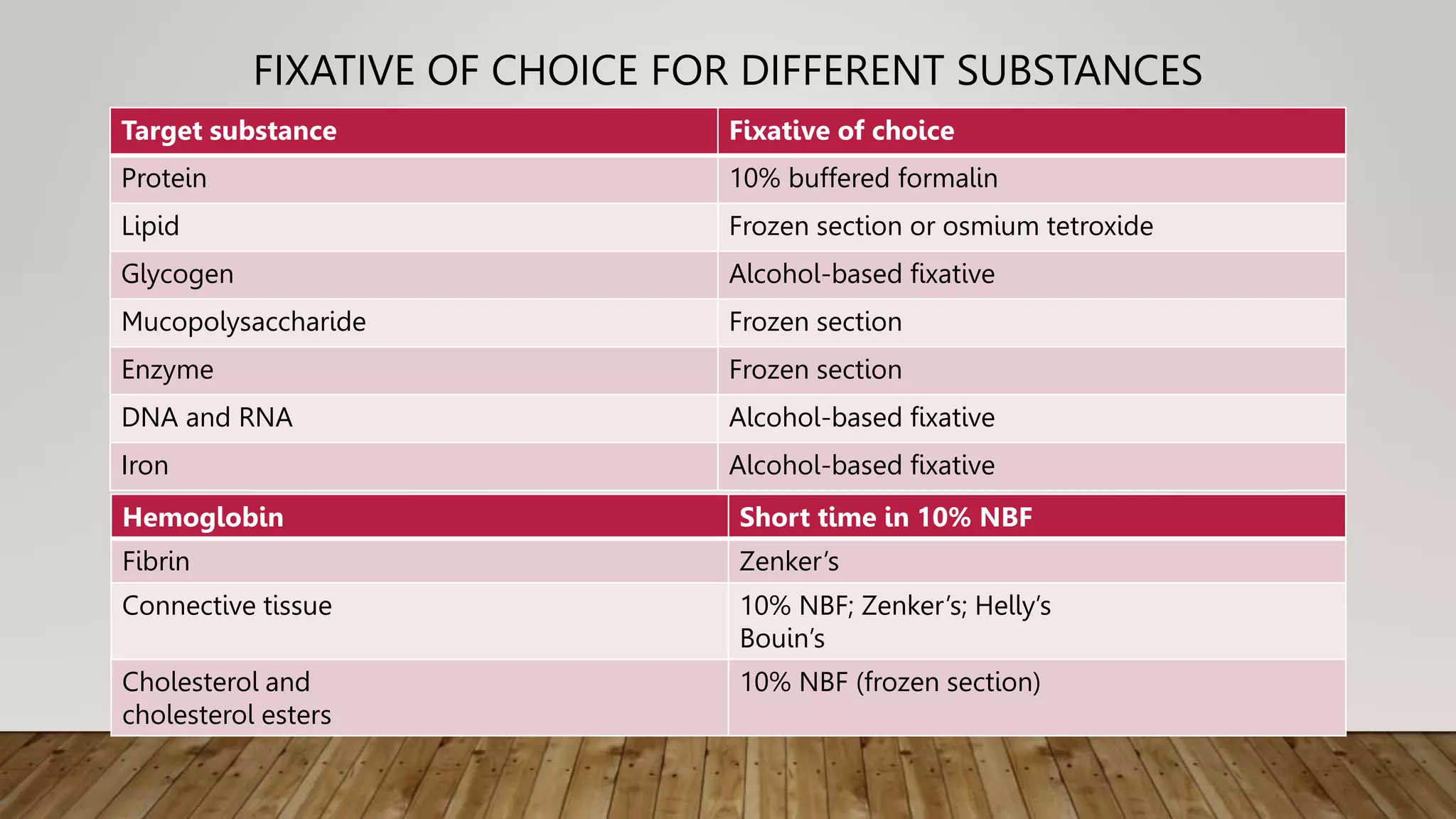
FIXATIVES
BY DR.S.SUNDARESAN
TAGORE MEDICAL COLLEGE
INTRODUCTION
• Fixation is the first step of any histological and cytological laboratory technique.
• The fixation helps to maintain the tissue nearest to its original state in the living
system.
AIMS OF FIXATION
The basic aims of fixation are the following:
 To preserve the tissue nearest to its living state
 To prevent any change in shape and size of the tissue at the time of processing
 To prevent any autolysis
 To make the tissue firm to hard
 To prevent any bacterial growth in the tissue
 To make it possible to have clear stain
 To have better optical quality of the cells
 To Prevent osmotic damage.
CHANGE IN TISSUE AFTER FIXATION
• VOLUME CHANGES
Shrinkage of the volume by formalin(33%). However the volume change may be due to (a) altered
membrane permeability, (b) inhibition of The enzymes responsible for respiration and (c) change of
transport Na+ ions. Glutaraldehyde and osmium tetroxide are used as fixations in epoxy resin then
70% increased of cell size is noted.
• HARDENING OF TISSUE
 Mild degree hardening may occur.
• INTERFERENCE OF STAINING
Formaldehyde inactivates 80% of ribonuclease enzyme. It has been noted that osmium tetroxide
inhibits haematoxylin and eosin staining.
• CHANGES OF OPTICAL DENSITY BY FIXATION
 Nuclei may look like hyperchromatic.
TYPES OF FIXATION
Types of fixative Classification
A. Nature of fixation • Immersion fixation
• Coating fixation
• Vapour fixation
• Perfusion fixation
• Freeze-drying
• Microwave fixation
B. Chemical properties Aldehyde: formaldehyde, glutaraldehyde
• Oxidising agent: osmium tetroxide
• Protein denaturing agent: ethyl alcohol, methyl alcohol
• Cross-linking agents: carbodiimide
• Miscellaneous: picric acid
C. Component present Simple(only one chemical present)
• Formaldehyde
• Ethyl alcohol
• Glutaraldehyde
• Picric acid
• Osmium tetroxide
Compound (more than one chemical present)
• Bouin’s fluid
• Carnoy’s solution
D. Action on tissue protein Coagulative: ethyl alcohol, picric acid
2. Noncoagulative: formaldehyde, osmium tetroxide, glutaraldehyde
DESCRIPTION OF NATURE OF FIXATION
1. Immersion fixation: This is the commonest way of fixation in the laboratories. 10% neutral buffered formalin or
cytology smear in 95% ethyl alcohol.
2. Coating fixation: This is commonly used in the cytology samples.
(a) Fixation of the cells (b) To impart a protective covering over the smear (c) No need to carry liquid fixative in bottle or jar
3. Vapour fixation: In this type of fixation, the vapour of chemical is used to fix either a smear or tissue section. The
vapour converts the soluble material to insoluble material, and these materials are retained when the smear comes in contact
with liquid solution.
4. Perfusion fixation: This is mainly used in research purpose. The organ such as the brain or spinal cord can also be fixed
by perfusion fixation.
5. Freeze-drying: In this technique the tissue is cut into thin sections and then rapidly frozen into a very low temperature.
Advantages: • Excellent for enzyme study, • No change of proteins • No shrinkage of tissue
• Preservation of glycogen
6. Microwave fixation -Microwave is a type of electromagnetic wave with frequencies between 300
MHz and 300 GHz. Preservation of the tissue antigen and good for immunohistochemistry.
FACTORS AFFECTING FIXATION
• PH of the fixative
Neutral pH is preferable.
pH 6–8 is the best range.
High acidity or alkalinity interferes fixation.
• Temperature
Room temperature suitable for routine work. no difference of cell morphology from 0 to 45 °C
High temperature facilitates fixation. (60–65 °C) antigen within the tissue may be destroyed
Low temperature (0–4 °C) suitable for enzyme histochemistry.
• Duration of fixation
Formalin fixes 1 mm/h.
Small tissue: 6 h in formalin is optimum.
Large tissue: 24 h is the optimum time,
Prolonged fixation in aldehyde: inhibition of enzymatic activity,
• Osmolarity of the fixative solution
Hypertonic: cell shrinkage
Hypotonic: cell swelling
Best: mild hypertonic (400–450 mOsm)
• Concentration
Mild lower concentration with neutral pH is preferable.
Very low concentration prolongs the time of fixation.
Higher concentration causes rapid fixation with undesirable effect.
CHOICE OF FIXATIVE BASED ON TECHNIQUE
Technique Fixative of choice
Routine histopathology 10% neutral buffered formalin
Electron microscopy Glutaraldehyde solution or osmium tetroxide
Immunohistochemistry 10% neutral buffered formalin, alcoholic formalin
Immunofluorescence Unfixed cryostat
Enzyme histochemistry Fresh frozen section
FIXATIVE OF CHOICE ACCORDING TO TISSUE
Tissue Fixative Time
Day-to-day sample (routine) 10% buffered formalin Small tissue: 6 h
Large tissue: 12–24 h
Lymph node B5 solution 18 h
Gastrointestinal tract 10% buffered formalin 6 h
Testis 10% buffered formalin
Or Bouin’s fluid
6 h
Bone marrow Bouin’s fluid 3 h
Spleen Zenker’s fluid 6 h
Eye 10% buffered formalin 48h
FIXATIVE OF CHOICE FOR DIFFERENT SUBSTANCES
Target substance Fixative of choice
Protein 10% buffered formalin
Lipid Frozen section or osmium tetroxide
Glycogen Alcohol-based fixative
Mucopolysaccharide Frozen section
Enzyme Frozen section
DNA and RNA Alcohol-based fixative
Iron Alcohol-based fixative
Hemoglobin Short time in 10% NBF
Fibrin Zenker’s
Connective tissue 10% NBF; Zenker’s; Helly’s
Bouin’s
Cholesterol and
cholesterol esters
10% NBF (frozen section)
USEFUL FORMULAE FOR FIXATIVES
• Neutral buffered 10% formalin
Tap water 900 ml
Formalin (37% formaldehyde solution) 100 ml
Sodium phosphate, monobasic, monohydrate 4 g
Sodium phosphate, dibasic, anhydrous 6.5 g
The pH should be 7.2–7.4
• Formal Saline
Formaldehyde (40%)-100 ml
Sodium Chloride- 9 gm
Distilled Water-900 ml

fixation.pptx

  • 1.
  • 2.
    INTRODUCTION • Fixation isthe first step of any histological and cytological laboratory technique. • The fixation helps to maintain the tissue nearest to its original state in the living system.
  • 3.
    AIMS OF FIXATION Thebasic aims of fixation are the following:  To preserve the tissue nearest to its living state  To prevent any change in shape and size of the tissue at the time of processing  To prevent any autolysis  To make the tissue firm to hard  To prevent any bacterial growth in the tissue  To make it possible to have clear stain  To have better optical quality of the cells  To Prevent osmotic damage.
  • 4.
    CHANGE IN TISSUEAFTER FIXATION • VOLUME CHANGES Shrinkage of the volume by formalin(33%). However the volume change may be due to (a) altered membrane permeability, (b) inhibition of The enzymes responsible for respiration and (c) change of transport Na+ ions. Glutaraldehyde and osmium tetroxide are used as fixations in epoxy resin then 70% increased of cell size is noted. • HARDENING OF TISSUE  Mild degree hardening may occur. • INTERFERENCE OF STAINING Formaldehyde inactivates 80% of ribonuclease enzyme. It has been noted that osmium tetroxide inhibits haematoxylin and eosin staining. • CHANGES OF OPTICAL DENSITY BY FIXATION  Nuclei may look like hyperchromatic.
  • 5.
    TYPES OF FIXATION Typesof fixative Classification A. Nature of fixation • Immersion fixation • Coating fixation • Vapour fixation • Perfusion fixation • Freeze-drying • Microwave fixation B. Chemical properties Aldehyde: formaldehyde, glutaraldehyde • Oxidising agent: osmium tetroxide • Protein denaturing agent: ethyl alcohol, methyl alcohol • Cross-linking agents: carbodiimide • Miscellaneous: picric acid C. Component present Simple(only one chemical present) • Formaldehyde • Ethyl alcohol • Glutaraldehyde • Picric acid • Osmium tetroxide Compound (more than one chemical present) • Bouin’s fluid • Carnoy’s solution D. Action on tissue protein Coagulative: ethyl alcohol, picric acid 2. Noncoagulative: formaldehyde, osmium tetroxide, glutaraldehyde
  • 6.
    DESCRIPTION OF NATUREOF FIXATION 1. Immersion fixation: This is the commonest way of fixation in the laboratories. 10% neutral buffered formalin or cytology smear in 95% ethyl alcohol. 2. Coating fixation: This is commonly used in the cytology samples. (a) Fixation of the cells (b) To impart a protective covering over the smear (c) No need to carry liquid fixative in bottle or jar 3. Vapour fixation: In this type of fixation, the vapour of chemical is used to fix either a smear or tissue section. The vapour converts the soluble material to insoluble material, and these materials are retained when the smear comes in contact with liquid solution. 4. Perfusion fixation: This is mainly used in research purpose. The organ such as the brain or spinal cord can also be fixed by perfusion fixation. 5. Freeze-drying: In this technique the tissue is cut into thin sections and then rapidly frozen into a very low temperature. Advantages: • Excellent for enzyme study, • No change of proteins • No shrinkage of tissue • Preservation of glycogen 6. Microwave fixation -Microwave is a type of electromagnetic wave with frequencies between 300 MHz and 300 GHz. Preservation of the tissue antigen and good for immunohistochemistry.
  • 10.
    FACTORS AFFECTING FIXATION •PH of the fixative Neutral pH is preferable. pH 6–8 is the best range. High acidity or alkalinity interferes fixation. • Temperature Room temperature suitable for routine work. no difference of cell morphology from 0 to 45 °C High temperature facilitates fixation. (60–65 °C) antigen within the tissue may be destroyed Low temperature (0–4 °C) suitable for enzyme histochemistry. • Duration of fixation Formalin fixes 1 mm/h. Small tissue: 6 h in formalin is optimum. Large tissue: 24 h is the optimum time, Prolonged fixation in aldehyde: inhibition of enzymatic activity,
  • 11.
    • Osmolarity ofthe fixative solution Hypertonic: cell shrinkage Hypotonic: cell swelling Best: mild hypertonic (400–450 mOsm) • Concentration Mild lower concentration with neutral pH is preferable. Very low concentration prolongs the time of fixation. Higher concentration causes rapid fixation with undesirable effect.
  • 12.
    CHOICE OF FIXATIVEBASED ON TECHNIQUE Technique Fixative of choice Routine histopathology 10% neutral buffered formalin Electron microscopy Glutaraldehyde solution or osmium tetroxide Immunohistochemistry 10% neutral buffered formalin, alcoholic formalin Immunofluorescence Unfixed cryostat Enzyme histochemistry Fresh frozen section
  • 13.
    FIXATIVE OF CHOICEACCORDING TO TISSUE Tissue Fixative Time Day-to-day sample (routine) 10% buffered formalin Small tissue: 6 h Large tissue: 12–24 h Lymph node B5 solution 18 h Gastrointestinal tract 10% buffered formalin 6 h Testis 10% buffered formalin Or Bouin’s fluid 6 h Bone marrow Bouin’s fluid 3 h Spleen Zenker’s fluid 6 h Eye 10% buffered formalin 48h
  • 14.
    FIXATIVE OF CHOICEFOR DIFFERENT SUBSTANCES Target substance Fixative of choice Protein 10% buffered formalin Lipid Frozen section or osmium tetroxide Glycogen Alcohol-based fixative Mucopolysaccharide Frozen section Enzyme Frozen section DNA and RNA Alcohol-based fixative Iron Alcohol-based fixative Hemoglobin Short time in 10% NBF Fibrin Zenker’s Connective tissue 10% NBF; Zenker’s; Helly’s Bouin’s Cholesterol and cholesterol esters 10% NBF (frozen section)
  • 15.
    USEFUL FORMULAE FORFIXATIVES • Neutral buffered 10% formalin Tap water 900 ml Formalin (37% formaldehyde solution) 100 ml Sodium phosphate, monobasic, monohydrate 4 g Sodium phosphate, dibasic, anhydrous 6.5 g The pH should be 7.2–7.4 • Formal Saline Formaldehyde (40%)-100 ml Sodium Chloride- 9 gm Distilled Water-900 ml