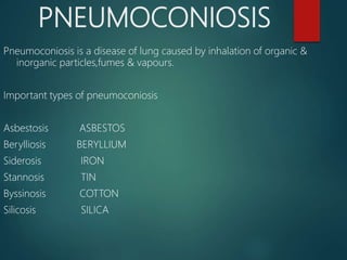
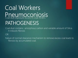
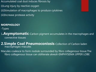
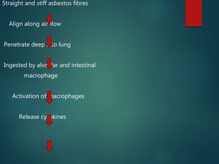
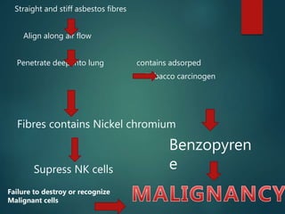
PNEUMOCONIOSIS
DR.SUNDARESAN
PATHOLOGY
LOYOLA COLLEGE
PNEUMOCONIOSIS
Pneumoconiosis is a disease of lung caused by inhalation of organic &
inorganic particles,fumes & vapours.
Important types of pneumoconiosis
Asbestosis ASBESTOS
Berylliosis BERYLLIUM
Siderosis IRON
Stannosis TIN
Byssinosis COTTON
Silicosis SILICA
Coal Workers
PneumoconiosisCoal dust exposure occurs in coal miners.
PATHOGENESIS
Coal dust contains amorphous carbon and variable amount of Silica.
It induces fibrosis
OR
Failure of normal clearance mechanism to remove excess coal leads to
fibrosis by accumulated coal.
Accumulated coal dust induces fibrosis by
(i)Lung injury by reactive oxygen
(ii)Stimulation of macrophages to produce cytokines
(iii)Increase protease activity
MORPHOLOGY
-Types
1.Asymptomatic-Carbon pigment accumulates in the macrophages and
connective tissues
2.Simple Coal Pneumoconiosis-Collection of Carbon laden
macrophages-macules
Macules coalesce to form nodule surrounded by fibro collagenous tissue.The
fibro collagenous tissue can obliterate alveoli-EMPHYSEMA UPPER LOBE
3. Progressive massive fibrosis
Over years simple coal workers pneumoconiosis progresses to progressive
massive fibrosis.
There are large areas of scarring and carbon pigmented areas.
There is lung dysfunction.
COMPLICATIONS
1.Simple Coal workers pneumoconiosis cause chronic
bronchiotis and Bronchiolitis.
2.Progressive Coal Workers pneumoconiosis cause
(a)Cor Pulmonale
(b)Caplan Syndrome-Coal worker pneumoconiosis along
with rheumatoid nodule.It can also be seen in
association with silicosis or asbestosis.The Lesions are
nodular with central necrosis surrounded by
fibroblasts and macrophages.
CLINICAL PICTURE
Coal workers pneumoconiosis is a
Benign asymptomatic disease.
Rarely progressive massive fibrosis
occurs patient develops dyspnoea
cough and respiratory dysfunction.
Silicosis
It is the most common occupational
disease caused by inhalation of
Silica.It is seen in sand Blasting
,foundry work and stone cutting.
PATHOGENESISInhalation of Silica
Engulfment by Release mediators
alveolar macrophages
Binds with membrane
protein and phospholipids
Damages cell membrane
Kills Macrophages
Free Silica particle
Following inhalation of Silica it is engulfed by alveolar macrophages
Silicon Hydroxide groups on the surface of particles form hydrogen
bond with membrane protein.
•Denaturation of membrane protein and
damage of microphage cell membrane.
•Macrophage is killed and silica particle is
released.
•The free silica is re-ingested by other
macrophages and cycle goes on.
•The silica ingested alveolar macrophages gets
activated and release mediators-IL1, TNF,O2
derived radicles, fibrogenic cytokines.
• These cause fibrosis.
MORPHOLOGY:
Early silicosis- Macroscopic features.
•Upper zone- small nodules
•Large areas of focal to confluent consolidation.
Microscopic features
•Chronic inflammation of alveoli and septa.
•Alveolar spaces filled with amorphous material.
•Long needle like space contain bifringent silica
particles
•Fibrinous pleuritis
CHRONIC NODULAR SILICOSIS
Macroscopic
• Tiny nodules like sand grains, initially in the
upper lobe, later spread to other areas.
• Tiny nodules coalesce to form hard visible
scars.
• Lung parenchyma between the scars is
compressed or emphysematous.
MICROSCOPIC FEATURES:
• Nodules are made up of concentric layers of
hyalinised collagen surrounded by a dense
collagenous capsule.
• Pleura shows dense plaques with adhesion
obliterating the pleural cavity.
• Hilar nodes - show thin calcification-
these appears like egg shell in xray chest
CLINICAL PICTURE:
 Dysnoea on exertion, later during rest.
 Respiratory dysfunction.
DIAGNOSIS
 X ray chest shows nodularity.
 Complications
 Caplan syndrome
 Chronic bronchitis
 Emphysema
 Cor pulmonale
 Pulmonary tuberculosis
ASBESTOSIS:
Inlation of asbestos fibres is called
asbestosis.
Asbestosis is seen in the following
occupation,
•Manufacturing of car components
•Flooring and roofing
•Mining
•Sewage
TYPE OF ASBESTOS FIBRES:
•Amphibole- straight and stiff fibres.
•Less soluble and mere pathogenic.
•Chrysotile- curled, flexible and more soluble
than amphibole
•Amphibole fibres are straight and stiff.
•They move along airflow and penetrate
deep into lung.
•Chrysotile fibres are curled and flexible.
•They get stuck in the upper respiratory
passages and are removed by mucociliary action
of air passages.
•They are more soluble than amphibole
fibres and are easily removed .
• They are less pathogenic.
Disease occurs after 10 years of exposure.
The asbestos fibres are indicators as well
as promotors of carcinogenesis.
Fibres contain trace metals such as nickel
and chromium which are carcinogenic.
Tobacco and asbestos act together and
produce malignancy.
Asbestos fibres are ingested by
macrophages they get activated and
release cytokines
Cytokines induce fibrosis.
Straight and stiff asbestos fibres
Align along air flow
Penetrate deep into lung
Ingested by alveolar and intestinal
macrophage
Activation of macrophages
Release cytokines
Straight and stiff asbestos fibres
Align along air flow
Penetrate deep into lung contains adsorped
Tobacco carcinogen
Failure to destroy or recognize
Malignant cells
Fibres contains Nickel chromium
Supress NK cells
Benzopyren
e
Morphology
 Diffuse pulmonary interstitial Fibrosis with
presence of asbestos bodies.
 Fibrosis starts around respiratory
and alveolar ducts.
 Fibrosis spreads to alveolar sacs and alveoli.
 Due to fibrosis the adjacent alveoli become
dilated- compensatory dilatation giving a
honey comb appearance to the lung.
 In the areas of fibrosis asbestos bodies are
seen.
Asbestos bodies or Ferrugenous
bodies
They are club shaped or dumble
shaped Golden brown in colour.
The case of the body contains
asbestos fibre.
The fibre is coated by glycoprotein
with haemosiderins.
The haemosiderin give its golden
brown colour and beaded
PLEURAL PLAQUES
-Pleural plaques are well demarcated.
Pearly white smooth or nodular areas in
pleura.
-These are dense hyaline tissue with
calcification.
-Diffuse pleural fibrosis may lead to
adhesion to chest wall.
COMPLICATIONS
- Emphysema
- Bronchiectasis
- Cor pulmonale
- Caplan syndrome
-Mesothelioma
-Bronchogenic carcinoma
-Gastrointestinal carcinoma
-Renal carcinoma
-Lymphoma

Pneumoconiosis

  • 1.
  • 2.
    PNEUMOCONIOSIS Pneumoconiosis is adisease of lung caused by inhalation of organic & inorganic particles,fumes & vapours. Important types of pneumoconiosis Asbestosis ASBESTOS Berylliosis BERYLLIUM Siderosis IRON Stannosis TIN Byssinosis COTTON Silicosis SILICA
  • 3.
    Coal Workers PneumoconiosisCoal dustexposure occurs in coal miners. PATHOGENESIS Coal dust contains amorphous carbon and variable amount of Silica. It induces fibrosis OR Failure of normal clearance mechanism to remove excess coal leads to fibrosis by accumulated coal.
  • 4.
    Accumulated coal dustinduces fibrosis by (i)Lung injury by reactive oxygen (ii)Stimulation of macrophages to produce cytokines (iii)Increase protease activity MORPHOLOGY -Types 1.Asymptomatic-Carbon pigment accumulates in the macrophages and connective tissues 2.Simple Coal Pneumoconiosis-Collection of Carbon laden macrophages-macules Macules coalesce to form nodule surrounded by fibro collagenous tissue.The fibro collagenous tissue can obliterate alveoli-EMPHYSEMA UPPER LOBE
  • 5.
    3. Progressive massivefibrosis Over years simple coal workers pneumoconiosis progresses to progressive massive fibrosis. There are large areas of scarring and carbon pigmented areas. There is lung dysfunction.
  • 6.
    COMPLICATIONS 1.Simple Coal workerspneumoconiosis cause chronic bronchiotis and Bronchiolitis. 2.Progressive Coal Workers pneumoconiosis cause (a)Cor Pulmonale (b)Caplan Syndrome-Coal worker pneumoconiosis along with rheumatoid nodule.It can also be seen in association with silicosis or asbestosis.The Lesions are nodular with central necrosis surrounded by fibroblasts and macrophages.
  • 7.
    CLINICAL PICTURE Coal workerspneumoconiosis is a Benign asymptomatic disease. Rarely progressive massive fibrosis occurs patient develops dyspnoea cough and respiratory dysfunction.
  • 8.
    Silicosis It is themost common occupational disease caused by inhalation of Silica.It is seen in sand Blasting ,foundry work and stone cutting.
  • 9.
    PATHOGENESISInhalation of Silica Engulfmentby Release mediators alveolar macrophages Binds with membrane protein and phospholipids Damages cell membrane Kills Macrophages Free Silica particle
  • 10.
    Following inhalation ofSilica it is engulfed by alveolar macrophages Silicon Hydroxide groups on the surface of particles form hydrogen bond with membrane protein.
  • 11.
    •Denaturation of membraneprotein and damage of microphage cell membrane. •Macrophage is killed and silica particle is released. •The free silica is re-ingested by other macrophages and cycle goes on. •The silica ingested alveolar macrophages gets activated and release mediators-IL1, TNF,O2 derived radicles, fibrogenic cytokines. • These cause fibrosis.
  • 12.
    MORPHOLOGY: Early silicosis- Macroscopicfeatures. •Upper zone- small nodules •Large areas of focal to confluent consolidation. Microscopic features •Chronic inflammation of alveoli and septa. •Alveolar spaces filled with amorphous material. •Long needle like space contain bifringent silica particles •Fibrinous pleuritis
  • 13.
    CHRONIC NODULAR SILICOSIS Macroscopic •Tiny nodules like sand grains, initially in the upper lobe, later spread to other areas. • Tiny nodules coalesce to form hard visible scars. • Lung parenchyma between the scars is compressed or emphysematous.
  • 14.
    MICROSCOPIC FEATURES: • Nodulesare made up of concentric layers of hyalinised collagen surrounded by a dense collagenous capsule. • Pleura shows dense plaques with adhesion obliterating the pleural cavity. • Hilar nodes - show thin calcification- these appears like egg shell in xray chest
  • 15.
    CLINICAL PICTURE:  Dysnoeaon exertion, later during rest.  Respiratory dysfunction. DIAGNOSIS  X ray chest shows nodularity.  Complications  Caplan syndrome  Chronic bronchitis  Emphysema  Cor pulmonale  Pulmonary tuberculosis
  • 16.
    ASBESTOSIS: Inlation of asbestosfibres is called asbestosis. Asbestosis is seen in the following occupation, •Manufacturing of car components •Flooring and roofing •Mining •Sewage
  • 17.
    TYPE OF ASBESTOSFIBRES: •Amphibole- straight and stiff fibres. •Less soluble and mere pathogenic. •Chrysotile- curled, flexible and more soluble than amphibole
  • 18.
    •Amphibole fibres arestraight and stiff. •They move along airflow and penetrate deep into lung. •Chrysotile fibres are curled and flexible. •They get stuck in the upper respiratory passages and are removed by mucociliary action of air passages. •They are more soluble than amphibole fibres and are easily removed . • They are less pathogenic.
  • 19.
    Disease occurs after10 years of exposure. The asbestos fibres are indicators as well as promotors of carcinogenesis. Fibres contain trace metals such as nickel and chromium which are carcinogenic. Tobacco and asbestos act together and produce malignancy. Asbestos fibres are ingested by macrophages they get activated and release cytokines Cytokines induce fibrosis.
  • 20.
    Straight and stiffasbestos fibres Align along air flow Penetrate deep into lung Ingested by alveolar and intestinal macrophage Activation of macrophages Release cytokines
  • 22.
    Straight and stiffasbestos fibres Align along air flow Penetrate deep into lung contains adsorped Tobacco carcinogen Failure to destroy or recognize Malignant cells Fibres contains Nickel chromium Supress NK cells Benzopyren e
  • 23.
    Morphology  Diffuse pulmonaryinterstitial Fibrosis with presence of asbestos bodies.  Fibrosis starts around respiratory and alveolar ducts.  Fibrosis spreads to alveolar sacs and alveoli.  Due to fibrosis the adjacent alveoli become dilated- compensatory dilatation giving a honey comb appearance to the lung.  In the areas of fibrosis asbestos bodies are seen.
  • 24.
    Asbestos bodies orFerrugenous bodies They are club shaped or dumble shaped Golden brown in colour. The case of the body contains asbestos fibre. The fibre is coated by glycoprotein with haemosiderins. The haemosiderin give its golden brown colour and beaded
  • 25.
    PLEURAL PLAQUES -Pleural plaquesare well demarcated. Pearly white smooth or nodular areas in pleura. -These are dense hyaline tissue with calcification. -Diffuse pleural fibrosis may lead to adhesion to chest wall.
  • 26.
    COMPLICATIONS - Emphysema - Bronchiectasis -Cor pulmonale - Caplan syndrome -Mesothelioma -Bronchogenic carcinoma -Gastrointestinal carcinoma -Renal carcinoma -Lymphoma