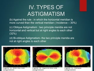
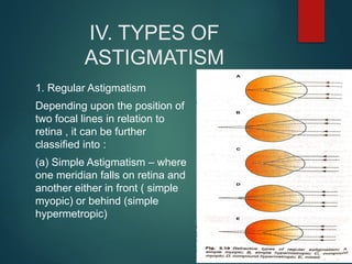
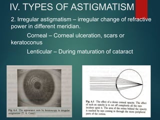
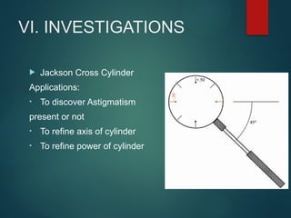
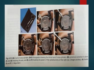
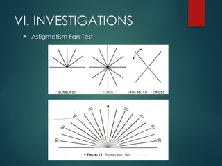
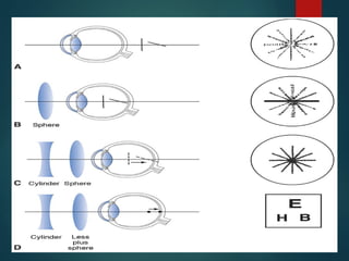
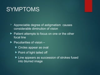
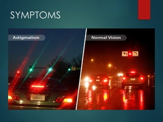
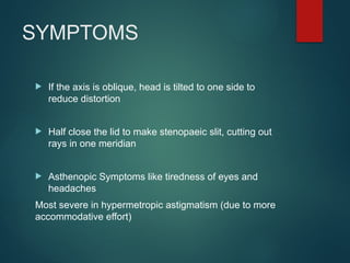
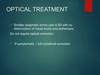
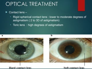
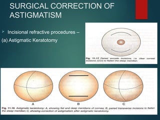
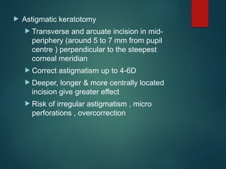
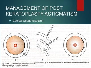
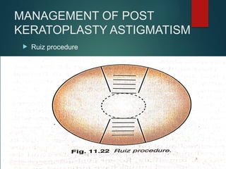
ASTIGMATISM
MAJ ABHISHEK SINGLA
RESIDENT OPHTHALMOLOGY
CHAF BANGALORE
BIBLIOGRAPHY
1. Parson’s Diseases of the Eye 23rd
edition
2. Duke-Elder’s Practice of Refraction 10th
edition
3. A.K Khurana Theory & Practice of Optics & Refraction 5th
edition
4. American Academy of Ophthalmology Vol 3 – Clinical Optics and
Vision Rehabilitation
Sl No. Topic
I. WHAT IS ASTIGMATISM ?
II. HISTORY
III. AETIOLOGY
IV. TYPES OF ASTIGMATISM
V. STRUM CONOID
VI. INVESTIGATIONS
VII. SYMPTOMS
VIII. TREATMENT OF ASTIGMATISM
INDEX
I. WHAT IS ASTIGMATISM ?
 Type of refractive error with
unequal refraction of light in
different meridian
 A point focus of light cannot
be formed upon the retina
instead a focal line is
formed.
III. AETIOLOGY
 Curvatural astigmatism of cornea –
 Curvatural astigmatism occurs more frequently in
cornea
 Usually congenital
 Physiological – direct astigmatism with up to
0.25D due to constant pressure of upper lid
III. AETIOLOGY
 Acquired astigmatism due
to corneal pathologies –
 Keratoconus
 Inflammation
 Ulceration
 Trauma
 Acquired astigmatism due
to external pressure -
 Chalazion
 Lid neoplasm
 Finger pressure on eye
 Contraction of lids
 Extra ocular muscle
action
III. AETIOLOGY
 Lenticular Astigmatism
 Curvatural: Marked astigmatism in
Lenticonus
 Positional: Decentering of IOL,
traumatic subluxation
 Index astigmatism: due to variable
refractive indices like in nuclear sclerosis
and cataract
STURM CONOID-
 Compound hypermetropic astigmatism
 Simple hypermetropic astigmatism
 Mixed astigmatism – distant vision is good
 Simple myopic astigmatism
 Compound myopic astigmatism
IV. TYPES OF ASTIGMATISM
1.Regular astigmatism :
Depending upon the axis and angle between the two meridian
can be classified as following types:
(a) With the rule : the two principle meridia are placed at right
angles to each other but the vertical meridian is curved more
than the horizontal ( Incidence – 38 %)
IV. TYPES OF
ASTIGMATISM
(b) Against the rule : in which the horizontal meridian is
more curved than the vertical meridian ( Incidence – 30%)
(c) Oblique Astigmatism : two principle meridia are not
horizontal and vertical but at right angles to each other
(32%)
(d) Bi-oblique Astigmatism- the two principle meridia are
not at right angles to each other
IV. TYPES OF
ASTIGMATISM
1. Regular Astigmatism
Depending upon the position of
two focal lines in relation to
retina , it can be further
classified into :
(a) Simple Astigmatism – where
one meridian falls on retina and
another either in front ( simple
myopic) or behind (simple
hypermetropic)
IV. TYPES OF
ASTIGMATISM
(b) Compound Astigmatism – the
rays of light in both the meridian
either focus in front (compound
myopic) or behind the retina
(compound hypermetropic)
(c) Mixed Astigmatism – the light
rays in one meridian are focused
in front and in other meridian
behind the retina
IV. TYPES OF ASTIGMATISM
2. Irregular astigmatism – irregular change of refractive
power in different meridian.
Corneal – Corneal ulceration, scars or
keratoconus
Lenticular – During maturation of cataract
CYLINDRICAL LENS
 Cut off from a cylinder
 Axis of a cylindrical lens - AB
CYLINDRICAL LENS
 Plano-convex vs plano-
concave lens
 Focal line is formed
 Refract rays of light in one
plane & leaves the rays in
the plane perpendicular to
this unaltered
IDENTIFICATION OF
CYLINDRICAL LENS
1. Lens Shape: Cylindrical lenses are not
spherical; they have a curvature in one
direction only (like a section of a cylinder). This
means that when you look at the lens from the
side, it may appear flat in one direction and
curved in another
2. Prescription Markings: The prescription for
cylindrical lenses usually includes a notation
for the cylinder power (indicated by "Cyl" or
"CYL") and the axis (measured in degrees from
0° to 180°)
3. Visual Distortion: When wearing cylindrical
glasses, individuals may notice that objects
appear distorted or blurred in certain
POWER CROSS
The rule is that the power meridian of a cylinder is
perpendicular to the axis of cylinder.
SPHERICAL EQUIVALENT
 It is the average power of spherocylinder(toric
lens) – Sum of sphere and half of cylinder
power
 Used to calculate the circle of least diffusion
 Used in fitting of contact lens
VI. INVESTIGATIONS
 Retinoscopy
 Keratometry- A Keratometer / Ophthalmometer is
a diagnostic instrument for measuring curvature
of anterior surface of the cornea
 Corneal topography
VI. INVESTIGATIONS
 Jackson Cross Cylinder
Applications:
• To discover Astigmatism
present or not
• To refine axis of cylinder
• To refine power of cylinder
VI. INVESTIGATIONS
 Astigmatism Fan Test
SYMPTOMS
 Appreciable degree of astigmatism causes
considerable diminution of vision
 Patient attempts to focus on one or the other
focal line
 Peculiarities of vision –
 Circles appear as oval
 Point of light tailed off
 Line appears as succession of strokes fused
into blurred image
SYMPTOMS
SYMPTOMS
 If the axis is oblique, head is tilted to one side to
reduce distortion
 Half close the lid to make stenopaeic slit, cutting out
rays in one meridian
 Asthenopic Symptoms like tiredness of eyes and
headaches
Most severe in hypermetropic astigmatism (due to more
accommodative effort)
OPTICAL TREATMENT
 Smaller astigmatic errors upto 0.5D with no
deterioration of visual acuity and asthenopia
Do not require optical correction
 If symptomatic – full cylindrical correction
OPTICAL TREATMENT
 Contact lens –
• Rigid spherical contact lens : lower to moderate degrees of
astigmatism ( 2 to 3D of astigmatism)
• Toric lens : high degrees of astigmatism
SURGICAL CORRECTION OF
ASTIGMATISM
 Incisional refractive procedures –
(a) Astigmatic Keratotomy
 Astigmatic keratotomy
 Transverse and arcuate incision in mid-
periphery (around 5 to 7 mm from pupil
centre ) perpendicular to the steepest
corneal meridian
 Correct astigmatism up to 4-6D
 Deeper, longer & more centrally located
incision give greater effect
 Risk of irregular astigmatism , micro
perforations , overcorrection
(b) Limbal relaxing incision
 To correct -1.0 to -2.0D astigmatism
 Incision made at limbus
 Advantages vs astigmatic keratectomy
Less glare
Incision heals faster
Optical quality of cornea preserved
Easier to incorporate with cataract
extraction
SURGICAL CORRECTION OF
ASTIGMATISM
Laser ablation corneal refractive
procedures –
 Photoastigmatic refractive
keratectomy
 Astigmatic LASIK – up to 0.5-
10.0D
 Astigmatic C-LASIK ( best
technique)
MANAGEMENT OF POST
KERATOPLASTY ASTIGMATISM
 Suture removal ( should be tried first of all other
procedures)
• In steep meridians
• Interrupted sutures after 03 months
• Continuous sutures after 01 year
 Relaxing incision
• Along steeper meridian in donor cornea
• Relaxing incision with compression sutures
MANAGEMENT OF POST
KERATOPLASTY ASTIGMATISM
 Astigmatic LASIK
 Wavefront guided
 Correct up to 6-8D
MANAGEMENT OF POST
KERATOPLASTY ASTIGMATISM
 Corneal wedge resection
MANAGEMENT OF POST
KERATOPLASTY ASTIGMATISM
 Ruiz procedure
THANK YOU

Astigmatism-Types and its Management.pptx

  • 1.
    ASTIGMATISM MAJ ABHISHEK SINGLA RESIDENTOPHTHALMOLOGY CHAF BANGALORE
  • 2.
    BIBLIOGRAPHY 1. Parson’s Diseasesof the Eye 23rd edition 2. Duke-Elder’s Practice of Refraction 10th edition 3. A.K Khurana Theory & Practice of Optics & Refraction 5th edition 4. American Academy of Ophthalmology Vol 3 – Clinical Optics and Vision Rehabilitation
  • 3.
    Sl No. Topic I.WHAT IS ASTIGMATISM ? II. HISTORY III. AETIOLOGY IV. TYPES OF ASTIGMATISM V. STRUM CONOID VI. INVESTIGATIONS VII. SYMPTOMS VIII. TREATMENT OF ASTIGMATISM INDEX
  • 4.
    I. WHAT ISASTIGMATISM ?  Type of refractive error with unequal refraction of light in different meridian  A point focus of light cannot be formed upon the retina instead a focal line is formed.
  • 5.
    III. AETIOLOGY  Curvaturalastigmatism of cornea –  Curvatural astigmatism occurs more frequently in cornea  Usually congenital  Physiological – direct astigmatism with up to 0.25D due to constant pressure of upper lid
  • 6.
    III. AETIOLOGY  Acquiredastigmatism due to corneal pathologies –  Keratoconus  Inflammation  Ulceration  Trauma  Acquired astigmatism due to external pressure -  Chalazion  Lid neoplasm  Finger pressure on eye  Contraction of lids  Extra ocular muscle action
  • 7.
    III. AETIOLOGY  LenticularAstigmatism  Curvatural: Marked astigmatism in Lenticonus  Positional: Decentering of IOL, traumatic subluxation  Index astigmatism: due to variable refractive indices like in nuclear sclerosis and cataract
  • 8.
    STURM CONOID-  Compoundhypermetropic astigmatism  Simple hypermetropic astigmatism  Mixed astigmatism – distant vision is good  Simple myopic astigmatism  Compound myopic astigmatism
  • 9.
    IV. TYPES OFASTIGMATISM 1.Regular astigmatism : Depending upon the axis and angle between the two meridian can be classified as following types: (a) With the rule : the two principle meridia are placed at right angles to each other but the vertical meridian is curved more than the horizontal ( Incidence – 38 %)
  • 10.
    IV. TYPES OF ASTIGMATISM (b)Against the rule : in which the horizontal meridian is more curved than the vertical meridian ( Incidence – 30%) (c) Oblique Astigmatism : two principle meridia are not horizontal and vertical but at right angles to each other (32%) (d) Bi-oblique Astigmatism- the two principle meridia are not at right angles to each other
  • 11.
    IV. TYPES OF ASTIGMATISM 1.Regular Astigmatism Depending upon the position of two focal lines in relation to retina , it can be further classified into : (a) Simple Astigmatism – where one meridian falls on retina and another either in front ( simple myopic) or behind (simple hypermetropic)
  • 12.
    IV. TYPES OF ASTIGMATISM (b)Compound Astigmatism – the rays of light in both the meridian either focus in front (compound myopic) or behind the retina (compound hypermetropic) (c) Mixed Astigmatism – the light rays in one meridian are focused in front and in other meridian behind the retina
  • 13.
    IV. TYPES OFASTIGMATISM 2. Irregular astigmatism – irregular change of refractive power in different meridian. Corneal – Corneal ulceration, scars or keratoconus Lenticular – During maturation of cataract
  • 14.
    CYLINDRICAL LENS  Cutoff from a cylinder  Axis of a cylindrical lens - AB
  • 15.
    CYLINDRICAL LENS  Plano-convexvs plano- concave lens  Focal line is formed  Refract rays of light in one plane & leaves the rays in the plane perpendicular to this unaltered
  • 16.
    IDENTIFICATION OF CYLINDRICAL LENS 1.Lens Shape: Cylindrical lenses are not spherical; they have a curvature in one direction only (like a section of a cylinder). This means that when you look at the lens from the side, it may appear flat in one direction and curved in another 2. Prescription Markings: The prescription for cylindrical lenses usually includes a notation for the cylinder power (indicated by "Cyl" or "CYL") and the axis (measured in degrees from 0° to 180°) 3. Visual Distortion: When wearing cylindrical glasses, individuals may notice that objects appear distorted or blurred in certain
  • 17.
    POWER CROSS The ruleis that the power meridian of a cylinder is perpendicular to the axis of cylinder.
  • 18.
    SPHERICAL EQUIVALENT  Itis the average power of spherocylinder(toric lens) – Sum of sphere and half of cylinder power  Used to calculate the circle of least diffusion  Used in fitting of contact lens
  • 19.
    VI. INVESTIGATIONS  Retinoscopy Keratometry- A Keratometer / Ophthalmometer is a diagnostic instrument for measuring curvature of anterior surface of the cornea  Corneal topography
  • 20.
    VI. INVESTIGATIONS  JacksonCross Cylinder Applications: • To discover Astigmatism present or not • To refine axis of cylinder • To refine power of cylinder
  • 22.
  • 24.
    SYMPTOMS  Appreciable degreeof astigmatism causes considerable diminution of vision  Patient attempts to focus on one or the other focal line  Peculiarities of vision –  Circles appear as oval  Point of light tailed off  Line appears as succession of strokes fused into blurred image
  • 25.
  • 26.
    SYMPTOMS  If theaxis is oblique, head is tilted to one side to reduce distortion  Half close the lid to make stenopaeic slit, cutting out rays in one meridian  Asthenopic Symptoms like tiredness of eyes and headaches Most severe in hypermetropic astigmatism (due to more accommodative effort)
  • 27.
    OPTICAL TREATMENT  Smallerastigmatic errors upto 0.5D with no deterioration of visual acuity and asthenopia Do not require optical correction  If symptomatic – full cylindrical correction
  • 28.
    OPTICAL TREATMENT  Contactlens – • Rigid spherical contact lens : lower to moderate degrees of astigmatism ( 2 to 3D of astigmatism) • Toric lens : high degrees of astigmatism
  • 29.
    SURGICAL CORRECTION OF ASTIGMATISM Incisional refractive procedures – (a) Astigmatic Keratotomy
  • 30.
     Astigmatic keratotomy Transverse and arcuate incision in mid- periphery (around 5 to 7 mm from pupil centre ) perpendicular to the steepest corneal meridian  Correct astigmatism up to 4-6D  Deeper, longer & more centrally located incision give greater effect  Risk of irregular astigmatism , micro perforations , overcorrection
  • 31.
    (b) Limbal relaxingincision  To correct -1.0 to -2.0D astigmatism  Incision made at limbus  Advantages vs astigmatic keratectomy Less glare Incision heals faster Optical quality of cornea preserved Easier to incorporate with cataract extraction
  • 32.
    SURGICAL CORRECTION OF ASTIGMATISM Laserablation corneal refractive procedures –  Photoastigmatic refractive keratectomy  Astigmatic LASIK – up to 0.5- 10.0D  Astigmatic C-LASIK ( best technique)
  • 33.
    MANAGEMENT OF POST KERATOPLASTYASTIGMATISM  Suture removal ( should be tried first of all other procedures) • In steep meridians • Interrupted sutures after 03 months • Continuous sutures after 01 year  Relaxing incision • Along steeper meridian in donor cornea • Relaxing incision with compression sutures
  • 35.
    MANAGEMENT OF POST KERATOPLASTYASTIGMATISM  Astigmatic LASIK  Wavefront guided  Correct up to 6-8D
  • 36.
    MANAGEMENT OF POST KERATOPLASTYASTIGMATISM  Corneal wedge resection
  • 37.
    MANAGEMENT OF POST KERATOPLASTYASTIGMATISM  Ruiz procedure
  • 38.

Editor's Notes

  • #7 Lenticonus present in Alport syn, Lowe Syndrome , macula coloboma
  • #8 Sturm conoid explains optics of regular Astigmatism Configuration of rays refracted through a toric surface is called Sturm Conoid
  • #20 Used for subjective verification of refraction It’s a spherocylinder Sphere = ½ of cylinder (opposite power) Axis and power of coreecting cylinder are perpendicular Handle is at 45 degrees to the meridian of Jackson cross Red depicts minus power and white/black depicts plus power
  • #23 Eg of compound hypermetropic astigmatism First check best visual acuity using snellens with only spherical correction Fogging – artificial blurring to 6/18 using convex spherical lenses and converted to compound myopic astigmatism as both of these focal lines are pulled in front of retina 3 Ask the patient which line is darkest and sharpest 4. Add a minus cylinder with axis perpendicular to the darkest and sharpest line so that the ant focal line makes a focal point with the horizontal line ( collapsing the sturm conoid ) 5. Move focal point to retina by adding minus spherical lens
  • #29 Incised meridian flatten and meridian 90 degrees gets steeper and corrects upto 4 to 6 D of astigmatism Incision b/w 5 to 7 mm from pupil using transverse or arcuate incisions