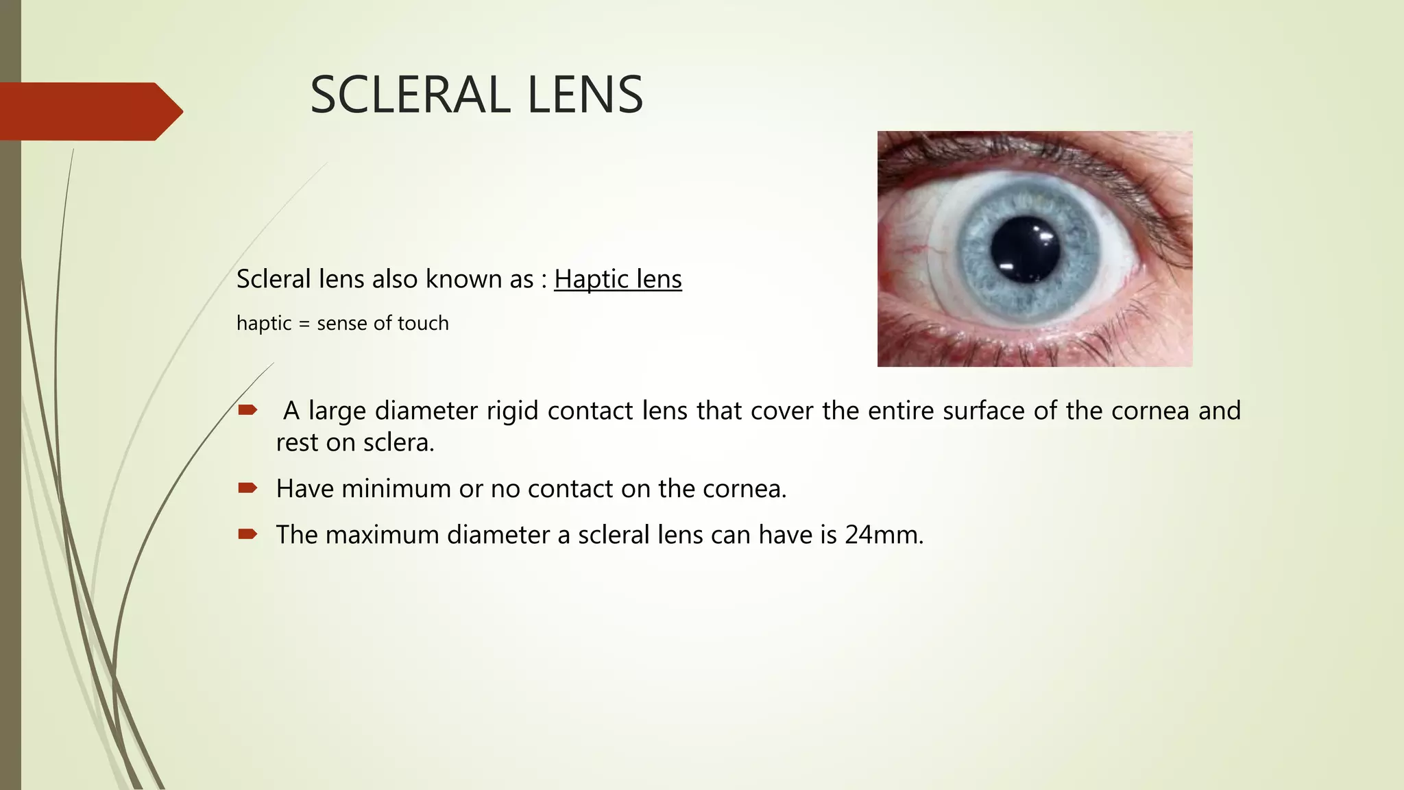
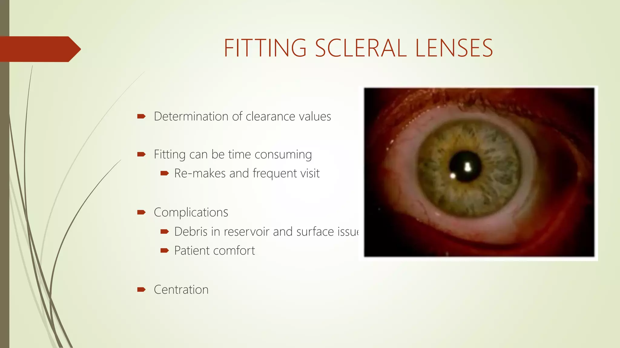
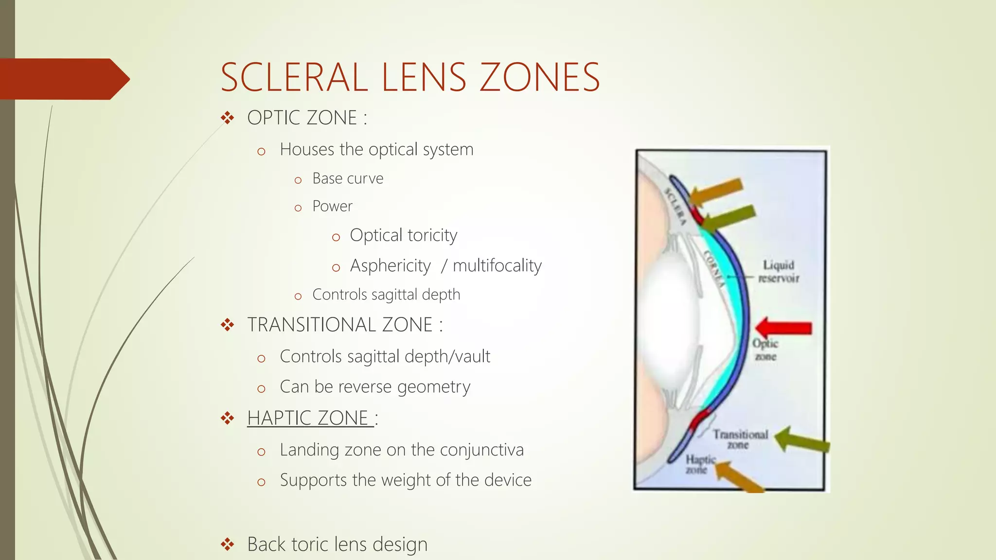
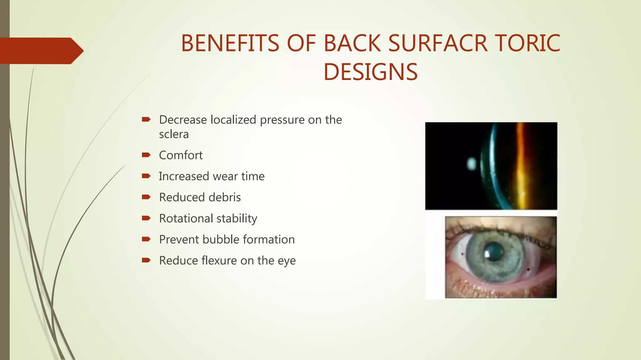
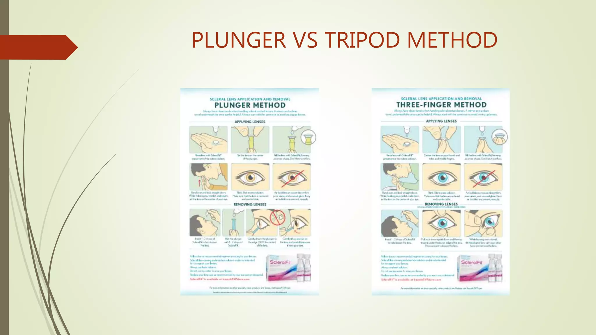
SCLERAL LENS FITTING
PHILOSOPHY
VIDYAMOLK
3RD YEAR OPTOMETRY
ABHAYA COLLEGE OF OPTOMETRY
SCLERAL LENS
Scleral lens also known as : Haptic lens
haptic = sense of touch
 A large diameter rigid contact lens that cover the entire surface of the cornea and
rest on sclera.
 Have minimum or no contact on the cornea.
 The maximum diameter a scleral lens can have is 24mm.
TYPES OF SCLERAL LENSES
Lens type Description Definition of bearing area
Corneal Lens rests entirely on the cornea
Corneo - scleral Lens rests partly on the cornea, partly
on the sclera
Scleral Mini scleral
Lens is up to 6mm larger than HVID
Large scleral
Lens is more than 6mm than HVID
Lens rests entirely on the sclera
SCLERAL LENS
INDICATIONS
 VISION CORRECTION
 PROTECTING THE OCULAR SURFACE
 PROVIDING COMFORT
VISION
CORRECTION keratoconus
Penetrating
keratoplasy
Radial and
Astigmatic
keratotomy
1. primary ectasias
2. Post-surgical and/or
secondary ectasias
3. Corneal irregularity
4. Refractive error
PROTECTING THE
OCULAR SURFACE
1. Exposure keratoplasy
2. Lagophthalmos
3. Trichiasis and entropion
4. Neorotrophic corneal
disease
5. Persistent epithelial defects
PROVIDING
COMFORT
Advanced ocular surface
disease:
a. sjogren’s
b. Graft versus host disease
c. Steven’s Johnson syndrome
d. Ocular cicatricial
pemphigioid
CONTRAINDICATIONS
 Expensive
 Large lens diameter
 Difficult to fit
 Fragile
 Lack of expertise to fit
 Complication when patient wear it overnight
FITTING SCLERAL LENSES
 Determination of clearance values
 Fitting can be time consuming
 Re-makes and frequent visit
 Complications
 Debris in reservoir and surface issue
 Patient comfort
 Centration
SCLERAL LENS ZONES
 OPTIC ZONE :
o Houses the optical system
o Base curve
o Power
o Optical toricity
o Asphericity / multifocality
o Controls sagittal depth
 TRANSITIONAL ZONE :
o Controls sagittal depth/vault
o Can be reverse geometry
 HAPTIC ZONE :
o Landing zone on the conjunctiva
o Supports the weight of the device
 Back toric lens design
FITTING PRINCIPLES: TORIC PERIPHERY
 Toric Peripheral Curves
o Better match natural toric nature
of the sclera.
o Better centration alignment, and
help decrease air/debris under
lens.
 Assess need for toric curves
o Instil NaFL and watch it enter the
lens chamber. If happens in one
meridian, consider toric
peripheral curves
o Chamber debris
o OCT evaluation
BENEFITS OF BACK SURFACR TORIC
DESIGNS
 Decrease localized pressure on the
sclera
 Comfort
 Increased wear time
 Reduced debris
 Rotational stability
 Prevent bubble formation
 Reduce flexure on the eye
CHALLENGES
 Determination of clearance values.
 Fitting can be time consuming
 Re-makes and frequent visits
 Complications
 debris in the reservoir and surface
issues
 Patient comfort
 centration
FITTING GUIDE
LENS PREPARATION
 Wash your hands
 Wash and clean lenses as with GP lenses
 Both plungers available
 Large DMV plungers-insertion
 Small DMV plunger-removal
PATIENT PREPARATION
CENTER LENS
SUCTION
FILLING THE LENS
NON PRESERVED SALINE
PLUNGER VS TRIPOD METHOD
INSERTION
 Place on a large or medium DMV suction cup up or use the tripod method using
the thumb, index finger and middle finger to hold the lens.
 Fill with non-preserved saline solution.
 Instruct the patient to lower head so that the face is parallel to the floor.
 Hold both upper and lower lids wide open.
 Patient may hold their lower lid
INSERTION
 Move quickly!
 Squeeze the suction cup so that the lens releases onto the eye.
 Have the patient close their eyes. Make sure the patient has a paper towel under
their eye to catch the overflow of saline and fluorescein during insertion.
 Check for bubbles
 If patient, remove the lens and reinsert again.
PATIENT POSITION
PRACTITIONER POSITION
MOVE QUICKLY
BUBBLES
LENS REMOVAL
 Small DMV suction device.
 Have patient look down with his head upright and against the headrest.
 Hold the patient’s upper eyelid.
 Place the suction cup on the superior portion of the lens, as close to the edge as
possible.
 Have the patient look up after suction is released to remove the lens off the eye.
LENS REMOVAL
THANK YOU

Scleral lens fitting

  • 1.
    SCLERAL LENS FITTING PHILOSOPHY VIDYAMOLK 3RDYEAR OPTOMETRY ABHAYA COLLEGE OF OPTOMETRY
  • 2.
    SCLERAL LENS Scleral lensalso known as : Haptic lens haptic = sense of touch  A large diameter rigid contact lens that cover the entire surface of the cornea and rest on sclera.  Have minimum or no contact on the cornea.  The maximum diameter a scleral lens can have is 24mm.
  • 3.
    TYPES OF SCLERALLENSES Lens type Description Definition of bearing area Corneal Lens rests entirely on the cornea Corneo - scleral Lens rests partly on the cornea, partly on the sclera Scleral Mini scleral Lens is up to 6mm larger than HVID Large scleral Lens is more than 6mm than HVID Lens rests entirely on the sclera
  • 4.
    SCLERAL LENS INDICATIONS  VISIONCORRECTION  PROTECTING THE OCULAR SURFACE  PROVIDING COMFORT
  • 5.
    VISION CORRECTION keratoconus Penetrating keratoplasy Radial and Astigmatic keratotomy 1.primary ectasias 2. Post-surgical and/or secondary ectasias 3. Corneal irregularity 4. Refractive error
  • 6.
    PROTECTING THE OCULAR SURFACE 1.Exposure keratoplasy 2. Lagophthalmos 3. Trichiasis and entropion 4. Neorotrophic corneal disease 5. Persistent epithelial defects
  • 7.
    PROVIDING COMFORT Advanced ocular surface disease: a.sjogren’s b. Graft versus host disease c. Steven’s Johnson syndrome d. Ocular cicatricial pemphigioid
  • 8.
    CONTRAINDICATIONS  Expensive  Largelens diameter  Difficult to fit  Fragile  Lack of expertise to fit  Complication when patient wear it overnight
  • 9.
    FITTING SCLERAL LENSES Determination of clearance values  Fitting can be time consuming  Re-makes and frequent visit  Complications  Debris in reservoir and surface issue  Patient comfort  Centration
  • 10.
    SCLERAL LENS ZONES OPTIC ZONE : o Houses the optical system o Base curve o Power o Optical toricity o Asphericity / multifocality o Controls sagittal depth  TRANSITIONAL ZONE : o Controls sagittal depth/vault o Can be reverse geometry  HAPTIC ZONE : o Landing zone on the conjunctiva o Supports the weight of the device  Back toric lens design
  • 11.
    FITTING PRINCIPLES: TORICPERIPHERY  Toric Peripheral Curves o Better match natural toric nature of the sclera. o Better centration alignment, and help decrease air/debris under lens.  Assess need for toric curves o Instil NaFL and watch it enter the lens chamber. If happens in one meridian, consider toric peripheral curves o Chamber debris o OCT evaluation
  • 12.
    BENEFITS OF BACKSURFACR TORIC DESIGNS  Decrease localized pressure on the sclera  Comfort  Increased wear time  Reduced debris  Rotational stability  Prevent bubble formation  Reduce flexure on the eye
  • 13.
    CHALLENGES  Determination ofclearance values.  Fitting can be time consuming  Re-makes and frequent visits  Complications  debris in the reservoir and surface issues  Patient comfort  centration
  • 14.
    FITTING GUIDE LENS PREPARATION Wash your hands  Wash and clean lenses as with GP lenses  Both plungers available  Large DMV plungers-insertion  Small DMV plunger-removal
  • 15.
  • 16.
  • 17.
  • 18.
  • 19.
  • 20.
  • 21.
    INSERTION  Place ona large or medium DMV suction cup up or use the tripod method using the thumb, index finger and middle finger to hold the lens.  Fill with non-preserved saline solution.  Instruct the patient to lower head so that the face is parallel to the floor.  Hold both upper and lower lids wide open.  Patient may hold their lower lid
  • 22.
    INSERTION  Move quickly! Squeeze the suction cup so that the lens releases onto the eye.  Have the patient close their eyes. Make sure the patient has a paper towel under their eye to catch the overflow of saline and fluorescein during insertion.  Check for bubbles  If patient, remove the lens and reinsert again.
  • 23.
  • 24.
  • 25.
  • 26.
  • 27.
    LENS REMOVAL  SmallDMV suction device.  Have patient look down with his head upright and against the headrest.  Hold the patient’s upper eyelid.  Place the suction cup on the superior portion of the lens, as close to the edge as possible.  Have the patient look up after suction is released to remove the lens off the eye.
  • 28.
  • 29.