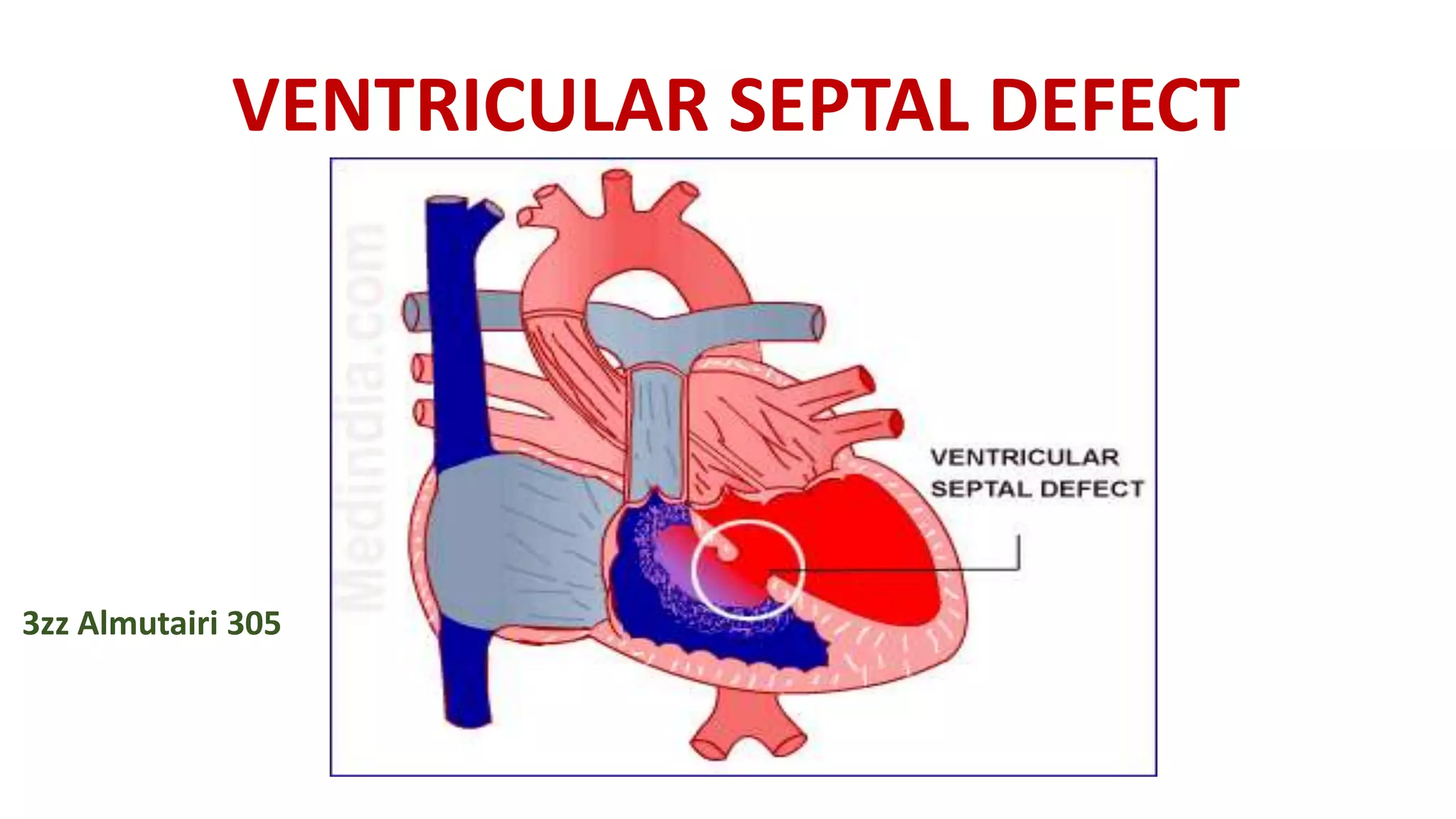
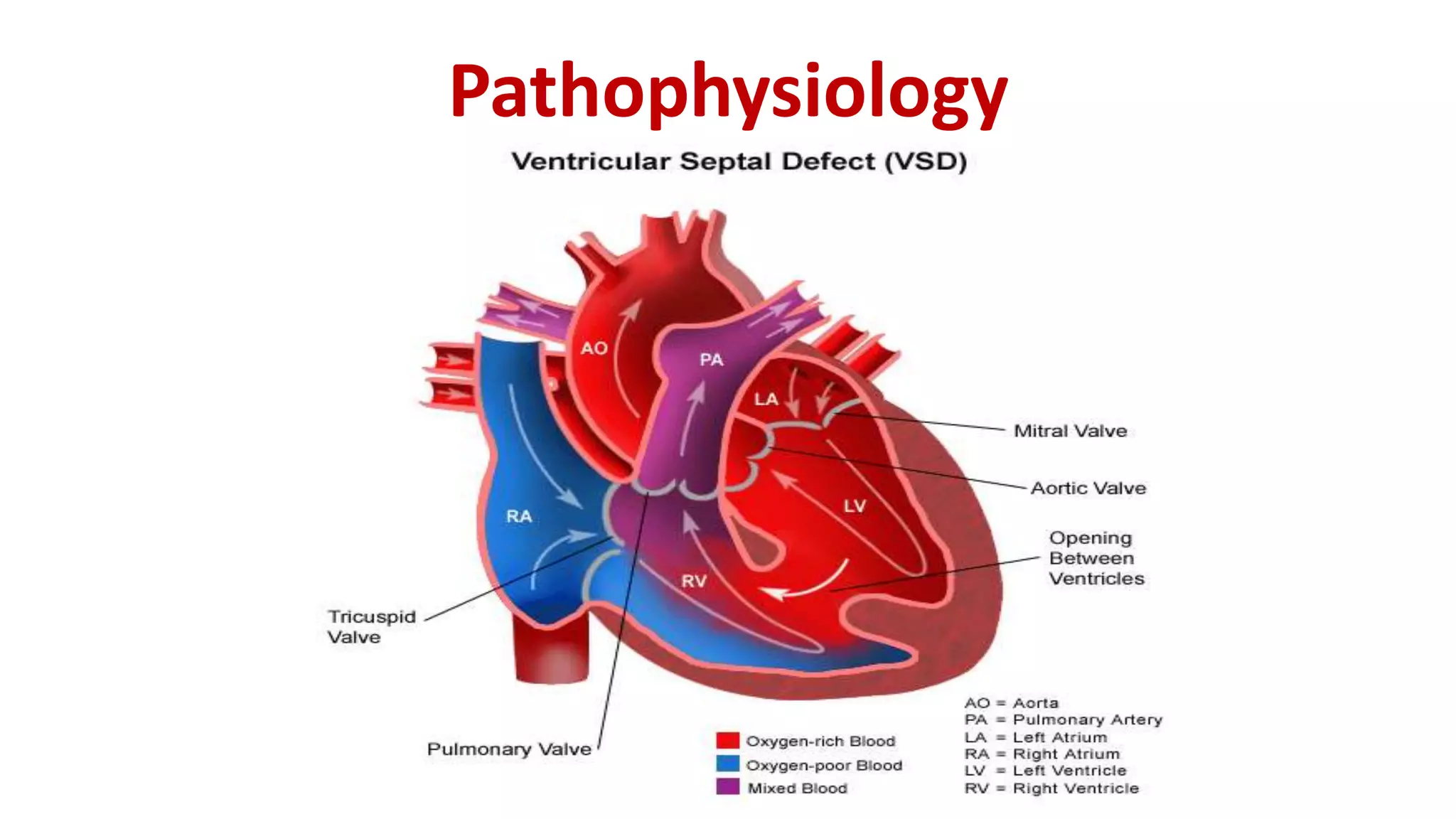
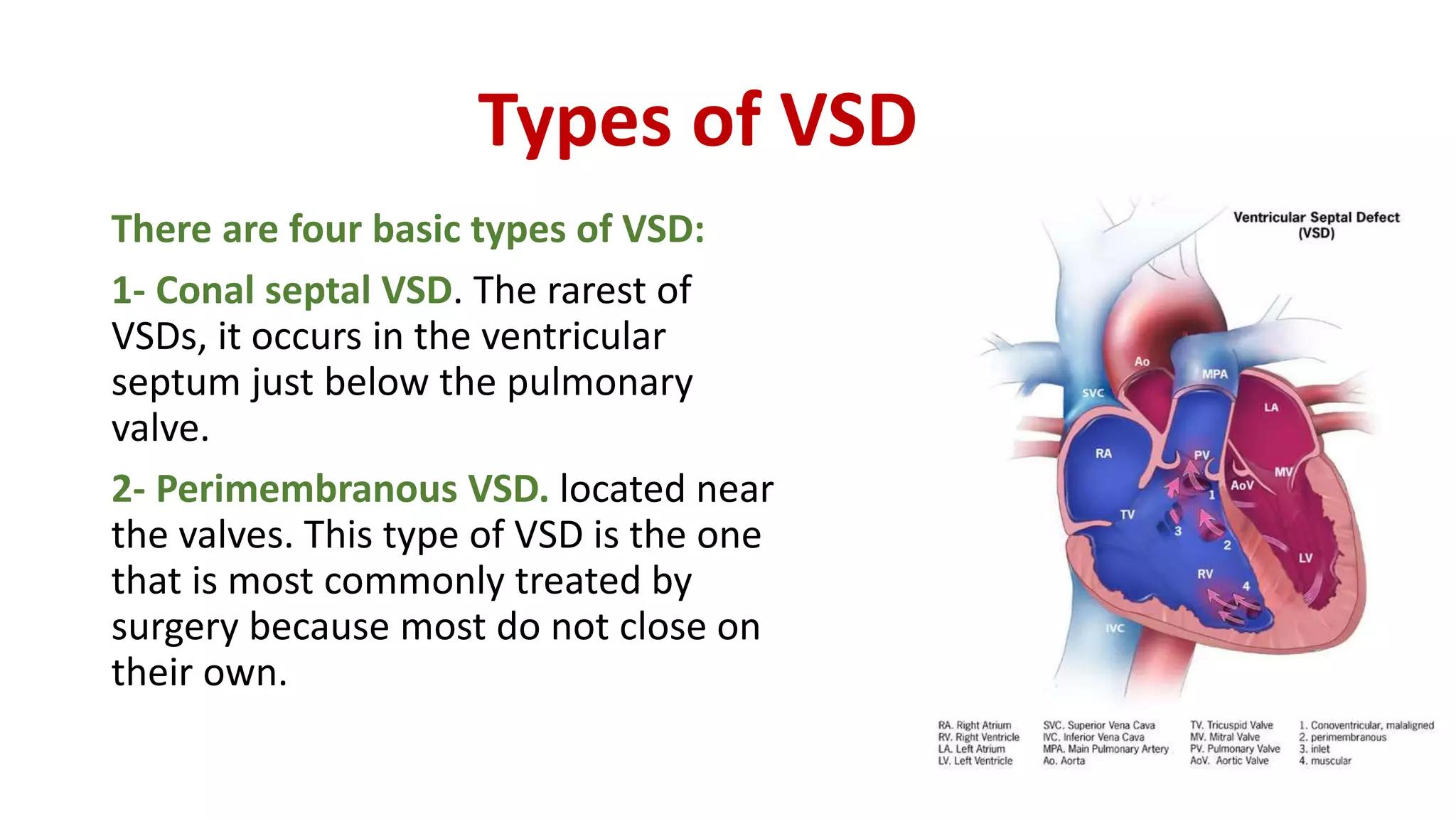
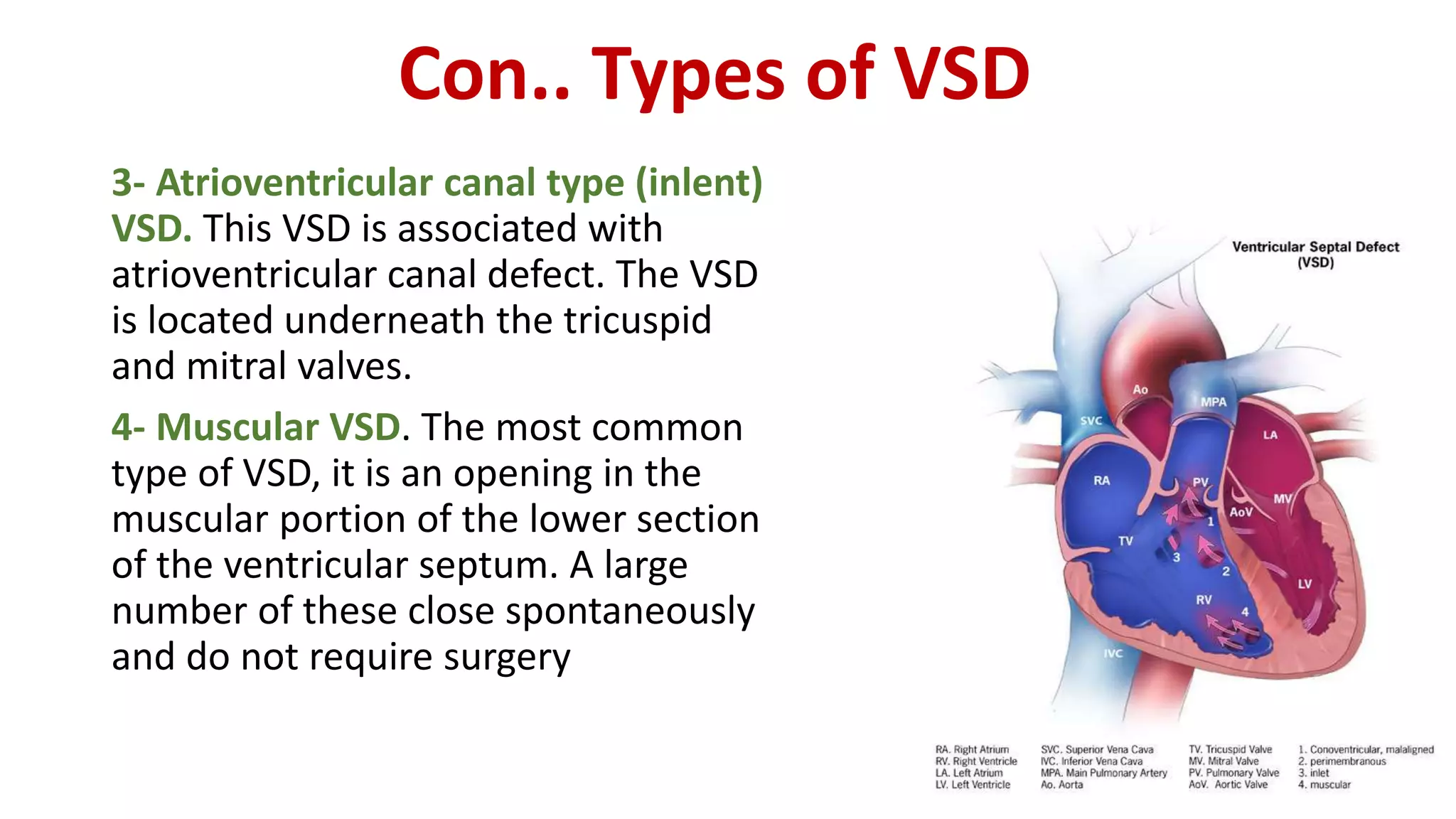
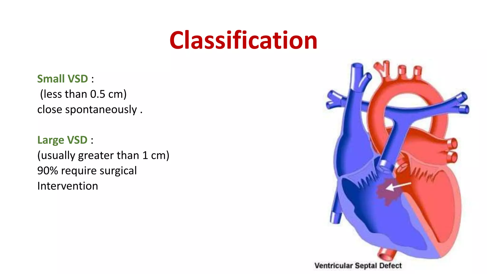
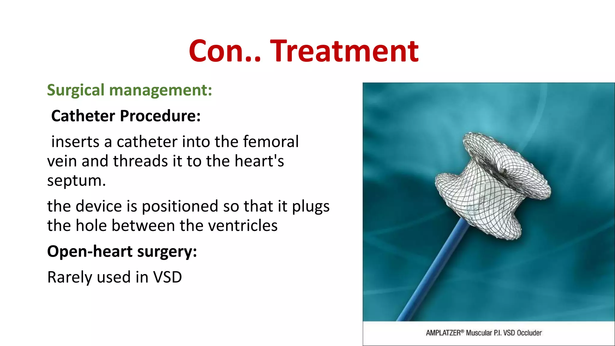
These are cardiac anomalies arising as a result of a defect in the structure or function of the heart and great vessels which is present at birth
These lesions either obstruct blood flow in the heart or vessels near it, or alter the pathway of blood circulating through the heart
commonly used for medical students, and helpful to use this ppt to study for them, and also a common man can understand very easily what is coarctation of aorta.
A cyanotic heart defect is a group-type of congenital heart defects (CHDs). The patient appears blue (cyanotic), due to deoxygenated blood bypassing the lungs and entering the systemic circulation. This can be caused by right-to-left or bidirectional shunting, or malposition of the great arteries.
Cyanotic heart defects, which account for approximately 25% of all CHDs, include:
Tetralogy of Fallot (ToF)
Total anomalous pulmonary venous connection
Hypoplastic left heart syndrome (HLHS)
Transposition of the great arteries (d-TGA)
Truncus arteriosus (Persistent)
Tricuspid atresia
Interrupted aortic arch
Pulmonary atresia (PA)
Pulmonary stenosis (critical)
Eisenmenger syndrome(Reversal of Shunt due to Pulmonary Hypertension) .
Patent ductus arteriosus may cause cyanosis in late stage.
Kindly leave your comment if you found this helpful ;)
Some of the slides, i hide it from my real presentations for my own reference. Download to see all of them.
CARDIAC TAMPONADE ( Cardiac emergency) • Cardiac Tamponade is a life threatening complication caused by excessive accumulation of fluid in the pericardium. Or • Compression of all cardiac chambers due to excessive accumulation of pericardial fluid leading to compromised cardiac out put.
Pulmonary stenosis (also called pulmonic stenosis) is when the pulmonary valve (the valve between the right ventricle and the pulmonary artery) is too small, narrow, or stiff. Symptoms of pulmonary stenosis depend on how small the narrowing of the pulmonary valve is
A "hole" in the wall that separates the top two chambers of the heart.
This defect allows oxygen-rich blood to leak into the oxygen-poor blood chambers in the heart. ASD is a defect in the septum between the heart's two upper chambers (atria). The septum is a wall that separates the heart's left and right sides
rare case of mycotic aneurysm in ventricular septal defect with infective endocarditis
Neurologic complications, dramatically changes the prognosis in IE
Such complications are clinically apparent in 20% -40% of cases
True incidence of acute brain embolization is not actually known.
Clinically silent embolisations occur in 35- 60% of the cases.
In general CNS takes up to 65% of the systemic embolization in IE
Mostly involve MCA
CNS emboli are mainly caused by Staph aureus
Early surgery is main treatment in embolism prevention
Antithrombotic drugs have no role
Patent ductus arteriosus (PDA) is a congenital disorder in the heart wherein a neonate's ductus arteriosus fails to close after birth. Early symptoms are uncommon, but in the first year of life include increased work of breathing and poor weight gain. With age, the PDA may lead to congestive heart failure if left uncorrected. The ductus arteriosus is a normal fetal blood vessel that closes soon after birth. In a patent ductus arteriosus (PDA) the vessel does not close and remains "patent" (open) resulting in irregular transmission of blood between two of the most important arteries close to the heart, the aorta and the pulmonary artery. PDA is common in neonates with persistent respiratory problems such as hypoxia, and has a high occurrence in premature children. In hypoxic newborns, too little oxygen reaches the lungs to produce sufficient levels of bradykinin and subsequent closing of the DA. Premature children are more likely to be hypoxic and thus have PDA because of their underdeveloped heart and lungs.
A patent ductus arteriosus allows a portion of the oxygenated blood from the left heart to flow back to the lungs by flowing from the aorta (which has higher pressure) to the pulmonary artery. If this shunt is substantial, the neonate becomes short of breath: the additional fluid returning to the lungs increases lung pressure to the point that the neonate has greater difficulty inflating the lungs. This uses more calories than normal and often interferes with feeding in infancy. This condition, as a constellation of findings, is called congestive heart failure.
In some cases, such as in transposition of the great vessels (the pulmonary artery and the aorta), a PDA may need to remain open. In this cardiovascular condition, the PDA is the only way that oxygenated blood can mix with deoxygenated blood. In these cases, prostaglandins are used to keep the patent ductus arteriosus open
congenital heart disease is a heart defect, it is caused by the exposure of teracogenic substances during pregnancy ,it may lead to the congenital defect .
The lecture is for medical student. It is from Dr RUSINGIZA Emmanuel, MD, senior lecture at UR( UNIVERSITY OF RWANDA) .
It will help to understand heart diseases in newborn, infants and children.
ACYANOTIC DISEASE- Non cyanotic heart diseasesNelsonNgulube
ETIOLOGY AND EPIDEMIOLOGY
Congenital heart disease occurs in 8 per 1,000 births. The spectrum of lesions ranges from asymptomatic to fatal. Although most cases of congenital heart disease are multifactorial, some lesions are associated with chromosomal disorders, single gene defects, teratogens, or maternal metabolic disease (see Table139-2).
Congenital heart defects can be divided into three pathophysiological groups (Table 143.1).
1. Left-to-right shunts
2. Right-to-left shunts
3. Obstructive, stenotic lesions
Acyanotic congenital heart disease includes left-to-right shunts resulting in an increase in pulmonary blood flow (patent ductus arteriosus [PDA], ventricular septal defect [VSD], atrial septal defect [ASD]) and obstructive lesions (aortic stenosis, pulmonary stenosis, coarctation of the aorta), which usually have normal pulmonary blood flow.
VENTRICULAR SEPTAL DEFECTEtiology and Epidemiology
The ventricular septum is a complex structure that can be divided
into four components. The largest component is the muscular
septum. The inlet or posterior septum comprises endocardial
cushion tissue. The subarterial or supracristal septum com
prises conotruncal tissue. The membranous septum is below
the aortic valve and is relatively small. VSDs occur when any of these components fail to develop normally (Fig. 143.1). VSD,
the most common congenital heart defect, accounts for 25% of all congenital heart disease. Perimembranous VSD
Title: Sense of Smell
Presenter: Dr. Faiza, Assistant Professor of Physiology
Qualifications:
MBBS (Best Graduate, AIMC Lahore)
FCPS Physiology
ICMT, CHPE, DHPE (STMU)
MPH (GC University, Faisalabad)
MBA (Virtual University of Pakistan)
Learning Objectives:
Describe the primary categories of smells and the concept of odor blindness.
Explain the structure and location of the olfactory membrane and mucosa, including the types and roles of cells involved in olfaction.
Describe the pathway and mechanisms of olfactory signal transmission from the olfactory receptors to the brain.
Illustrate the biochemical cascade triggered by odorant binding to olfactory receptors, including the role of G-proteins and second messengers in generating an action potential.
Identify different types of olfactory disorders such as anosmia, hyposmia, hyperosmia, and dysosmia, including their potential causes.
Key Topics:
Olfactory Genes:
3% of the human genome accounts for olfactory genes.
400 genes for odorant receptors.
Olfactory Membrane:
Located in the superior part of the nasal cavity.
Medially: Folds downward along the superior septum.
Laterally: Folds over the superior turbinate and upper surface of the middle turbinate.
Total surface area: 5-10 square centimeters.
Olfactory Mucosa:
Olfactory Cells: Bipolar nerve cells derived from the CNS (100 million), with 4-25 olfactory cilia per cell.
Sustentacular Cells: Produce mucus and maintain ionic and molecular environment.
Basal Cells: Replace worn-out olfactory cells with an average lifespan of 1-2 months.
Bowman’s Gland: Secretes mucus.
Stimulation of Olfactory Cells:
Odorant dissolves in mucus and attaches to receptors on olfactory cilia.
Involves a cascade effect through G-proteins and second messengers, leading to depolarization and action potential generation in the olfactory nerve.
Quality of a Good Odorant:
Small (3-20 Carbon atoms), volatile, water-soluble, and lipid-soluble.
Facilitated by odorant-binding proteins in mucus.
Membrane Potential and Action Potential:
Resting membrane potential: -55mV.
Action potential frequency in the olfactory nerve increases with odorant strength.
Adaptation Towards the Sense of Smell:
Rapid adaptation within the first second, with further slow adaptation.
Psychological adaptation greater than receptor adaptation, involving feedback inhibition from the central nervous system.
Primary Sensations of Smell:
Camphoraceous, Musky, Floral, Pepperminty, Ethereal, Pungent, Putrid.
Odor Detection Threshold:
Examples: Hydrogen sulfide (0.0005 ppm), Methyl-mercaptan (0.002 ppm).
Some toxic substances are odorless at lethal concentrations.
Characteristics of Smell:
Odor blindness for single substances due to lack of appropriate receptor protein.
Behavioral and emotional influences of smell.
Transmission of Olfactory Signals:
From olfactory cells to glomeruli in the olfactory bulb, involving lateral inhibition.
Primitive, less old, and new olfactory systems with different path
Ozempic: Preoperative Management of Patients on GLP-1 Receptor Agonists Saeid Safari
Preoperative Management of Patients on GLP-1 Receptor Agonists like Ozempic and Semiglutide
ASA GUIDELINE
NYSORA Guideline
2 Case Reports of Gastric Ultrasound
micro teaching on communication m.sc nursing.pdfAnurag Sharma
Microteaching is a unique model of practice teaching. It is a viable instrument for the. desired change in the teaching behavior or the behavior potential which, in specified types of real. classroom situations, tends to facilitate the achievement of specified types of objectives.
Pulmonary Thromboembolism - etilogy, types, medical- Surgical and nursing man...VarunMahajani
Disruption of blood supply to lung alveoli due to blockage of one or more pulmonary blood vessels is called as Pulmonary thromboembolism. In this presentation we will discuss its causes, types and its management in depth.
Report Back from SGO 2024: What’s the Latest in Cervical Cancer?bkling
Are you curious about what’s new in cervical cancer research or unsure what the findings mean? Join Dr. Emily Ko, a gynecologic oncologist at Penn Medicine, to learn about the latest updates from the Society of Gynecologic Oncology (SGO) 2024 Annual Meeting on Women’s Cancer. Dr. Ko will discuss what the research presented at the conference means for you and answer your questions about the new developments.
ARTIFICIAL INTELLIGENCE IN HEALTHCARE.pdfAnujkumaranit
Artificial intelligence (AI) refers to the simulation of human intelligence processes by machines, especially computer systems. It encompasses tasks such as learning, reasoning, problem-solving, perception, and language understanding. AI technologies are revolutionizing various fields, from healthcare to finance, by enabling machines to perform tasks that typically require human intelligence.
New Directions in Targeted Therapeutic Approaches for Older Adults With Mantl...i3 Health
i3 Health is pleased to make the speaker slides from this activity available for use as a non-accredited self-study or teaching resource.
This slide deck presented by Dr. Kami Maddocks, Professor-Clinical in the Division of Hematology and
Associate Division Director for Ambulatory Operations
The Ohio State University Comprehensive Cancer Center, will provide insight into new directions in targeted therapeutic approaches for older adults with mantle cell lymphoma.
STATEMENT OF NEED
Mantle cell lymphoma (MCL) is a rare, aggressive B-cell non-Hodgkin lymphoma (NHL) accounting for 5% to 7% of all lymphomas. Its prognosis ranges from indolent disease that does not require treatment for years to very aggressive disease, which is associated with poor survival (Silkenstedt et al, 2021). Typically, MCL is diagnosed at advanced stage and in older patients who cannot tolerate intensive therapy (NCCN, 2022). Although recent advances have slightly increased remission rates, recurrence and relapse remain very common, leading to a median overall survival between 3 and 6 years (LLS, 2021). Though there are several effective options, progress is still needed towards establishing an accepted frontline approach for MCL (Castellino et al, 2022). Treatment selection and management of MCL are complicated by the heterogeneity of prognosis, advanced age and comorbidities of patients, and lack of an established standard approach for treatment, making it vital that clinicians be familiar with the latest research and advances in this area. In this activity chaired by Michael Wang, MD, Professor in the Department of Lymphoma & Myeloma at MD Anderson Cancer Center, expert faculty will discuss prognostic factors informing treatment, the promising results of recent trials in new therapeutic approaches, and the implications of treatment resistance in therapeutic selection for MCL.
Target Audience
Hematology/oncology fellows, attending faculty, and other health care professionals involved in the treatment of patients with mantle cell lymphoma (MCL).
Learning Objectives
1.) Identify clinical and biological prognostic factors that can guide treatment decision making for older adults with MCL
2.) Evaluate emerging data on targeted therapeutic approaches for treatment-naive and relapsed/refractory MCL and their applicability to older adults
3.) Assess mechanisms of resistance to targeted therapies for MCL and their implications for treatment selection
TEST BANK for Operations Management, 14th Edition by William J. Stevenson, Ve...kevinkariuki227
TEST BANK for Operations Management, 14th Edition by William J. Stevenson, Verified Chapters 1 - 19, Complete Newest Version.pdf
Flu Vaccine Alert in Bangalore Karnatakaaddon Scans
As flu season approaches, health officials in Bangalore, Karnataka, are urging residents to get their flu vaccinations. The seasonal flu, while common, can lead to severe health complications, particularly for vulnerable populations such as young children, the elderly, and those with underlying health conditions.
Dr. Vidisha Kumari, a leading epidemiologist in Bangalore, emphasizes the importance of getting vaccinated. "The flu vaccine is our best defense against the influenza virus. It not only protects individuals but also helps prevent the spread of the virus in our communities," he says.
This year, the flu season is expected to coincide with a potential increase in other respiratory illnesses. The Karnataka Health Department has launched an awareness campaign highlighting the significance of flu vaccinations. They have set up multiple vaccination centers across Bangalore, making it convenient for residents to receive their shots.
To encourage widespread vaccination, the government is also collaborating with local schools, workplaces, and community centers to facilitate vaccination drives. Special attention is being given to ensuring that the vaccine is accessible to all, including marginalized communities who may have limited access to healthcare.
Residents are reminded that the flu vaccine is safe and effective. Common side effects are mild and may include soreness at the injection site, mild fever, or muscle aches. These side effects are generally short-lived and far less severe than the flu itself.
Healthcare providers are also stressing the importance of continuing COVID-19 precautions. Wearing masks, practicing good hand hygiene, and maintaining social distancing are still crucial, especially in crowded places.
Protect yourself and your loved ones by getting vaccinated. Together, we can help keep Bangalore healthy and safe this flu season. For more information on vaccination centers and schedules, residents can visit the Karnataka Health Department’s official website or follow their social media pages.
Stay informed, stay safe, and get your flu shot today!
These simplified slides by Dr. Sidra Arshad present an overview of the non-respiratory functions of the respiratory tract.
Learning objectives:
1. Enlist the non-respiratory functions of the respiratory tract
2. Briefly explain how these functions are carried out
3. Discuss the significance of dead space
4. Differentiate between minute ventilation and alveolar ventilation
5. Describe the cough and sneeze reflexes
Study Resources:
1. Chapter 39, Guyton and Hall Textbook of Medical Physiology, 14th edition
2. Chapter 34, Ganong’s Review of Medical Physiology, 26th edition
3. Chapter 17, Human Physiology by Lauralee Sherwood, 9th edition
4. Non-respiratory functions of the lungs https://academic.oup.com/bjaed/article/13/3/98/278874
3. What is VSD..?
A ventricular septal defect (VSD)
is a congenital defects in the inter-
ventricular septum that allow
shunting of blood between the left
and right ventricles.
4. Introduction
VSD: are the most common congenital heart defects in infants and
children, and VSD is seen in up to 3.5 infants per 1000 live births
Most of these close spontaneously in childhood.
VSD may also accompany other congenital defects.
6. Types of VSD
There are four basic types of VSD:
1- Conal septal VSD. The rarest of
VSDs, it occurs in the ventricular
septum just below the pulmonary
valve.
2- Perimembranous VSD. located near
the valves. This type of VSD is the one
that is most commonly treated by
surgery because most do not close on
their own.
7. Con.. Types of VSD
3- Atrioventricular canal type (inlent)
VSD. This VSD is associated with
atrioventricular canal defect. The VSD
is located underneath the tricuspid
and mitral valves.
4- Muscular VSD. The most common
type of VSD, it is an opening in the
muscular portion of the lower section
of the ventricular septum. A large
number of these close spontaneously
and do not require surgery
8. Classification
Small VSD :
(less than 0.5 cm)
close spontaneously .
Large VSD :
(usually greater than 1 cm)
90% require surgical
Intervention
9. Clinical Manifestation
-Murmur sound during auscultation
-Fatigue
-Sweating
-Rapid breathing
-Congested breathing
-Anorexia
-Poor weight gain
-Cyanosis
11. Con.. Diagnostic Test’s
ECG : - Right ventricular hypertrophy (RVH).
- large biphasic (equiphasic) QRS complexes.
-biventricular hypertrophy developes.
12. Treatment
Medical management:
Digoxin (Lanoxin).
Increase the strength of the
heart's contractions.
furosemide (Lasix).
Decrease the amount of fluid in
circulation and in the lungs.
Bisoprolol (concor)
Keep the heartbeat regular.
13. Con.. Treatment
Surgical management:
Catheter Procedure:
inserts a catheter into the femoral
vein and threads it to the heart's
septum.
the device is positioned so that it plugs
the hole between the ventricles
Open-heart surgery:
Rarely used in VSD
14. Preoperative & postoperative care
- Prophylactic antibiotic are often required to prevent infectious
endocarditis.
- The child should be assessed postoperatively for dysrhythmia, since
edema in the septum may interfere with condition.
15. Nursing managment
Nursing Diagnosis:
Decrease in cardiac output associated with heart malformations.
Nursing goal:
to improve cardiac output.
Nursing Intervention:
Observe the quality and strength of the heartbeat,
peripheral pulses, skin color and warmth.
Assess the degree of cyanosis (mucous membranes, clubbing).
Monitor signs of CHF (anxiety, tachycardia,
tachipnea, shortness of breath, periorbital edema,
oliguria and hepatomegaly.
16. Con.. Nursing managment
Nursing Diagnosis:
Impaired gas exchange related to pulmonary congestion.
Nursing goal:
improved gas exchange.
Nursing Intervention:
Monitor the quality and rhythm of breathing.
Adjust the position of the child with Fowler position.
Avoid child of an infected person.
Give adequate rest.
Give oxygen as indicated.