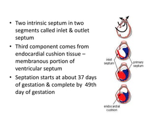
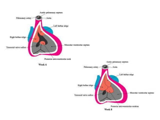
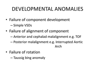
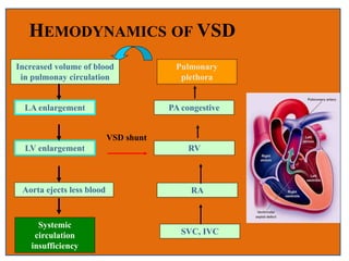
Ventricular Septal Defects
-Raja Lahiri
Introduction
• A VSD is a defect in the ventricular septum
• Most common congenital cardiac anomalies.
• 3-3.8 per 1000 live births
• 30-60% of all newborns with a CHD
• Prospective studies give a prevalence of 2-5
per 100 births of trabecular VSDs that closes
shortly after birth in 80-90% of the cases
MALADIE DE ROGER
• “A developmental defect of the heart occurs from
which cyanosis does not ensue inspite of the fact
that a communication exists between the cavities
of the two ventricles and inspite of the fact that
admixture of venous blood and arterial blood
occurs. This congenital defect , which is even
compatible with a long life, is a simple one. It
comprises a defect in the interventricular
septum.”
Henri Roger, 1879
Morphology – The Ventricular Septum
 Complex non – planar structure; 4
components
 Inlet septum – lightly
trabeculated; extends from
tricuspid annulus to attachments
of tricuspid valve
 Trabecular septum – heavily
trabeculated; trabecular septum
extends from inlet out to apex and
up to smooth-walled outlet
 Outlet septum – non trabeculated;
extends up to pulmonary valve&
diverge from small membranous
septum
 Membranous septum only
fibrous component of IVS, wedged
btwn AV, MV, TV.
Development of IVS
• Ventricles derived from 2 important
components of primitive heart – inlet & outlet
septum
• Three septal components are necessary for
septation
• Expansion of inlet & outlet components leads
to formation of partial septum between two –
primary interventricular septum
• Two intrinsic septum in two
segments called inlet & outlet
septum
• Third component comes from
endocardial cushion tissue –
membranous portion of
ventricular septum
• Septation starts at about 37 days
of gestation & complete by 49th
day of gestation
Prevelance
• Most common congenital heart lesion
• Occurs in 50% of children with heart lesions
• 15-20% in isolation
• 5-50 per 1000 live births
• 56% female
DEVELOPMENTAL ANOMALIES
• Failure of component development
– Simple VSDs
• Failure of alignment of component
– Anterior and cephalad malalignment e.g. TOF
– Posterior malalignment e.g. Interrupted Aortic
Arch
• Failure of rotation
– Taussig bing anomaly
Associated Defects
• Left Heart Defects
– Aortic stenosis
– Coarctation of the aorta
• Right Heart Defects
– Tetrology of Fallot
– Double Outlet Right Ventricle
• Truncus Arteriosus
• Some single ventricle (e.g. Tricuspid atresia)
Chromosomal Disorders associated
with VSD
• Trisomy 21: 40% of T21 will have VSD
• Trisomy 13, 18: 18% of T13, 31% of T18 will have VSD
• 22q11 deletion:
– Tetrology of Fallot is most common anomaly
– VSD with or without aortic arch anomaly is second most common
• Holt-Oram (Hand-heart syndrome): TBX5 gene found on
Chromosome 12
• Recurrence risk for VSD based on parental VSD
– Paternal 2%
– Maternal 6-10%
CLASSIFICATION
• CLASSICAL
• SOTO
• VAN PRAAGH
• ANDERSON
• MORPHOLOGICAL (congenital heart surgery
nomenclature & database project)
Classic classification
CONAL
PARAMEMBRA
NOUS
ATRIOVENTRIC
ULAR CANAL
MUSCULAR
Van praagh classification
CONAL
CONOVENTRIC
ULAR
AV CANAL
MUSCULAR
Anderson classification
PERIMEMBR
ANOUS
DOUBLY
COMMTTTE
D
MUSCULAR
EXPANDED classification (KIRKLIN)
 Classification Extension
1. Perimembranous inlet
outlet
anterior
2. Muscular outlet/conal
trabecular
inlet
anterior
apical
3. Doubly committed subarterial
4. Inlet septal AV septal type
5. Malalinged Ant (TOF)
Post(INTERRUPTED AORTIC ARCH)
Rotational (Taussig bing)
Hemodynamic classification
• Restrictive
• Moderately restrictive
• Non restrictive
– Restrictive- resistance that limits the shunt at the
site of vsd
– < 0.5 cm2 (Smaller than Ao valve orifice area)
– Small L to R shunt
– Normal RV output
– 75% spontaneously close < 2yrs
LVSP > RVSP
pulm /aortic systolic pressure ratio < 0.3
Qp / Qs<1.4--1
• Moderately restrictive - RVSP high, but
less than LVSP
• Qp/Qs 1.4--2.2
 Non restrictive -Shunt not limited at the site of
defect
– > 1.0 cm2 (Equal to or greater than to Ao valve
orifice area)
– Large hemodynamically significant L to R shunt
– Rarely close spontaneously
-RVSP , LVSP, PA , Aortic systolic
pressures equal
- Qp/Qs >2.2
- Flow determined by PVR
CLASSIFICATION OF VSD
According to the size of lesions
Small Size Medium Size Large Size
Diameter (mm)
Size (cm2)
<5
<0.5
5~15
0.5~1.5
>15
>1.0
Shunt small medium large
Symptom Non or little some Obvious
Pul. Vessel No affection affected Pul. Arterial
Hypertension
Eisenmenger’s
syndrome
• Defect size is often compared to aortic
annulus
– Large: > 75% of annulus size
– Medium: 75-33% of annulus size
– Small: <33% of annulus size
• Atrioventricular canal type VSD
• Muscular VSDs: midventricular
(1), apical (2), anterior (3), and
posterior (4)
• Conoventricular septal defect,
which includes
perimembranous and
malalignment conoventricular
septal defects
• Conal septal defects.
Conoventricular (Membranous)
Defects
• Conoventricular defects are located between the conal
septum and the ventricular septum.
• They are centered in or around the membranous
septum and comprise 80% of all VSDs.
• Located exclusively within the membranous septum, or
can extend beyond the boundaries of the membranous
septum toward inferior, posterior, or anterior
directions, and are then sometimes called
“perimembranous” or “paramembranous” VSDs.
• The prefix “peri-,” appearing in loan words from the Greek,
means “surrounding” (i.e., perimeter). As such, a truly
perimembranous ventricular septal defect would surround
the membranous septum.
• In contrast, the prefix “para-,” also from the Greek, means
“adjacent to” or “beside” and more accurately reflects the
notion of a defect adjacent to the membranous septum.
• Neither perimembranous nor paramembranous correctly
describes the typical defect involving the membranous
septum and extending into the adjacent septum.
• The current recommendation is to call these defects either
membranous VSDs or conoventricular defects.
• Malalignment of the conal septal plane vis-à-vis the
ventricular septal plane results in the typical
conoventricular defect.
• The malalignment can be anterior, as seen in tetralogy of
Fallot, or posterior, as seen in interrupted aortic arch.
• Anterior conal septal malalignment also results in RVOT
• Posterior malalignment of the conal septum results in LVOT.
• Important landmarks in conoventricular septal defects are
the anteroseptal commissure of the tricuspid valve
inferiorly and the noncoronary cusp of the aortic valve.
• When the ventricular portion of the membranous septum
is entirely absent, the VSD extends to the base of the aortic
valve (sometimes called “subaortic” VSD).
• The medial papillary muscle (muscle of Lancisi) located at
the inferior–posterior border of the defect is also an
important landmark.
• Both the septal and anterior tricuspid valve leaflets are
attached to it.
Conal Septal Defects
• Approximately 8% of VSDs are located in the
conal (infundibulum or outlet) septum.
• They also are called supracristal VSDs.
• They are either entirely surrounded by muscle
(muscular conal VSDs) or limited upstream by
the aortic or pulmonary annuli (sometimes
called subarterial VSDs).
Inlet (Atrioventricular Canal Type) VSD
• This defect is characterized by the absence of
part or all of the inlet septum.
• The VSD is located immediately underneath
the septal leaflet of the tricuspid valve with no
tissue in between.
• Approximately 6% of all VSDs are inlet-type
VSDs.
Muscular VSDs
• Muscular VSDs (10% of all VSDs) are entirely surrounded by
muscle.
• They can occur anywhere in the trabecular portion of the
septum and can be isolated or multiple.
• They are described by their location, that is, anterior,
midventricular, posterior, or apical.
• When inspected through the left side of the septum, what
appeared to be multiple muscular defects often converge
into either a single hole or two separate holes.
PATHOPHYSIOLOGY
• Blood flow dependent on multiple factors
–Small and restrictive
• Lesion size
–Large and non-restrictive
• Balance between pulmonary and
systemic vascular resistance
size
Pulmonary vascular
resistance
systemic vascular
resistance
HEMODYNAMICS OF VSD
VSD shunt
Pulmonary
plethora
PA congestive
RV
RA
SVC, IVC
Increased volume of blood
in pulmonay circulation
LA enlargement
LV enlargement
Aorta ejects less blood
Systemic
circulation
insufficiency
Natural history
 Spontaneous closure :75-85 % all VSDs
• :35% perimemb ( 1st 6/12)
• Spontaneous closure by age 1month—80%
• 3 months--- 60%
• 6 months--- 50%
• 12 months__ 25%
 More frequent in small defects
 Decrease in size with age
 Inlet & outlet defects do not become smaller /close spont
 Large & nonrestrictive defects : 10- 15%
o Endocarditis – risk of endocarditis 4-10% for the first 30 years of life
→high velocity turbulent jet into RV
CHF - Large VSDs
Mod sized VSDs survive into adulthood
Increased rt sided flow  pulmonary vascular
disease  Eisenmenger’s physiology if left
untreated
Mechanisms of closure
Growth & hypertrophy of septum around the
defect
By development of subacute bacterial endocarditis
Adherence of leaflet tissue to the margins
• (Negative pressure effect exerted by a high
velocity stream flowing through the defect )
Ventricular septal aneurysm
Prolapse of aortic cusp
Intrusion of a sinus of valsalva aneurysm
VSD THAT DON’T CLOSE
1. perimembranous (malaligned)
2.juxta aortic,
3. inlet:av septal type,
4.perimembranous (Gerbode defect)
Commonly Associated Defects
• Patent Ductus Arteriosus
• Aortic Coarctation
• Left Ventricular Outflow tract Obstruction
• Large atrial septal defects
• Right ventricular outflow tract obstruction
• Persistent left superior vena cava
Patent Ductus Arteriosus
• In symptomatic neonates or infants with VSDs, a large patent
ductus arteriosus (PDA) is present in about 25% of cases.
• Preoperative echocardiography may fail to show a PDA in the
presence of a large amount of left-to-right shunting.
• Intraoperative TEE is notoriously unreliable in excluding PDAs.
• Therefore the possibility of a PDA should be kept in mind while
approaching a VSD, and if there is any doubt, or if there is a large
amount of backflow through the pulmonary arteries on
cardiopulmonary bypass
• The PDA should be ligated or clipped.
Aortic Coarctation
• A hemodynamically significant aortic
coarctation is present in approximately 10% of
cases.
• Because of the unique pathophysiology here
(more left-to-right shunting across the VSD
because of increased afterload caused by the
coarctation), these patients usually have
presenting symptoms before 3 months of age.
Left Ventricular Outflow Tract
Obstruction
• Congenital valvar or subvalvar aortic stenosis is
seen in approximately 4% of patients requiring an
operation for VSD.
• The most common type of subaortic stenosis
associated with VSDs is the discrete
fibromuscular membrane of the VSD that is
located inferior or upstream to it.
• Congenital mitral valve stenosis is rare and occurs
in about 2% of patients.
vsd

vsd

  • 1.
  • 2.
    Introduction • A VSDis a defect in the ventricular septum • Most common congenital cardiac anomalies. • 3-3.8 per 1000 live births • 30-60% of all newborns with a CHD • Prospective studies give a prevalence of 2-5 per 100 births of trabecular VSDs that closes shortly after birth in 80-90% of the cases
  • 3.
    MALADIE DE ROGER •“A developmental defect of the heart occurs from which cyanosis does not ensue inspite of the fact that a communication exists between the cavities of the two ventricles and inspite of the fact that admixture of venous blood and arterial blood occurs. This congenital defect , which is even compatible with a long life, is a simple one. It comprises a defect in the interventricular septum.” Henri Roger, 1879
  • 4.
    Morphology – TheVentricular Septum  Complex non – planar structure; 4 components  Inlet septum – lightly trabeculated; extends from tricuspid annulus to attachments of tricuspid valve  Trabecular septum – heavily trabeculated; trabecular septum extends from inlet out to apex and up to smooth-walled outlet  Outlet septum – non trabeculated; extends up to pulmonary valve& diverge from small membranous septum  Membranous septum only fibrous component of IVS, wedged btwn AV, MV, TV.
  • 6.
    Development of IVS •Ventricles derived from 2 important components of primitive heart – inlet & outlet septum • Three septal components are necessary for septation • Expansion of inlet & outlet components leads to formation of partial septum between two – primary interventricular septum
  • 7.
    • Two intrinsicseptum in two segments called inlet & outlet septum • Third component comes from endocardial cushion tissue – membranous portion of ventricular septum • Septation starts at about 37 days of gestation & complete by 49th day of gestation
  • 9.
    Prevelance • Most commoncongenital heart lesion • Occurs in 50% of children with heart lesions • 15-20% in isolation • 5-50 per 1000 live births • 56% female
  • 10.
    DEVELOPMENTAL ANOMALIES • Failureof component development – Simple VSDs • Failure of alignment of component – Anterior and cephalad malalignment e.g. TOF – Posterior malalignment e.g. Interrupted Aortic Arch • Failure of rotation – Taussig bing anomaly
  • 11.
    Associated Defects • LeftHeart Defects – Aortic stenosis – Coarctation of the aorta • Right Heart Defects – Tetrology of Fallot – Double Outlet Right Ventricle • Truncus Arteriosus • Some single ventricle (e.g. Tricuspid atresia)
  • 12.
    Chromosomal Disorders associated withVSD • Trisomy 21: 40% of T21 will have VSD • Trisomy 13, 18: 18% of T13, 31% of T18 will have VSD • 22q11 deletion: – Tetrology of Fallot is most common anomaly – VSD with or without aortic arch anomaly is second most common • Holt-Oram (Hand-heart syndrome): TBX5 gene found on Chromosome 12 • Recurrence risk for VSD based on parental VSD – Paternal 2% – Maternal 6-10%
  • 13.
    CLASSIFICATION • CLASSICAL • SOTO •VAN PRAAGH • ANDERSON • MORPHOLOGICAL (congenital heart surgery nomenclature & database project)
  • 14.
  • 15.
  • 16.
  • 17.
    EXPANDED classification (KIRKLIN) Classification Extension 1. Perimembranous inlet outlet anterior 2. Muscular outlet/conal trabecular inlet anterior apical 3. Doubly committed subarterial 4. Inlet septal AV septal type 5. Malalinged Ant (TOF) Post(INTERRUPTED AORTIC ARCH) Rotational (Taussig bing)
  • 18.
    Hemodynamic classification • Restrictive •Moderately restrictive • Non restrictive
  • 19.
    – Restrictive- resistancethat limits the shunt at the site of vsd – < 0.5 cm2 (Smaller than Ao valve orifice area) – Small L to R shunt – Normal RV output – 75% spontaneously close < 2yrs LVSP > RVSP pulm /aortic systolic pressure ratio < 0.3 Qp / Qs<1.4--1
  • 20.
    • Moderately restrictive- RVSP high, but less than LVSP • Qp/Qs 1.4--2.2
  • 21.
     Non restrictive-Shunt not limited at the site of defect – > 1.0 cm2 (Equal to or greater than to Ao valve orifice area) – Large hemodynamically significant L to R shunt – Rarely close spontaneously -RVSP , LVSP, PA , Aortic systolic pressures equal - Qp/Qs >2.2 - Flow determined by PVR
  • 22.
    CLASSIFICATION OF VSD Accordingto the size of lesions Small Size Medium Size Large Size Diameter (mm) Size (cm2) <5 <0.5 5~15 0.5~1.5 >15 >1.0 Shunt small medium large Symptom Non or little some Obvious Pul. Vessel No affection affected Pul. Arterial Hypertension Eisenmenger’s syndrome
  • 23.
    • Defect sizeis often compared to aortic annulus – Large: > 75% of annulus size – Medium: 75-33% of annulus size – Small: <33% of annulus size
  • 24.
    • Atrioventricular canaltype VSD • Muscular VSDs: midventricular (1), apical (2), anterior (3), and posterior (4) • Conoventricular septal defect, which includes perimembranous and malalignment conoventricular septal defects • Conal septal defects.
  • 25.
    Conoventricular (Membranous) Defects • Conoventriculardefects are located between the conal septum and the ventricular septum. • They are centered in or around the membranous septum and comprise 80% of all VSDs. • Located exclusively within the membranous septum, or can extend beyond the boundaries of the membranous septum toward inferior, posterior, or anterior directions, and are then sometimes called “perimembranous” or “paramembranous” VSDs.
  • 26.
    • The prefix“peri-,” appearing in loan words from the Greek, means “surrounding” (i.e., perimeter). As such, a truly perimembranous ventricular septal defect would surround the membranous septum. • In contrast, the prefix “para-,” also from the Greek, means “adjacent to” or “beside” and more accurately reflects the notion of a defect adjacent to the membranous septum. • Neither perimembranous nor paramembranous correctly describes the typical defect involving the membranous septum and extending into the adjacent septum.
  • 27.
    • The currentrecommendation is to call these defects either membranous VSDs or conoventricular defects. • Malalignment of the conal septal plane vis-à-vis the ventricular septal plane results in the typical conoventricular defect. • The malalignment can be anterior, as seen in tetralogy of Fallot, or posterior, as seen in interrupted aortic arch. • Anterior conal septal malalignment also results in RVOT • Posterior malalignment of the conal septum results in LVOT.
  • 28.
    • Important landmarksin conoventricular septal defects are the anteroseptal commissure of the tricuspid valve inferiorly and the noncoronary cusp of the aortic valve. • When the ventricular portion of the membranous septum is entirely absent, the VSD extends to the base of the aortic valve (sometimes called “subaortic” VSD). • The medial papillary muscle (muscle of Lancisi) located at the inferior–posterior border of the defect is also an important landmark. • Both the septal and anterior tricuspid valve leaflets are attached to it.
  • 29.
    Conal Septal Defects •Approximately 8% of VSDs are located in the conal (infundibulum or outlet) septum. • They also are called supracristal VSDs. • They are either entirely surrounded by muscle (muscular conal VSDs) or limited upstream by the aortic or pulmonary annuli (sometimes called subarterial VSDs).
  • 30.
    Inlet (Atrioventricular CanalType) VSD • This defect is characterized by the absence of part or all of the inlet septum. • The VSD is located immediately underneath the septal leaflet of the tricuspid valve with no tissue in between. • Approximately 6% of all VSDs are inlet-type VSDs.
  • 31.
    Muscular VSDs • MuscularVSDs (10% of all VSDs) are entirely surrounded by muscle. • They can occur anywhere in the trabecular portion of the septum and can be isolated or multiple. • They are described by their location, that is, anterior, midventricular, posterior, or apical. • When inspected through the left side of the septum, what appeared to be multiple muscular defects often converge into either a single hole or two separate holes.
  • 32.
    PATHOPHYSIOLOGY • Blood flowdependent on multiple factors –Small and restrictive • Lesion size –Large and non-restrictive • Balance between pulmonary and systemic vascular resistance size Pulmonary vascular resistance systemic vascular resistance
  • 33.
    HEMODYNAMICS OF VSD VSDshunt Pulmonary plethora PA congestive RV RA SVC, IVC Increased volume of blood in pulmonay circulation LA enlargement LV enlargement Aorta ejects less blood Systemic circulation insufficiency
  • 34.
    Natural history  Spontaneousclosure :75-85 % all VSDs • :35% perimemb ( 1st 6/12) • Spontaneous closure by age 1month—80% • 3 months--- 60% • 6 months--- 50% • 12 months__ 25%  More frequent in small defects  Decrease in size with age  Inlet & outlet defects do not become smaller /close spont  Large & nonrestrictive defects : 10- 15% o Endocarditis – risk of endocarditis 4-10% for the first 30 years of life →high velocity turbulent jet into RV
  • 35.
    CHF - LargeVSDs Mod sized VSDs survive into adulthood Increased rt sided flow  pulmonary vascular disease  Eisenmenger’s physiology if left untreated
  • 36.
    Mechanisms of closure Growth& hypertrophy of septum around the defect By development of subacute bacterial endocarditis Adherence of leaflet tissue to the margins • (Negative pressure effect exerted by a high velocity stream flowing through the defect ) Ventricular septal aneurysm Prolapse of aortic cusp Intrusion of a sinus of valsalva aneurysm
  • 37.
    VSD THAT DON’TCLOSE 1. perimembranous (malaligned) 2.juxta aortic, 3. inlet:av septal type, 4.perimembranous (Gerbode defect)
  • 38.
    Commonly Associated Defects •Patent Ductus Arteriosus • Aortic Coarctation • Left Ventricular Outflow tract Obstruction • Large atrial septal defects • Right ventricular outflow tract obstruction • Persistent left superior vena cava
  • 39.
    Patent Ductus Arteriosus •In symptomatic neonates or infants with VSDs, a large patent ductus arteriosus (PDA) is present in about 25% of cases. • Preoperative echocardiography may fail to show a PDA in the presence of a large amount of left-to-right shunting. • Intraoperative TEE is notoriously unreliable in excluding PDAs. • Therefore the possibility of a PDA should be kept in mind while approaching a VSD, and if there is any doubt, or if there is a large amount of backflow through the pulmonary arteries on cardiopulmonary bypass • The PDA should be ligated or clipped.
  • 40.
    Aortic Coarctation • Ahemodynamically significant aortic coarctation is present in approximately 10% of cases. • Because of the unique pathophysiology here (more left-to-right shunting across the VSD because of increased afterload caused by the coarctation), these patients usually have presenting symptoms before 3 months of age.
  • 41.
    Left Ventricular OutflowTract Obstruction • Congenital valvar or subvalvar aortic stenosis is seen in approximately 4% of patients requiring an operation for VSD. • The most common type of subaortic stenosis associated with VSDs is the discrete fibromuscular membrane of the VSD that is located inferior or upstream to it. • Congenital mitral valve stenosis is rare and occurs in about 2% of patients.