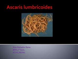
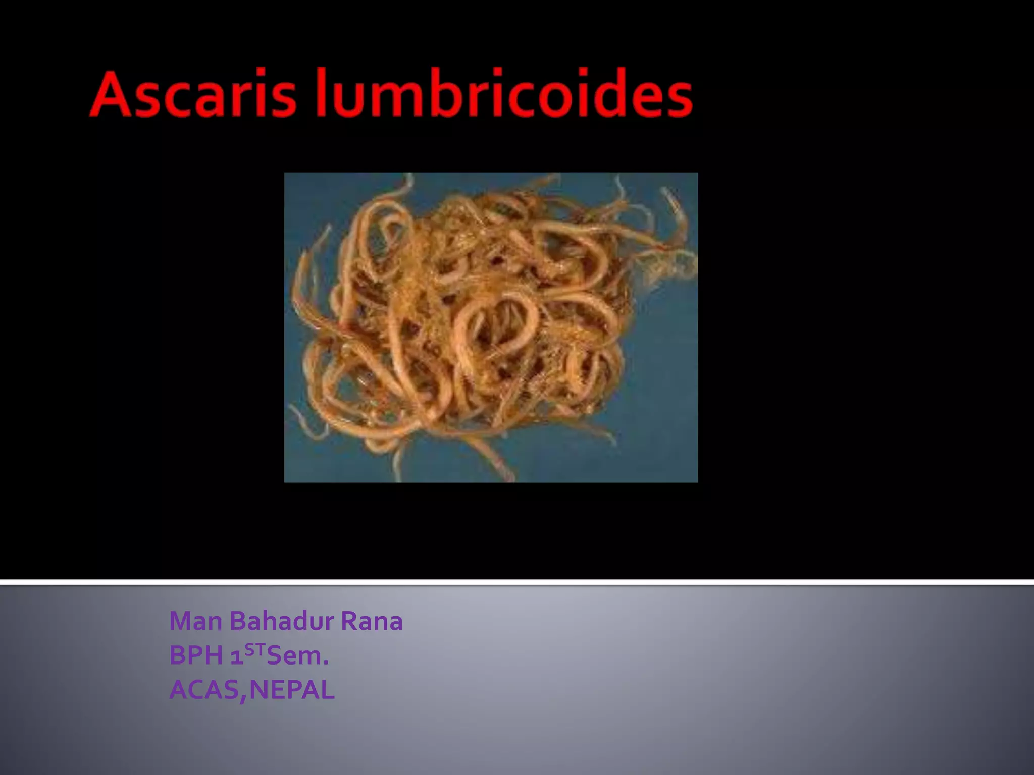
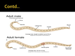
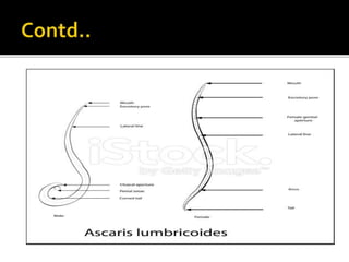
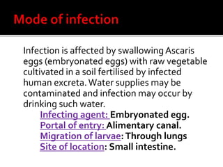
1. Ascaris lumbricoides, commonly known as the large roundworm, infects humans and lives in the small intestine.
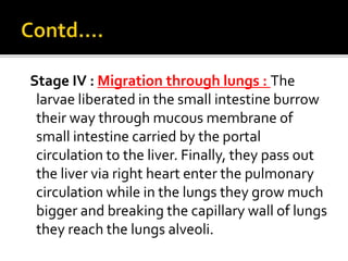
2. The life cycle involves eggs passing in feces and developing in soil, which are then ingested and hatch in the small intestine. Larvae migrate through the lungs before reaching maturity in the small intestine.
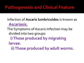
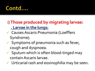
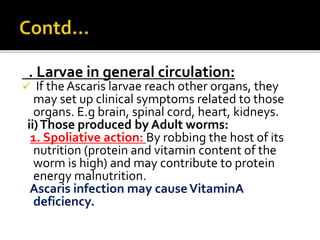
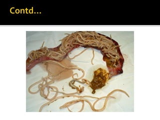
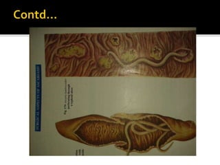
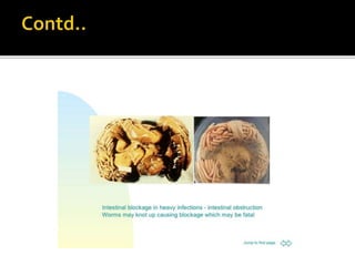
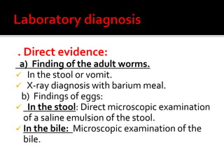
3. Symptoms can include pneumonia during larval migration through the lungs, and intestinal obstruction, malnutrition, and allergic reactions from adult worms in the small intestine. Diagnosis involves finding eggs in stool or adult worms after treatment.


![[Micro] hymenolepis nana](https://cdn.slidesharecdn.com/ss_thumbnails/3rxjz7ekrwinb1sq3uxs-signature-2127a2ca5368c7fdfd023e8d90dde3fc0b9fe7d91346a4189562c9f63dc0d19d-poli-150819190755-lva1-app6892-thumbnail.jpg?width=640&height=640&fit=bounds)


![CASE_PRESENTATION_ON_subdural_hematoma(SDH)[1 FINAL PPT]-1.pptx](https://cdn.slidesharecdn.com/ss_thumbnails/casepresentationonsubduralhematomasdh1finalppt-1-260129172522-d405d375-thumbnail.jpg?width=640&height=640&fit=bounds)


