Downloaded 132 times


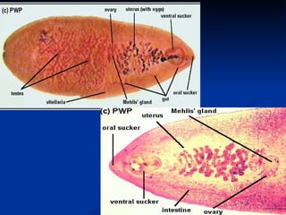
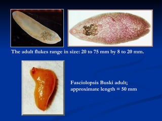
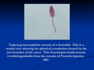
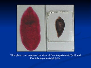


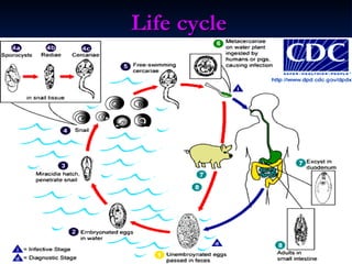


Fasciolopsis buski is a large intestinal fluke that infects humans and pigs. It lives in the small intestine of its hosts and can measure up to 80 mm in length, making it the largest fluke that infects humans. The lifecycle involves eggs passed in feces that hatch in water and infect snail intermediate hosts, developing through sporocyst and redia stages before releasing cercariae that encyst on aquatic plants. Humans and pigs become infected by eating these metacercariae.


![Trematode parasites of man[1]. A detailed lecturepptx](https://cdn.slidesharecdn.com/ss_thumbnails/trematodeparasitesofman1-250501020756-a3a59859-thumbnail.jpg?width=640&height=640&fit=bounds)


