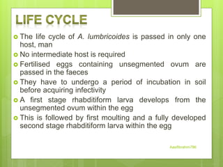
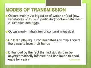
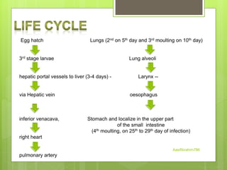
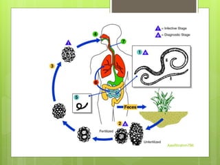
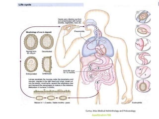
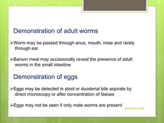
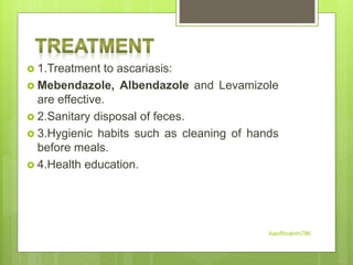
Ascariasis
PGT BIOLOGY JAWAHAR NAVODAYA VIDYALAYA
RAKH JAGANOO UDHAMPUR(J&K)
 Ascaris lumbricoides is the
largest nematode
(roundworm) parasitizing the
human intestine.
 Ascaris lumbricoides is an
intestinal worm found in the
small intestine of man (mainly
in the jejunum and upper part
of the ileum).
 They are more common in
children than in adults
 As many as 500 to 5000 adult
worms may inhabit a single
host. Aasifibrahim786
Geographical distribution
 Worldwide
 High prevalence in underdeveloped countries
that have poor sanitation (parts of Asia, South
America and Africa)
 Occurs during rainy months, tropical and
subtropical countries
 Even occurs in rural areas in the United
States
Aasifibrahim786
 It is a elongated, cylindrical and
tapering at both ends.
 Sexes are separate
 The female is longer than male 25 –
40 cm long, 4-6 mm in diameter.
 Male is smaller being 15-30 cm
long, 2-4 mm in diameter.
 The posterior end of male is curved
ventrally in the form of a hook
 The digestive and respiratory
organs of the worm float inside the
body cavity possessing a toxic fluid
known as ascaron
Aasifibrahim786
 The mouth opens at the anterior
end.
 It is surrounded by three finely
toothed lips.
 The lips are one dorsal and two
ventrolateral.
 These lips bear sensory
structures called labial papillae
Aasifibrahim786
Adult worms of A. lumbricoides
Aasifibrahim786
A mature female A. lumbricoides lays enormous
number of eggs
(nearly 2,00,000 eggs daily) which are passed in the
faeces
 There are two kinds of the eggs. They are fertilized
eggs, and unfertilized eggs
 We usually describe an egg in 5 aspects: size, color,
shape,
shell and content
 Decorticated eggs: Both fertilized and unfertilized
eggs
sometimes may lack their outer albuminous
coats and are colorless
Aasifibrahim786
 Broad oval in shape, brown in color,
an average size 60× 45µm.
 The shell is thicker and consists of
chitinous layer, and mammillated
albuminous coat stained brown by
bile.
 The content is a fertilized ovum.
 There is a new-moon(crescent)
shaped clear space at the each end
inside the shell. Aasifibrahim786
 Narrower and longer and measure
90 µm in length and 55 µm in
breadth
 They are bile stained and brown in
colour
 The chitinous layer and albuminous
coat are thinner and irregular than
those of the fertilized eggs
 The content is made of small
atrophied ovum suurounded by
many refractable granules of various
size.
 Heaviest of all the helminthic eggs
Aasifibrahim786
Both fertilized and unfertilized
eggs sometimes may lack their
outer albuminous coats and are
colorless.
Aasifibrahim786
 The life cycle of A. lumbricoides is passed in only one
host, man
 No intermediate host is required
 Fertilised eggs containing unsegmented ovum are
passed in the faeces
 They have to undergo a period of incubation in soil
before acquiring infectivity
 A first stage rhabditiform larva develops from the
unsegmented ovum within the egg
 This is followed by first moulting and a fully developed
second stage rhabditiform larva within the egg
Aasifibrahim786
 Occurs mainly via ingestion of water or food (raw
vegetables or fruits in particular) contaminated with
A. lumbricoides eggs.
 Occasionally inhalation of contaminated dust
 Children playing in contaminated soil may acquire
the parasite from their hands
 Enhanced by the fact that individuals can be
asymptomatically infected and continues to shed
eggs for years Aasifibrahim786
Egg hatch Lungs (2nd on 5th day and 3rd moulting on 10th day)
3rd stage larvae Lung alveoli
hepatic portal vessels to liver (3-4 days) - Larynx --
via Hepatic vein oesophagus
inferior venacava, Stomach and localize in the upper part
of the small intestine
(4th moulting, on 25th to 29th day of infection)
right heart
pulmonary artery
Aasifibrahim786
Aasifibrahim786
Aasifibrahim786
Disease produced by A. lumbricoides is known as
ascariasis and
is caused by both adult worms and migrating larvae
There are two phases in ascariasis:
1. The blood-lung migration phase of the larvae
2. The intestinal phase of the adults
Aasifibrahim786
1. The blood-lung migration phase of the larvae: During
the migration through the lungs, the larvae may cause a
pneumonia. The symptoms of the pneumonia are low
fever, cough, blood-tinged sputum, asthma. Large
numbers of worms may give rise to allergic symptoms.
Eosionophilia is generally present. These clinical
manifestation is also called Loeffler’s syndrome.
Aasifibrahim786
2. The intestinal phase of the adults. The presence of a
few adult worms in the lumen of the small intestine usually
produces no symptoms, but may give rise to vague
abdominal pains or intermittent colic, especially in children.
A heavy worm burden can result in malnutrition. More
serious manifestations have been observed. Wandering
adults may block the appendical lumen or the common
bile duct and even perforate the intestinal wall. Thus
complications of ascariasis, such as intestinal obstruction,
appendicitis, biliary ascariasis, perforation of the intestine,
cholecystitis, pancreatitis and peritonitis, etc., may occur,
in which biliary ascariasis is the most common
complication.
Aasifibrahim786
Laboratory diagnosis
Done by following methods
1. Parasitic diagnosis
a) Demonstration of adult worm
b) Demonstration of eggs
c) Demonstration of larvae
2. Serodiagnosis
3. Eosinophilia
Aasifibrahim786
Demonstration of adult worms
Worm may be passed through anus, mouth, nose and rarely
through ear
Barium meal may occasionally reveal the presence of adult
worms in the small intestine
Demonstration of eggs
Eggs may be detected in stool or duodenal bile aspirate by
direct microscopy or after concentration of faeces
Eggs may not be seen if only male worms are present
Aasifibrahim786
Ascaris larvae may be detected in the sputum during the stage
of migration
2. Serodiagnosis
Ascaris antibody can be detected by indirect haemagglutination
(IHA) And immunofluorescence antibody (IFA) test
These tests are useful for the diagnosis of extraintestinal –
ascariasis like Loeffler’s syndrome
3. Eosinophilia
It is seen in larval invasion stage
Aasifibrahim786
 1.Treatment to ascariasis:
 Mebendazole, Albendazole and Levamizole
are effective.
 2.Sanitary disposal of feces.
 3.Hygienic habits such as cleaning of hands
before meals.
 4.Health education.
Aasifibrahim786
Ascariasis can be prevented by
 Proper disposal of human faeces
 Avoidance of eating raw vegetables and
salads
 Periodic treatment with an effective
anthelminthic, in communities that lack
sanitary facilities
Aasifibrahim786
Aasifibrahim786

Ascariasis by Asif Ibrahim

  • 1.
    Ascariasis PGT BIOLOGY JAWAHARNAVODAYA VIDYALAYA RAKH JAGANOO UDHAMPUR(J&K)
  • 2.
     Ascaris lumbricoidesis the largest nematode (roundworm) parasitizing the human intestine.  Ascaris lumbricoides is an intestinal worm found in the small intestine of man (mainly in the jejunum and upper part of the ileum).  They are more common in children than in adults  As many as 500 to 5000 adult worms may inhabit a single host. Aasifibrahim786
  • 3.
    Geographical distribution  Worldwide High prevalence in underdeveloped countries that have poor sanitation (parts of Asia, South America and Africa)  Occurs during rainy months, tropical and subtropical countries  Even occurs in rural areas in the United States Aasifibrahim786
  • 4.
     It isa elongated, cylindrical and tapering at both ends.  Sexes are separate  The female is longer than male 25 – 40 cm long, 4-6 mm in diameter.  Male is smaller being 15-30 cm long, 2-4 mm in diameter.  The posterior end of male is curved ventrally in the form of a hook  The digestive and respiratory organs of the worm float inside the body cavity possessing a toxic fluid known as ascaron Aasifibrahim786
  • 5.
     The mouthopens at the anterior end.  It is surrounded by three finely toothed lips.  The lips are one dorsal and two ventrolateral.  These lips bear sensory structures called labial papillae Aasifibrahim786
  • 6.
    Adult worms ofA. lumbricoides Aasifibrahim786
  • 7.
    A mature femaleA. lumbricoides lays enormous number of eggs (nearly 2,00,000 eggs daily) which are passed in the faeces  There are two kinds of the eggs. They are fertilized eggs, and unfertilized eggs  We usually describe an egg in 5 aspects: size, color, shape, shell and content  Decorticated eggs: Both fertilized and unfertilized eggs sometimes may lack their outer albuminous coats and are colorless Aasifibrahim786
  • 8.
     Broad ovalin shape, brown in color, an average size 60× 45µm.  The shell is thicker and consists of chitinous layer, and mammillated albuminous coat stained brown by bile.  The content is a fertilized ovum.  There is a new-moon(crescent) shaped clear space at the each end inside the shell. Aasifibrahim786
  • 9.
     Narrower andlonger and measure 90 µm in length and 55 µm in breadth  They are bile stained and brown in colour  The chitinous layer and albuminous coat are thinner and irregular than those of the fertilized eggs  The content is made of small atrophied ovum suurounded by many refractable granules of various size.  Heaviest of all the helminthic eggs Aasifibrahim786
  • 10.
    Both fertilized andunfertilized eggs sometimes may lack their outer albuminous coats and are colorless. Aasifibrahim786
  • 11.
     The lifecycle of A. lumbricoides is passed in only one host, man  No intermediate host is required  Fertilised eggs containing unsegmented ovum are passed in the faeces  They have to undergo a period of incubation in soil before acquiring infectivity  A first stage rhabditiform larva develops from the unsegmented ovum within the egg  This is followed by first moulting and a fully developed second stage rhabditiform larva within the egg Aasifibrahim786
  • 12.
     Occurs mainlyvia ingestion of water or food (raw vegetables or fruits in particular) contaminated with A. lumbricoides eggs.  Occasionally inhalation of contaminated dust  Children playing in contaminated soil may acquire the parasite from their hands  Enhanced by the fact that individuals can be asymptomatically infected and continues to shed eggs for years Aasifibrahim786
  • 13.
    Egg hatch Lungs(2nd on 5th day and 3rd moulting on 10th day) 3rd stage larvae Lung alveoli hepatic portal vessels to liver (3-4 days) - Larynx -- via Hepatic vein oesophagus inferior venacava, Stomach and localize in the upper part of the small intestine (4th moulting, on 25th to 29th day of infection) right heart pulmonary artery Aasifibrahim786
  • 14.
  • 15.
  • 16.
    Disease produced byA. lumbricoides is known as ascariasis and is caused by both adult worms and migrating larvae There are two phases in ascariasis: 1. The blood-lung migration phase of the larvae 2. The intestinal phase of the adults Aasifibrahim786
  • 17.
    1. The blood-lungmigration phase of the larvae: During the migration through the lungs, the larvae may cause a pneumonia. The symptoms of the pneumonia are low fever, cough, blood-tinged sputum, asthma. Large numbers of worms may give rise to allergic symptoms. Eosionophilia is generally present. These clinical manifestation is also called Loeffler’s syndrome. Aasifibrahim786
  • 18.
    2. The intestinalphase of the adults. The presence of a few adult worms in the lumen of the small intestine usually produces no symptoms, but may give rise to vague abdominal pains or intermittent colic, especially in children. A heavy worm burden can result in malnutrition. More serious manifestations have been observed. Wandering adults may block the appendical lumen or the common bile duct and even perforate the intestinal wall. Thus complications of ascariasis, such as intestinal obstruction, appendicitis, biliary ascariasis, perforation of the intestine, cholecystitis, pancreatitis and peritonitis, etc., may occur, in which biliary ascariasis is the most common complication. Aasifibrahim786
  • 19.
    Laboratory diagnosis Done byfollowing methods 1. Parasitic diagnosis a) Demonstration of adult worm b) Demonstration of eggs c) Demonstration of larvae 2. Serodiagnosis 3. Eosinophilia Aasifibrahim786
  • 20.
    Demonstration of adultworms Worm may be passed through anus, mouth, nose and rarely through ear Barium meal may occasionally reveal the presence of adult worms in the small intestine Demonstration of eggs Eggs may be detected in stool or duodenal bile aspirate by direct microscopy or after concentration of faeces Eggs may not be seen if only male worms are present Aasifibrahim786
  • 21.
    Ascaris larvae maybe detected in the sputum during the stage of migration 2. Serodiagnosis Ascaris antibody can be detected by indirect haemagglutination (IHA) And immunofluorescence antibody (IFA) test These tests are useful for the diagnosis of extraintestinal – ascariasis like Loeffler’s syndrome 3. Eosinophilia It is seen in larval invasion stage Aasifibrahim786
  • 22.
     1.Treatment toascariasis:  Mebendazole, Albendazole and Levamizole are effective.  2.Sanitary disposal of feces.  3.Hygienic habits such as cleaning of hands before meals.  4.Health education. Aasifibrahim786
  • 23.
    Ascariasis can beprevented by  Proper disposal of human faeces  Avoidance of eating raw vegetables and salads  Periodic treatment with an effective anthelminthic, in communities that lack sanitary facilities Aasifibrahim786
  • 24.