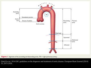
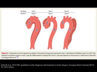
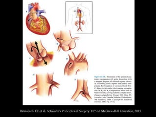
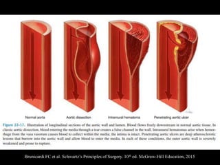
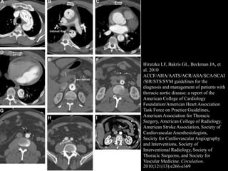
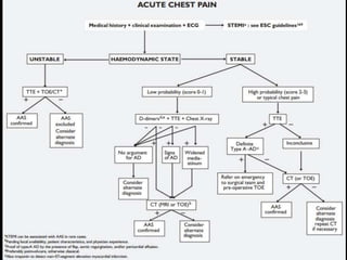
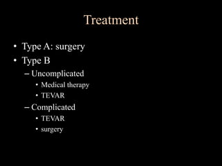
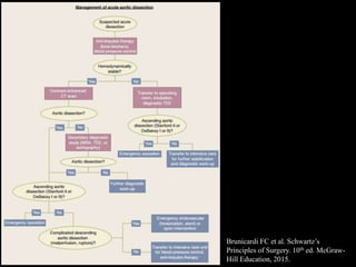
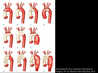
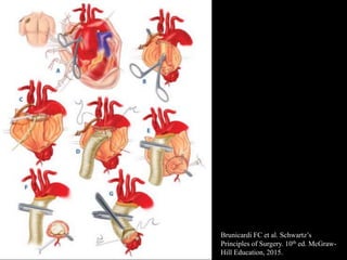
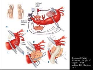
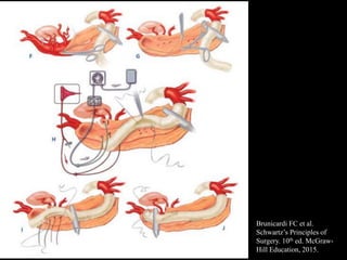
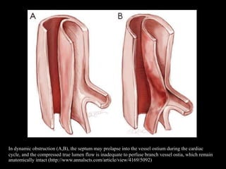
Aortic Dissection
Facebook: Happy Friday Knight
Erbel R et al. 2014 ESC guidelines on the diagnosis and treatment of aortic disease. European Heart Journal.(2014)
35, 2873-2926.
• Normal aortic diameter: less than 40 mm and
taper gradually downstream
• Variation influenced by factors: age, gender,
body size, blood pressure
• The rate of aortic expansion per year
– In men: 0.9 mm
– In women: 0.7 mm
Definition and Classification
• Disruption of medial layer provoked by
intramural bleeding resulting in separation of
aortic wall layers and formation of true and
false lumen
• Intimal tear in the initiating condition 
tracking the blood in a dissection plane within
the media
• Acute AD: < 14 days
• Subacute AD: 15 – 90 days
• Chronic AD: > 90 days
Erbel R et al. 2014 ESC guidelines on the diagnosis and treatment of aortic disease. European Heart Journal.(2014)
35, 2873-2926.c
Brunicardi FC et al. Schwartz’s Principles of Surgery. 10th ed. McGraw-Hill Education, 2015
Clinical Presentation
• Severe Chest or back pain:
– Sharp, ripping, tearing, knife-like
– abruptness
• Aortic regurgitation and congestive heart failure
• Pericardial tamponade
• Myocardial ischemia: mislead to ACS
• Pleural effusion
• Syncope
• Coma/stoke
• Mesenteric ischemia and aortoenteric fistula
• Renal failure
Brunicardi FC et al. Schwartz’s Principles of Surgery. 10th ed. McGraw-Hill Education, 2015
Erbel R et al. 2014 ESC guidelines on the diagnosis and treatment of aortic disease. European Heart Journal.(2014)
35, 2873-2926.c
Erbel R et al. 2014 ESC guidelines on the diagnosis and treatment of aortic disease. European Heart Journal.(2014)
35, 2873-2926.c
Diagnostic Imaging
• Assessment entire aorta
– Diameter
– Shape and extension of the dissection membrane
– Aortic valve
– Aortic branches
– Relationship with the adjacent structures
– Mural thrombus
Echocardiography
• Detecting
– intimal flap in aorta
– complete obstruction of false lumen
– central displacement of intimal calcification
– separation of intimal layers from thrombus
– shearing of different wall layers during aortic
pulsation.
• Sensitivity 77-80% specificity 93-96%
Computed Tomography
• Key: intimal flap separating 2 lumens
– Non-contrast: detect medially displaced aortic
calcifications or intimal flap
– Determining extension including aortic branch vessels
– length and diameter of the aorta
– TL and FL
– involvement of vital vasculature
– distance from the intimal tear to the vital vascular
branches
– Convex surface toward FL
– FL: slower flow, larger diameter, contained thrombi
Computed Tomography
• Spiral fashion: FL from Rt anterolateral wall of
ascending aorta extend to Lt posterolateral
wall of descending aorta
• ECG-gated 64-detector CT
– Triple rule out: evaluate patient with chest pain
with potential causes: AD, PE, ACS
– Pulsation artifact: most common cause of misdx
Hiratzka LF, Bakris GL, Beckman JA, et
al. 2010
ACCF/AHA/AATS/ACR/ASA/SCA/SCAI
/SIR/STS/SVM guidelines for the
diagnosis and management of patients with
thoracic aortic disease: a report of the
American College of Cardiology
Foundation/American Heart Association
Task Force on Practice Guidelines,
American Association for Thoracic
Surgery, American College of Radiology,
American Stroke Association, Society of
Cardiovascular Anesthesiologists,
Society for Cardiovascular Angiography
and Interventions, Society of
Interventional Radiology, Society of
Thoracic Surgeons, and Society for
Vascular Medicine. Circulation.
2010;121(13):e266-e369
• Magnetic Resonance Imaging (MRI)
• Aortography
Treatment
• Type A: surgery
• Type B
– Uncomplicated
• Medical therapy
• TEVAR
– Complicated
• TEVAR
• surgery
Brunicardi FC et al. Schwartz’s
Principles of Surgery. 10th ed. McGraw-
Hill Education, 2015.
Initial Managment
• Aggressive pharmacologic treatment
• The goals are to stabilize the dissection and
prevent rupture
• Monitor radial pressure in the arm with better
pulse
• Monitor: neurological status, peripheral pulse,
urine output
Initial Management
• Anti-impulse therapy or blood pressure control
– Reducing aortic wall stress, the force of left
ventricular ejection, chronotropy, and the rate of
change in blood pressure (dP/dT)
– dP/dT: Achieve by lowering cardiac contractility
and blood pressure
– Adequate pain control: opioids
Intial managment
• Controlled:
– Heart rate 60 – 80 bpm
– Systolic blood pressure 100 -110 mmHg
– Mean arterial pressure 60 – 75 mmHg
• Drugs:
– IV beta-adrenergic blocker: esmolol, labetalol
– Direct vasodilators
– Calcium channel blocker: diltiazem
– Angiotensin-converting enzyme inhibitors: enalapril
Initial management
• Drugs: contraindications
– Severe heart failure
– Bradyarrthythmia
– High-grade atrioventricular conduction block
– Bronchospastic disease
Treatment of type A dissection
• Surgery = treatment of choice in both acute and
chronic dissection
– Prevent aortic rupture, cardiac tamponade, relieve
aortic regurgitation
• Mortality 50% in 48 hr if not operated
• Perioperative mortality 25%
• Neurologic complication 18%
• 1-month mortality
– Nonsurgery 90%
– Surgery 30%
• Absolute indication for emergency surgical
repair: graft replacement of ascending aorta
• Delayed repair should be considered in:
– Severe acute stroke
– Mesenteric ischemia
– Elderly and comorbidity
– Stable condition and may benefit from transfer to
specialized center
– Have undergone cardiac surgery less than 3 weeks
Treatment of type A dissection
Brunicardi FC et al. Schwartz’s Principles of
Surgery. 10th ed. McGraw-Hill Education, 2015.
Brunicardi FC et al. Schwartz’s
Principles of Surgery. 10th ed. McGraw-
Hill Education, 2015.
Treatment of Type A Dissection
• The majority, the dissection persists distal to
the repaired site and susceptible to dilatation:
25% - 40% of survivors  need further aortic
repair
• Rupture of the dilated distal aorta is common
cause of death
• Alternate strategies: total arch replacement and
hybrid arch strategies
Treatment of Type A Dissection
• Chronic dissection:
– Surgery
– Tissue is stronger: aggressive repair
Treatment of type B Dissection:
Uncomplicated
• Uncomplicated = absence of malperfusion or
signs of disease progression
• Safely stabilize patients with medical alone
• Medical treatment results in lower morbidity
and mortality rates than traditional surgical
repair
• 2 CT scan on day 2,3 and 8,9 used to rule out
significant aortic expansion
• Drug of choice: beta blocker
• Follow up CT:
– Q3months x 1 year, then
– Q6montns x 1 year, then
– annually
Treatment of type B Dissection:
Uncomplicated
• TEVAR can be the choice: no significant lower
total mortality
Treatment of type B Dissection:
Uncomplicated
Treatment of Complicated Type B Dissection
• Complicated:
– Persistent or recurrent pain
– Uncontrolled hypertension despite full medication
– Early aortic expansion
– Malperfusion
– Signs of rupture (hemothorax, increasing periaortic
and mediastinal hematoma)
• Surgery
• TEVAR
Complicated Type B Dissection: Surgery
• Acute dissection
– High morbidity and mortality rates
– Primary goals are to prevent fatal rupture and restore
branch vessel perfusion: limited graft repair
– Common site of rupture: proximal third  upper half
of descending aorta usually repaired
– Distal half may be replaced if it exceeds 4 cm in
diameter
– Indications: lower extremity disease, severe tortuosity
of iliac arteries, a sharp angulation of aortic arch,
absence of proximal landing zone
• Malperfusion syndrome
– Second line therapy
– Surgical extra-anatomic revascularization
– Femoral-to-femoral bypass for lower-extremity
ischemia
– Open aortic fenestration
– Visceral or renal artery bypass
Complicated Type B Dissection: Surgery
http://www.annalscts.com/article/view/3872/4786
http://www.annalscts.com/article/view/4169/5092
• Chronic dissection
– More aggressive replacement
– Elective surgery
– Difference: excise as much dissecting membrane
as possible to identify true and false lumens and to
locate all important vessel branches
Complicated Type B Dissection: Surgery
Brunicardi FC et al.
Schwartz’s Principles of
Surgery. 10th ed.
McGraw-Hill Education,
2015.
Brunicardi FC et al.
Schwartz’s Principles of
Surgery. 10th ed. McGraw-
Hill Education, 2015.
Complicated Type B Dissection: TEVAR
• Malperfusion syndrome
– Routinely used
– Endovascular fenestration: a balloon is used to
create a tear in the dissection flap which allows
blood to flow in both true and false lumen
– Placement of stent graft in the true lumen resolves
dynamic malperfusion
– Placement of stent graft in visceral artery resolves
static malperfusion
In dynamic obstruction (A,B), the septum may prolapse into the vessel ostium during the cardiac
cycle, and the compressed true lumen flow is inadequate to perfuse branch vessel ostia, which remain
anatomically intact (http://www.annalscts.com/article/view/4169/5092)
Mechanisms of static obstruction. (A) Compression of the vessel by blind ends of the false lumen; (B)
presence of true and false lumen in the vessel causing further compression; (C) thrombosis of the
vessel distal to the compromised ostia (http://www.annalscts.com/article/view/4169/5092)
• Acute dissection
– The goal is to
• Use stent graft to cover the intimal tear
• Seal the entry site of dissection
• Cause thrombosis of false lumen to aid aortic
remodeling and reduce late aortic expansion
– Not recommend in patients with connective tissue
disorder but can be used as a bridge to later
definitive repair
Complicated Type B Dissection: TEVAR
• Chronic dissection
– Controversial and remains under investigation
– Rigidity of dissecting membrane and multiple re-
entry sites make it difficult to exclude false lumen
Complicated Type B Dissection: TEVAR
TEVAR
• New emerging treatment modality
• Far less invasive approach than open surgery
and ease of application has extended
management options especially in patients
unfit for surgery
• Not recommend in patients with connective
tissue disease
TEVAR
• Technical success: closure of primary entry
tear and induction of false lumen thrombosis
• Size of graft: diameter of aorta proximal to the
dissected segment
• Ballooning is not recommended: rupture and
retrograde dissection has been reported
TEVAR
• Intraprocedural monitoring
– Invasive blood pressure monitoring
– Pharmacological lowering systolic blood pressure
to <80 mmHg during stent graft deployment 
avoid displacement of the device
• Hybrid procedure:
– 2 different approaches to extensive disease
• Frozen elephant trunk with antegrade stent grafting
• Re-routing supraaortic branches
http://www.annalscts.com/article/view/2014/2744
TEVAR: complications
• Vascular and vessel-related injuries
(thrombosis, bleeding, retrograde type A
dissection, stroke)
• Myocardial injury
• Erosion of left main bronchus or esophagus
• Spinal cord injury
TEVAR: techniques
• Patient selection:
– Asymptomatic descending aortic aneurysm > 5cm
without evidence of connective tissue disorder
– Symptomatic aneurysm
– Proximal and distal neck ≥ 2cm without significant
thrombus or calcification
– Access vessel diameter 8 mm without extreme
tortuosity
TEVAR: techniques
• Supine position. Preparation of skin
• CSF drainage in:
– Previous AAA repair
– Iliac conduit or subclavian artery covered is planned
– TAA with extensive graft coverage
• Femoral artery cutdown. Patient is heparinized
with ACT ≥ 300 secs and maintain throughout the
procedure
• 12F sheath is used for femoral access
TEVAR: techniques
• And angled catheter and guidewire are used to
access abdominal aorta under fluoroscope
guidance to ascending aorta
• Watch for arrhythmia
• A percutaneous 6F sheath is placed in the
contralateral femoral artery and a second
guidewire positioned in ascending aorta
TEVAR: techniques
• The pigtail catheter is inserted and positioned
at ascending aorta
• Perform aortogram
• Evaluate proximal neck
• Measure length and diameter of proximal and
distal neck
• Choose stent graft, heparinize, and advance
into proximal neck
http://www.annalscts.com/article/view/2014/2744
TEVAR: techniques
• Repeat angiogram
• Sheath and wire are removed
• Repair femoral artery by standard fashion
• Check distal pulse
• Reverse heparin
http://www.hkma.org/english/cme/onlinecme/cme201605main.htm
https://sites.google.com/site/hepaticmri/endoleak
http://evtoday.com/2015/04/endoleak-
and-the-role-of-embolization/
References
Erbel R et al. 2014 ESC guidelines on the diagnosis and treatment of aortic disease. European
Heart Journal.(2014) 35, 2873-2926.
Erbel R et al. Diagnosis and management of aortic dissection: recommendation of the task force
on aortic dissection, european society of cardiology. European Heart Journal.(2001) 22, 1642-81.
Kaiser LR et al. Mastery of cardiothoracic surgery. 3rd ed. Philadelphia: Wolters kluwer, 2014.
Brunicardi FC et al. Schwartz’s Principles of Surgery. 10th ed. McGraw-Hill Education, 2015.
Grabenwoger M et al. Thoracic Endovascular Aortic Repair (TEVAR) for the treatment of aortic
diseases: a positstatement from the European Association for Cardio-Thoracic Surgery (EACTS)
and the European Society of Cardiology (ESC), in collaboration with the European Association of
Percutaneous Cardiovascular Interventions (EAPCI) .European Heart Journal. (2012)
References
Hiratzka LF, Bakris GL, Beckman JA, et al. 2010
ACCF/AHA/AATS/ACR/ASA/SCA/SCAI/SIR/STS/SVM guidelines for the diagnosis
and management of patients with thoracic aortic disease: a report of the American
College of Cardiology Foundation/American Heart Association Task Force on Practice
Guidelines, American Association for Thoracic Surgery, American College of
Radiology, American Stroke Association, Society of Cardiovascular Anesthesiologists,
Society for Cardiovascular Angiography and Interventions, Society of Interventional
Radiology, Society of Thoracic Surgeons, and Society for Vascular Medicine.
Circulation. 2010;121(13):e266-e369
http://www.annalscts.com/article/view/4169/5092
http://www.hkma.org/english/cme/onlinecme/cme201605main.htm
http://www.annalscts.com/article/view/2014/2744

Aortic dissection

  • 1.
  • 2.
    Erbel R etal. 2014 ESC guidelines on the diagnosis and treatment of aortic disease. European Heart Journal.(2014) 35, 2873-2926.
  • 3.
    • Normal aorticdiameter: less than 40 mm and taper gradually downstream • Variation influenced by factors: age, gender, body size, blood pressure • The rate of aortic expansion per year – In men: 0.9 mm – In women: 0.7 mm
  • 4.
    Definition and Classification •Disruption of medial layer provoked by intramural bleeding resulting in separation of aortic wall layers and formation of true and false lumen • Intimal tear in the initiating condition  tracking the blood in a dissection plane within the media
  • 5.
    • Acute AD:< 14 days • Subacute AD: 15 – 90 days • Chronic AD: > 90 days
  • 6.
    Erbel R etal. 2014 ESC guidelines on the diagnosis and treatment of aortic disease. European Heart Journal.(2014) 35, 2873-2926.c
  • 7.
    Brunicardi FC etal. Schwartz’s Principles of Surgery. 10th ed. McGraw-Hill Education, 2015
  • 8.
    Clinical Presentation • SevereChest or back pain: – Sharp, ripping, tearing, knife-like – abruptness • Aortic regurgitation and congestive heart failure • Pericardial tamponade • Myocardial ischemia: mislead to ACS • Pleural effusion • Syncope • Coma/stoke • Mesenteric ischemia and aortoenteric fistula • Renal failure
  • 9.
    Brunicardi FC etal. Schwartz’s Principles of Surgery. 10th ed. McGraw-Hill Education, 2015
  • 10.
    Erbel R etal. 2014 ESC guidelines on the diagnosis and treatment of aortic disease. European Heart Journal.(2014) 35, 2873-2926.c
  • 11.
    Erbel R etal. 2014 ESC guidelines on the diagnosis and treatment of aortic disease. European Heart Journal.(2014) 35, 2873-2926.c
  • 12.
    Diagnostic Imaging • Assessmententire aorta – Diameter – Shape and extension of the dissection membrane – Aortic valve – Aortic branches – Relationship with the adjacent structures – Mural thrombus
  • 13.
    Echocardiography • Detecting – intimalflap in aorta – complete obstruction of false lumen – central displacement of intimal calcification – separation of intimal layers from thrombus – shearing of different wall layers during aortic pulsation. • Sensitivity 77-80% specificity 93-96%
  • 14.
    Computed Tomography • Key:intimal flap separating 2 lumens – Non-contrast: detect medially displaced aortic calcifications or intimal flap – Determining extension including aortic branch vessels – length and diameter of the aorta – TL and FL – involvement of vital vasculature – distance from the intimal tear to the vital vascular branches – Convex surface toward FL – FL: slower flow, larger diameter, contained thrombi
  • 15.
    Computed Tomography • Spiralfashion: FL from Rt anterolateral wall of ascending aorta extend to Lt posterolateral wall of descending aorta • ECG-gated 64-detector CT – Triple rule out: evaluate patient with chest pain with potential causes: AD, PE, ACS – Pulsation artifact: most common cause of misdx
  • 16.
    Hiratzka LF, BakrisGL, Beckman JA, et al. 2010 ACCF/AHA/AATS/ACR/ASA/SCA/SCAI /SIR/STS/SVM guidelines for the diagnosis and management of patients with thoracic aortic disease: a report of the American College of Cardiology Foundation/American Heart Association Task Force on Practice Guidelines, American Association for Thoracic Surgery, American College of Radiology, American Stroke Association, Society of Cardiovascular Anesthesiologists, Society for Cardiovascular Angiography and Interventions, Society of Interventional Radiology, Society of Thoracic Surgeons, and Society for Vascular Medicine. Circulation. 2010;121(13):e266-e369
  • 17.
    • Magnetic ResonanceImaging (MRI) • Aortography
  • 19.
    Treatment • Type A:surgery • Type B – Uncomplicated • Medical therapy • TEVAR – Complicated • TEVAR • surgery
  • 20.
    Brunicardi FC etal. Schwartz’s Principles of Surgery. 10th ed. McGraw- Hill Education, 2015.
  • 21.
    Initial Managment • Aggressivepharmacologic treatment • The goals are to stabilize the dissection and prevent rupture • Monitor radial pressure in the arm with better pulse • Monitor: neurological status, peripheral pulse, urine output
  • 22.
    Initial Management • Anti-impulsetherapy or blood pressure control – Reducing aortic wall stress, the force of left ventricular ejection, chronotropy, and the rate of change in blood pressure (dP/dT) – dP/dT: Achieve by lowering cardiac contractility and blood pressure – Adequate pain control: opioids
  • 23.
    Intial managment • Controlled: –Heart rate 60 – 80 bpm – Systolic blood pressure 100 -110 mmHg – Mean arterial pressure 60 – 75 mmHg • Drugs: – IV beta-adrenergic blocker: esmolol, labetalol – Direct vasodilators – Calcium channel blocker: diltiazem – Angiotensin-converting enzyme inhibitors: enalapril
  • 24.
    Initial management • Drugs:contraindications – Severe heart failure – Bradyarrthythmia – High-grade atrioventricular conduction block – Bronchospastic disease
  • 25.
    Treatment of typeA dissection • Surgery = treatment of choice in both acute and chronic dissection – Prevent aortic rupture, cardiac tamponade, relieve aortic regurgitation • Mortality 50% in 48 hr if not operated • Perioperative mortality 25% • Neurologic complication 18% • 1-month mortality – Nonsurgery 90% – Surgery 30%
  • 26.
    • Absolute indicationfor emergency surgical repair: graft replacement of ascending aorta • Delayed repair should be considered in: – Severe acute stroke – Mesenteric ischemia – Elderly and comorbidity – Stable condition and may benefit from transfer to specialized center – Have undergone cardiac surgery less than 3 weeks Treatment of type A dissection
  • 27.
    Brunicardi FC etal. Schwartz’s Principles of Surgery. 10th ed. McGraw-Hill Education, 2015.
  • 28.
    Brunicardi FC etal. Schwartz’s Principles of Surgery. 10th ed. McGraw- Hill Education, 2015.
  • 29.
    Treatment of TypeA Dissection • The majority, the dissection persists distal to the repaired site and susceptible to dilatation: 25% - 40% of survivors  need further aortic repair • Rupture of the dilated distal aorta is common cause of death • Alternate strategies: total arch replacement and hybrid arch strategies
  • 30.
    Treatment of TypeA Dissection • Chronic dissection: – Surgery – Tissue is stronger: aggressive repair
  • 31.
    Treatment of typeB Dissection: Uncomplicated • Uncomplicated = absence of malperfusion or signs of disease progression • Safely stabilize patients with medical alone • Medical treatment results in lower morbidity and mortality rates than traditional surgical repair • 2 CT scan on day 2,3 and 8,9 used to rule out significant aortic expansion
  • 32.
    • Drug ofchoice: beta blocker • Follow up CT: – Q3months x 1 year, then – Q6montns x 1 year, then – annually Treatment of type B Dissection: Uncomplicated
  • 33.
    • TEVAR canbe the choice: no significant lower total mortality Treatment of type B Dissection: Uncomplicated
  • 34.
    Treatment of ComplicatedType B Dissection • Complicated: – Persistent or recurrent pain – Uncontrolled hypertension despite full medication – Early aortic expansion – Malperfusion – Signs of rupture (hemothorax, increasing periaortic and mediastinal hematoma) • Surgery • TEVAR
  • 35.
    Complicated Type BDissection: Surgery • Acute dissection – High morbidity and mortality rates – Primary goals are to prevent fatal rupture and restore branch vessel perfusion: limited graft repair – Common site of rupture: proximal third  upper half of descending aorta usually repaired – Distal half may be replaced if it exceeds 4 cm in diameter – Indications: lower extremity disease, severe tortuosity of iliac arteries, a sharp angulation of aortic arch, absence of proximal landing zone
  • 36.
    • Malperfusion syndrome –Second line therapy – Surgical extra-anatomic revascularization – Femoral-to-femoral bypass for lower-extremity ischemia – Open aortic fenestration – Visceral or renal artery bypass Complicated Type B Dissection: Surgery
  • 38.
  • 39.
    • Chronic dissection –More aggressive replacement – Elective surgery – Difference: excise as much dissecting membrane as possible to identify true and false lumens and to locate all important vessel branches Complicated Type B Dissection: Surgery
  • 40.
    Brunicardi FC etal. Schwartz’s Principles of Surgery. 10th ed. McGraw-Hill Education, 2015.
  • 41.
    Brunicardi FC etal. Schwartz’s Principles of Surgery. 10th ed. McGraw- Hill Education, 2015.
  • 42.
    Complicated Type BDissection: TEVAR • Malperfusion syndrome – Routinely used – Endovascular fenestration: a balloon is used to create a tear in the dissection flap which allows blood to flow in both true and false lumen – Placement of stent graft in the true lumen resolves dynamic malperfusion – Placement of stent graft in visceral artery resolves static malperfusion
  • 43.
    In dynamic obstruction(A,B), the septum may prolapse into the vessel ostium during the cardiac cycle, and the compressed true lumen flow is inadequate to perfuse branch vessel ostia, which remain anatomically intact (http://www.annalscts.com/article/view/4169/5092)
  • 44.
    Mechanisms of staticobstruction. (A) Compression of the vessel by blind ends of the false lumen; (B) presence of true and false lumen in the vessel causing further compression; (C) thrombosis of the vessel distal to the compromised ostia (http://www.annalscts.com/article/view/4169/5092)
  • 45.
    • Acute dissection –The goal is to • Use stent graft to cover the intimal tear • Seal the entry site of dissection • Cause thrombosis of false lumen to aid aortic remodeling and reduce late aortic expansion – Not recommend in patients with connective tissue disorder but can be used as a bridge to later definitive repair Complicated Type B Dissection: TEVAR
  • 46.
    • Chronic dissection –Controversial and remains under investigation – Rigidity of dissecting membrane and multiple re- entry sites make it difficult to exclude false lumen Complicated Type B Dissection: TEVAR
  • 47.
    TEVAR • New emergingtreatment modality • Far less invasive approach than open surgery and ease of application has extended management options especially in patients unfit for surgery • Not recommend in patients with connective tissue disease
  • 48.
    TEVAR • Technical success:closure of primary entry tear and induction of false lumen thrombosis • Size of graft: diameter of aorta proximal to the dissected segment • Ballooning is not recommended: rupture and retrograde dissection has been reported
  • 49.
    TEVAR • Intraprocedural monitoring –Invasive blood pressure monitoring – Pharmacological lowering systolic blood pressure to <80 mmHg during stent graft deployment  avoid displacement of the device • Hybrid procedure: – 2 different approaches to extensive disease • Frozen elephant trunk with antegrade stent grafting • Re-routing supraaortic branches
  • 50.
  • 51.
    TEVAR: complications • Vascularand vessel-related injuries (thrombosis, bleeding, retrograde type A dissection, stroke) • Myocardial injury • Erosion of left main bronchus or esophagus • Spinal cord injury
  • 52.
    TEVAR: techniques • Patientselection: – Asymptomatic descending aortic aneurysm > 5cm without evidence of connective tissue disorder – Symptomatic aneurysm – Proximal and distal neck ≥ 2cm without significant thrombus or calcification – Access vessel diameter 8 mm without extreme tortuosity
  • 53.
    TEVAR: techniques • Supineposition. Preparation of skin • CSF drainage in: – Previous AAA repair – Iliac conduit or subclavian artery covered is planned – TAA with extensive graft coverage • Femoral artery cutdown. Patient is heparinized with ACT ≥ 300 secs and maintain throughout the procedure • 12F sheath is used for femoral access
  • 54.
    TEVAR: techniques • Andangled catheter and guidewire are used to access abdominal aorta under fluoroscope guidance to ascending aorta • Watch for arrhythmia • A percutaneous 6F sheath is placed in the contralateral femoral artery and a second guidewire positioned in ascending aorta
  • 55.
    TEVAR: techniques • Thepigtail catheter is inserted and positioned at ascending aorta • Perform aortogram • Evaluate proximal neck • Measure length and diameter of proximal and distal neck • Choose stent graft, heparinize, and advance into proximal neck
  • 56.
  • 57.
    TEVAR: techniques • Repeatangiogram • Sheath and wire are removed • Repair femoral artery by standard fashion • Check distal pulse • Reverse heparin
  • 58.
  • 59.
  • 60.
    References Erbel R etal. 2014 ESC guidelines on the diagnosis and treatment of aortic disease. European Heart Journal.(2014) 35, 2873-2926. Erbel R et al. Diagnosis and management of aortic dissection: recommendation of the task force on aortic dissection, european society of cardiology. European Heart Journal.(2001) 22, 1642-81. Kaiser LR et al. Mastery of cardiothoracic surgery. 3rd ed. Philadelphia: Wolters kluwer, 2014. Brunicardi FC et al. Schwartz’s Principles of Surgery. 10th ed. McGraw-Hill Education, 2015. Grabenwoger M et al. Thoracic Endovascular Aortic Repair (TEVAR) for the treatment of aortic diseases: a positstatement from the European Association for Cardio-Thoracic Surgery (EACTS) and the European Society of Cardiology (ESC), in collaboration with the European Association of Percutaneous Cardiovascular Interventions (EAPCI) .European Heart Journal. (2012)
  • 61.
    References Hiratzka LF, BakrisGL, Beckman JA, et al. 2010 ACCF/AHA/AATS/ACR/ASA/SCA/SCAI/SIR/STS/SVM guidelines for the diagnosis and management of patients with thoracic aortic disease: a report of the American College of Cardiology Foundation/American Heart Association Task Force on Practice Guidelines, American Association for Thoracic Surgery, American College of Radiology, American Stroke Association, Society of Cardiovascular Anesthesiologists, Society for Cardiovascular Angiography and Interventions, Society of Interventional Radiology, Society of Thoracic Surgeons, and Society for Vascular Medicine. Circulation. 2010;121(13):e266-e369 http://www.annalscts.com/article/view/4169/5092 http://www.hkma.org/english/cme/onlinecme/cme201605main.htm http://www.annalscts.com/article/view/2014/2744

Editor's Notes

  • #52 Retrograde Associated factors may include radial force of uncovered struts, diagnosis of TAD, extensive oversizing and ballooning