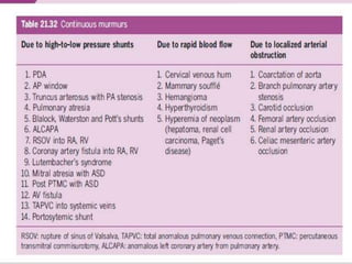
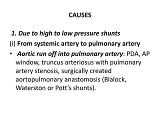
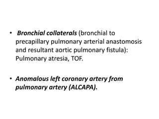
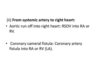
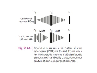
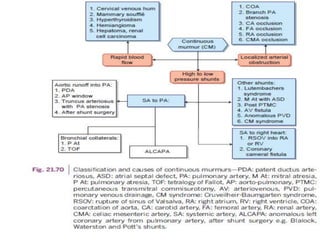
CONTINUOUS MURMURS
DR AMZATHKHAN
PG GENERAL MEDICINE
 A murmur that begins in systole and continues
without interruption through S2 into all or a part of
diastole without change in the character of the
murmur is defined as a continuous murmur.
 Continuous murmurs are usually generated by
uninterrupted flow from a high pressure vascular
bed into a low pressure vascular bed without phasic
interruption between systole and diastole.
CAUSES
1. Due to high to low pressure shunts
(i) From systemic artery to pulmonary artery
• Aortic run off into pulmonary artery: PDA, AP
window, truncus arteriosus with pulmonary
artery stenosis, surgically created
aortopulmonary anastomosis (Blalock,
Waterston or Pott’s shunts).
• Bronchial collaterals (bronchial to
precapillary pulmonary arterial anastomosis
and resultant aortic pulmonary fistula):
Pulmonary atresia, TOF.
• Anomalous left coronary artery from
pulmonary artery (ALCAPA).
(ii) From systemic artery to right heart:
• Aortic run off into right heart: RSOV into RA or
RV.
• Coronary cameral fistula: Coronary artery
fistula into RA or RV (LA).
Patent ductus arteriosus (PDA)
• Gibsons murmur (George Gibson in 1900)
• rough machinery murmur due to the
combination of high and low frequency
vibrations and associated thrill is common
.
• It peaks at S2, after which it gradually
wanes until it terminates before S1
(crescendo -decrescendo murmur).
LOCATION
• The murmur is best heard in the left infra
clavicular and pulmonary areas during
expiration.
• The systolic component is widely audible,
while the diastolic component is restricted to
pulmonary and infra clavicular areas.
INTENSITY
• Isometric hand grip increases the intensity and
duration of the murmur and may bring out the
diastolic component when it is not heard.
• The murmur disappears or decreases with
Valsalva maneuver.
• Amylnitrate inhalation decreases the diastolic
component.
• 2) Continuous murmur associated with
bronchial collaterals: It is heard in the same
location as that of PDA, but radiates widely
especially over the posterior thorax.
• 3) Continuous murmur in anomalous origin
of left coronary artery from pulmonary artery
(ALCAPA): The murmur is continuous when
the left-to-right shunt is large and is usually
best heard at left sternal border.
4) Continuous murmur in RSOV (rupture of sinus
of valsalva)
• The murmur is usually continuous when RSOV
occurs into RA or RV.
• But it is early diastolic when sinus of Valsalva
aneurysm ruptures into LV. It also becomes early
diastolic when PH supervenes with high RVSP
which shortens or abolishes systolic flow and
systolic component of the murmur.
• Superficial murmur, with prominent diastolic
thrill.
• “Purring of a cat”
• SITE – Lower left sternal border (or) xiphoid
process.
5) Continuous murmur in coronary cameral fistula
• soft, superficial and high pitched.
• In 90%, coronary artery (CA) fistula drains into
right heart and resultant murmur is continuous.
But in case of CA fistula draining into RV, the
murmur softens during systole (when the systolic
flow decreases due to compression of the fistula
during RV contraction).
CHARACTER
• The murmur is louder in systole as the pressure
gradient is more in systole in case of CA fistula
draining into RA or LA.
• The murmur is not continuous if it empties into
LV. It may be purely diastolic or systolic and
diastolic.
• Continuous murmur is also absent and may be
silent if multiple coronary artery fistulas empty
into pulmonary artery.
LOCATION
• The murmur is best heard either left or right
of the lower sternal area when CA drains into
RA.
• It is maximally heard at left mid to lower
sternal border or in sub xiphoid region in CA
draining into RV.
• In CA draining into LA, it is best heard in upper
to mid left sternal border and may radiate
leftward as far as the anterior axillary line
Other shunts
• Left to right shunts (mitral valve obstruction)
 Lutembachers syndrome
 Mitral atresia with ASD
 Post PTMC
• AV fistula
• Venovenous shunts
 Portosystemic shunts Curveiller Baumgartens
syndrome)
2. Due to rapid blood flow
• Cervical venous hum
• Mammary souffle
• Hyperthyroidism
• Hemiangioma
• Hyperemia of neoplasm: Hepatoma, renal cell
carcinoma, Paget’s disease.
Venous Hum
First recognized by Potain in 1867.
• The normal flow of blood across the normal veins in the neck
is noiseless. But increased velocity of blood flow gives rise to a
continuous bruit over the neck veins which is known as
cervical venous hum.
• CHARACTER - It may be rough and noisy and typically louder
in diastole.
MECHANISM
• The laminar flow in internal jugular vein may
be disturbed by the deformation of the vein at
the level of the transverse process of the atlas
during head rotation designed to elicit the
hum.
POSITION
• The venous hum is best heard in sitting posture with head
rotated to the opposite side and chin upwards, placing the
bell of the stethoscope at the base of the neck in between the
two heads of the sternocleidomastoid muscle and may be
more prominent on the right side. Sometimes it may radiate
below the clavicles and may be confused with the continuous
murmur of PDA if not evaluated carefully
• The murmur is abolished by the digital
compression of the internal jugular vein with
head in neutral position
– It is poorly heard in supine position, while
– Anemia and thyrotoxicosis initiate or
reinforce the venous hum.
Mammary souffle
• This innocent continuous arterial murmur
occurs in 10–15% of pregnant women during
2nd and 3rd trimesters and in early
postpartum period in lactating mothers.
• MECHANISM - It is due to increased blood
flow to the breast tissue.
PITCH AND CHARACTER
• This medium to high pitched murmur is best
heard over the breast on either side between 2nd
and 6th anterior intercostal spaces with no
significant change with respiration and may be
confused with the continuous murmur of PDA or
AV fistula.
• The mammary soufflé usually begins after S1
with a distinct gap and systolic component is the
loudest.
INTENSITY
• Light pressure with the stethoscope
augments the murmur, whereas the firm
pressure with the stethoscope or digital
compression abolishes the murmur.
• Valsalva maneuver has no significant effect on
the murmur. It disappears after the
termination of lactation.
Localized arterial obstruction
• Continous murmur (obstruction >80%) with
no adequate collaterals pressure gradient
produced throughout the cardiac cycle with
systolic accentuation.
• Systolic gradient +/ No diastolic gradient
• Coarctation of aorta
• Pulmonary artery stenosis
• Carotid/femoral/renal/celiac mesenteric
artery occulusion
COARCTATION OF AORTA
• Continuous murmur in coarctation of aorta
(COA) heard over the thorax and over the back
in the midline between the scapulae.
• rapid blood flow through tortuous intercostal
collaterals.
ADDED SOUNDS
• Pacemaker sounds
• Pericardial rub
• Mediastinal crunch
PACEMAKER SOUNDS
• These sounds are brief and high frequency
sounds occasionally produced by transvenous
pacemakers implanted in RV apex, due to
stimulation of the intercostal muscles(through
intercostal nerves) by the endocardial
electrodes.
• Pectoral muscles and diaphragmatic
stimulation can also occur.
• They occur synchronously with pacemaker
spike and often associated with twitching of
the muscles.
• The audible high frequency pacemaker sounds
always suggest myocardial perforation by the
endocardial lead, although it is not always
present.
PERICARDIAL RUB
• Pericardial rub is the hallmark of pericardial
inflammation.
a) Mechanism of production of pericardial rub
(PR): It is due to the movement of the parietal
and visceral surfaces of the pericardium
moving against each other.
b) Components of PR: The classic rub is triple
phased: systolic, mid-diastolic and late
diastolic (or presystolic/atrial systole).
• However, it is often biphasic, i.e. to and fro rubs due to
ventricular systolic and atrial systolic components.
• The diagnosis is the simplest when all the three phases
are present, which occurs only in 50% of the cases. It is
commonly audible in uremic pericarditis especially
when associated with hypertension.
• The systolic phase is most consistent followed by the
presystolic phase.
• In the setting of atrial fibrillation presystolic
component disappears.
c) Causes:
• Acute pericarditis of any etiology: especially tubercular
in origin
• Following open heart surgery: post cardiotomy
pericarditis
• Uremic pericarditis
• Rheumatic pancarditis
• Acute phase of transmural MI giving rise to post MI
pericarditis, which dramatically responds to steroids
• Infective endocarditis due to ring abscess.
d) Clinical recognition:
• These friction rubs are high-pitched, leathery
and scratchy in nature.
• They are best heard over the 2nd and 3rd left
intercostal spaces over the ‘bare area’ of the
heart.
• They seem close to the ear and are auscultated
best with diaphragm of the stethoscope with the
patient leaning forward or in knee-chest position
and holding the breath after forced expiration.
• Unlike pleural rub, pericardial rub can be
present even with large pericardial effusion
and cardiac tamponade.
• Occasionally, the short scratchy pulmonic
ejection systolic murmur heard in
hyperthyroidism (Means-Lerman sign) is
misinterpreted as pericardial rub.
MEDIASTINAL CRUNCH
• It is a series of high-pitched scratchy sounds heard in
mediastinal emphysema, which is designated as
Hamman’s sign.
• These sounds are common following cardiac surgery.
• They occur most frequently during ventricular systole
in a random fashion.
• They may be prominent during different phases of
respiration or different postures.
• The mediastinal emphysema is clinically confirmed by
noting the crepitations in the neck secondary to
subcutaneous air.
OPENING SNAP
• The opening of the normal AV valves is
noiseless, but with thickening and deformity
of the leaflets, a high frequency clicky sound is
generated in early diastole, which is called as
‘opening snap.
• This term ‘opening snap’ was coined by
Thayer WS in 1908.
CHARACTER AND INTENSITY
• OS is an early diastolic crisp, sharp sound, which
correlates with the mobility of the AV valve i.e. anterior
mitral leaflet (AML) in MS and septal leaflet in TS.
• The intensity of OS parallels the intensity of mitral
component (M1) of S1. The mobile valves in MS have a
loud OS and an accentuated M1, while immobile valves
have an attenuated M1 and a decreased or absent OS,
though OS may be found in 50–60% of the calcified MS
patients, since mere presence of valvular calcium does
not preclude the mobility of valve leaflets.
• OS follows A2 by an interval of 0.03 to 0.15 s
and A2–OS interval has been used to predict
the level of left atrial pressure (LAP) and the
severity of MS
MECHANISM
• Thickened but mobile AV leaflets: immobile
valve precludes OS as in severely calcified MS
• High atrial pressure ( LAP or RAP): OS may
occur early or late depending upon the atrial
pressure
• High velocity flow across AV valves causes
rapid excursion of leaflets producing OS in the
absence of MS or TS.
• Margolies and Wolferth theory: It is due to
sudden stopping of the opening movement of
the valve. This has been confirmed by
hemodynamic and angiographic studies.
Absent OS in MS is noted in:
• Severly calcified (immobile) mitral valve
• Associated with significant MR
• severe AR
• severe AS
• CAD and LV dysfunction.
MITRAL AREA
• As OS is a high frequency sound, it is best heard with
diaphragm of the stethoscope in the midprecordium
between the left sternal border and just inside the apex
without any significant change with respiration.
• It is often well heard at the base of the heart (pulmonary
area) and may be confused with S2 split.
• The MDM follows the OS after a short interval.
• It becomes more prominent with exercise.
• A2–OS interval: It sounds like a split S2 with the shortest
interval (about 40 ms), simulates wide fixed split of ASD
with moderate A2–OS interval and it may be mistaken for
S3 of MR or severe heart failure with widest A2–OS interval
(120 ms).
TRICUSPID AREA
• It is frequently not detected due to
prominence of the findings of coexisting MS.
• It is best heard with diaphragm of the
stethoscope with the maximum intensity
closer to the left sternal border during
inspiration.
• When audible, it generally follows the mitral
OS.
EJECTION CLICK
• Occurs in early systole after S1.
• Heard in aortic and pulmonary stenosis.
MID SYSTOLIC CLICK
• Mitral valve prolapse.
• High pitched.
• Heard at apex.
• Abrupt halting of prolapsing mitral leaflets
into the chordae.

Continuous Murmurs

  • 1.
  • 2.
     A murmurthat begins in systole and continues without interruption through S2 into all or a part of diastole without change in the character of the murmur is defined as a continuous murmur.  Continuous murmurs are usually generated by uninterrupted flow from a high pressure vascular bed into a low pressure vascular bed without phasic interruption between systole and diastole.
  • 4.
    CAUSES 1. Due tohigh to low pressure shunts (i) From systemic artery to pulmonary artery • Aortic run off into pulmonary artery: PDA, AP window, truncus arteriosus with pulmonary artery stenosis, surgically created aortopulmonary anastomosis (Blalock, Waterston or Pott’s shunts).
  • 5.
    • Bronchial collaterals(bronchial to precapillary pulmonary arterial anastomosis and resultant aortic pulmonary fistula): Pulmonary atresia, TOF. • Anomalous left coronary artery from pulmonary artery (ALCAPA).
  • 6.
    (ii) From systemicartery to right heart: • Aortic run off into right heart: RSOV into RA or RV. • Coronary cameral fistula: Coronary artery fistula into RA or RV (LA).
  • 7.
    Patent ductus arteriosus(PDA) • Gibsons murmur (George Gibson in 1900) • rough machinery murmur due to the combination of high and low frequency vibrations and associated thrill is common .
  • 8.
    • It peaksat S2, after which it gradually wanes until it terminates before S1 (crescendo -decrescendo murmur).
  • 10.
    LOCATION • The murmuris best heard in the left infra clavicular and pulmonary areas during expiration. • The systolic component is widely audible, while the diastolic component is restricted to pulmonary and infra clavicular areas.
  • 12.
    INTENSITY • Isometric handgrip increases the intensity and duration of the murmur and may bring out the diastolic component when it is not heard. • The murmur disappears or decreases with Valsalva maneuver. • Amylnitrate inhalation decreases the diastolic component.
  • 13.
    • 2) Continuousmurmur associated with bronchial collaterals: It is heard in the same location as that of PDA, but radiates widely especially over the posterior thorax.
  • 14.
    • 3) Continuousmurmur in anomalous origin of left coronary artery from pulmonary artery (ALCAPA): The murmur is continuous when the left-to-right shunt is large and is usually best heard at left sternal border.
  • 15.
    4) Continuous murmurin RSOV (rupture of sinus of valsalva) • The murmur is usually continuous when RSOV occurs into RA or RV. • But it is early diastolic when sinus of Valsalva aneurysm ruptures into LV. It also becomes early diastolic when PH supervenes with high RVSP which shortens or abolishes systolic flow and systolic component of the murmur.
  • 16.
    • Superficial murmur,with prominent diastolic thrill. • “Purring of a cat” • SITE – Lower left sternal border (or) xiphoid process.
  • 17.
    5) Continuous murmurin coronary cameral fistula • soft, superficial and high pitched. • In 90%, coronary artery (CA) fistula drains into right heart and resultant murmur is continuous. But in case of CA fistula draining into RV, the murmur softens during systole (when the systolic flow decreases due to compression of the fistula during RV contraction).
  • 18.
    CHARACTER • The murmuris louder in systole as the pressure gradient is more in systole in case of CA fistula draining into RA or LA. • The murmur is not continuous if it empties into LV. It may be purely diastolic or systolic and diastolic. • Continuous murmur is also absent and may be silent if multiple coronary artery fistulas empty into pulmonary artery.
  • 19.
    LOCATION • The murmuris best heard either left or right of the lower sternal area when CA drains into RA. • It is maximally heard at left mid to lower sternal border or in sub xiphoid region in CA draining into RV. • In CA draining into LA, it is best heard in upper to mid left sternal border and may radiate leftward as far as the anterior axillary line
  • 20.
    Other shunts • Leftto right shunts (mitral valve obstruction)  Lutembachers syndrome  Mitral atresia with ASD  Post PTMC • AV fistula • Venovenous shunts  Portosystemic shunts Curveiller Baumgartens syndrome)
  • 22.
    2. Due torapid blood flow • Cervical venous hum • Mammary souffle • Hyperthyroidism • Hemiangioma • Hyperemia of neoplasm: Hepatoma, renal cell carcinoma, Paget’s disease.
  • 23.
    Venous Hum First recognizedby Potain in 1867. • The normal flow of blood across the normal veins in the neck is noiseless. But increased velocity of blood flow gives rise to a continuous bruit over the neck veins which is known as cervical venous hum. • CHARACTER - It may be rough and noisy and typically louder in diastole.
  • 24.
    MECHANISM • The laminarflow in internal jugular vein may be disturbed by the deformation of the vein at the level of the transverse process of the atlas during head rotation designed to elicit the hum.
  • 25.
    POSITION • The venoushum is best heard in sitting posture with head rotated to the opposite side and chin upwards, placing the bell of the stethoscope at the base of the neck in between the two heads of the sternocleidomastoid muscle and may be more prominent on the right side. Sometimes it may radiate below the clavicles and may be confused with the continuous murmur of PDA if not evaluated carefully
  • 26.
    • The murmuris abolished by the digital compression of the internal jugular vein with head in neutral position – It is poorly heard in supine position, while – Anemia and thyrotoxicosis initiate or reinforce the venous hum.
  • 28.
    Mammary souffle • Thisinnocent continuous arterial murmur occurs in 10–15% of pregnant women during 2nd and 3rd trimesters and in early postpartum period in lactating mothers. • MECHANISM - It is due to increased blood flow to the breast tissue.
  • 29.
    PITCH AND CHARACTER •This medium to high pitched murmur is best heard over the breast on either side between 2nd and 6th anterior intercostal spaces with no significant change with respiration and may be confused with the continuous murmur of PDA or AV fistula. • The mammary soufflé usually begins after S1 with a distinct gap and systolic component is the loudest.
  • 30.
    INTENSITY • Light pressurewith the stethoscope augments the murmur, whereas the firm pressure with the stethoscope or digital compression abolishes the murmur. • Valsalva maneuver has no significant effect on the murmur. It disappears after the termination of lactation.
  • 31.
    Localized arterial obstruction •Continous murmur (obstruction >80%) with no adequate collaterals pressure gradient produced throughout the cardiac cycle with systolic accentuation. • Systolic gradient +/ No diastolic gradient
  • 32.
    • Coarctation ofaorta • Pulmonary artery stenosis • Carotid/femoral/renal/celiac mesenteric artery occulusion
  • 33.
    COARCTATION OF AORTA •Continuous murmur in coarctation of aorta (COA) heard over the thorax and over the back in the midline between the scapulae. • rapid blood flow through tortuous intercostal collaterals.
  • 35.
  • 36.
    • Pacemaker sounds •Pericardial rub • Mediastinal crunch
  • 37.
    PACEMAKER SOUNDS • Thesesounds are brief and high frequency sounds occasionally produced by transvenous pacemakers implanted in RV apex, due to stimulation of the intercostal muscles(through intercostal nerves) by the endocardial electrodes.
  • 38.
    • Pectoral musclesand diaphragmatic stimulation can also occur. • They occur synchronously with pacemaker spike and often associated with twitching of the muscles. • The audible high frequency pacemaker sounds always suggest myocardial perforation by the endocardial lead, although it is not always present.
  • 39.
    PERICARDIAL RUB • Pericardialrub is the hallmark of pericardial inflammation. a) Mechanism of production of pericardial rub (PR): It is due to the movement of the parietal and visceral surfaces of the pericardium moving against each other. b) Components of PR: The classic rub is triple phased: systolic, mid-diastolic and late diastolic (or presystolic/atrial systole).
  • 40.
    • However, itis often biphasic, i.e. to and fro rubs due to ventricular systolic and atrial systolic components. • The diagnosis is the simplest when all the three phases are present, which occurs only in 50% of the cases. It is commonly audible in uremic pericarditis especially when associated with hypertension. • The systolic phase is most consistent followed by the presystolic phase. • In the setting of atrial fibrillation presystolic component disappears.
  • 41.
    c) Causes: • Acutepericarditis of any etiology: especially tubercular in origin • Following open heart surgery: post cardiotomy pericarditis • Uremic pericarditis • Rheumatic pancarditis • Acute phase of transmural MI giving rise to post MI pericarditis, which dramatically responds to steroids • Infective endocarditis due to ring abscess.
  • 42.
    d) Clinical recognition: •These friction rubs are high-pitched, leathery and scratchy in nature. • They are best heard over the 2nd and 3rd left intercostal spaces over the ‘bare area’ of the heart. • They seem close to the ear and are auscultated best with diaphragm of the stethoscope with the patient leaning forward or in knee-chest position and holding the breath after forced expiration.
  • 43.
    • Unlike pleuralrub, pericardial rub can be present even with large pericardial effusion and cardiac tamponade. • Occasionally, the short scratchy pulmonic ejection systolic murmur heard in hyperthyroidism (Means-Lerman sign) is misinterpreted as pericardial rub.
  • 45.
    MEDIASTINAL CRUNCH • Itis a series of high-pitched scratchy sounds heard in mediastinal emphysema, which is designated as Hamman’s sign. • These sounds are common following cardiac surgery. • They occur most frequently during ventricular systole in a random fashion. • They may be prominent during different phases of respiration or different postures. • The mediastinal emphysema is clinically confirmed by noting the crepitations in the neck secondary to subcutaneous air.
  • 46.
    OPENING SNAP • Theopening of the normal AV valves is noiseless, but with thickening and deformity of the leaflets, a high frequency clicky sound is generated in early diastole, which is called as ‘opening snap. • This term ‘opening snap’ was coined by Thayer WS in 1908.
  • 47.
    CHARACTER AND INTENSITY •OS is an early diastolic crisp, sharp sound, which correlates with the mobility of the AV valve i.e. anterior mitral leaflet (AML) in MS and septal leaflet in TS. • The intensity of OS parallels the intensity of mitral component (M1) of S1. The mobile valves in MS have a loud OS and an accentuated M1, while immobile valves have an attenuated M1 and a decreased or absent OS, though OS may be found in 50–60% of the calcified MS patients, since mere presence of valvular calcium does not preclude the mobility of valve leaflets.
  • 48.
    • OS followsA2 by an interval of 0.03 to 0.15 s and A2–OS interval has been used to predict the level of left atrial pressure (LAP) and the severity of MS
  • 49.
    MECHANISM • Thickened butmobile AV leaflets: immobile valve precludes OS as in severely calcified MS • High atrial pressure ( LAP or RAP): OS may occur early or late depending upon the atrial pressure • High velocity flow across AV valves causes rapid excursion of leaflets producing OS in the absence of MS or TS.
  • 50.
    • Margolies andWolferth theory: It is due to sudden stopping of the opening movement of the valve. This has been confirmed by hemodynamic and angiographic studies.
  • 52.
    Absent OS inMS is noted in: • Severly calcified (immobile) mitral valve • Associated with significant MR • severe AR • severe AS • CAD and LV dysfunction.
  • 53.
    MITRAL AREA • AsOS is a high frequency sound, it is best heard with diaphragm of the stethoscope in the midprecordium between the left sternal border and just inside the apex without any significant change with respiration. • It is often well heard at the base of the heart (pulmonary area) and may be confused with S2 split. • The MDM follows the OS after a short interval. • It becomes more prominent with exercise. • A2–OS interval: It sounds like a split S2 with the shortest interval (about 40 ms), simulates wide fixed split of ASD with moderate A2–OS interval and it may be mistaken for S3 of MR or severe heart failure with widest A2–OS interval (120 ms).
  • 54.
    TRICUSPID AREA • Itis frequently not detected due to prominence of the findings of coexisting MS. • It is best heard with diaphragm of the stethoscope with the maximum intensity closer to the left sternal border during inspiration. • When audible, it generally follows the mitral OS.
  • 55.
    EJECTION CLICK • Occursin early systole after S1. • Heard in aortic and pulmonary stenosis.
  • 56.
    MID SYSTOLIC CLICK •Mitral valve prolapse. • High pitched. • Heard at apex. • Abrupt halting of prolapsing mitral leaflets into the chordae.