1) Cardiac catheterization allows measurement of pressures within the heart by inserting catheters connected to transducers.
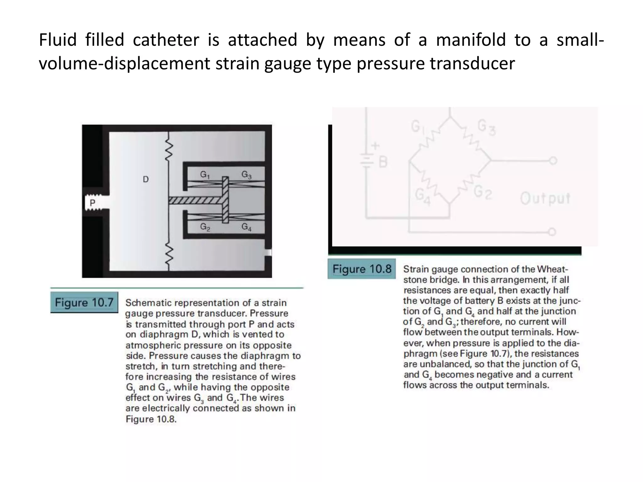
2) Proper equipment selection and setup is important to minimize artifacts and obtain accurate pressure tracings.
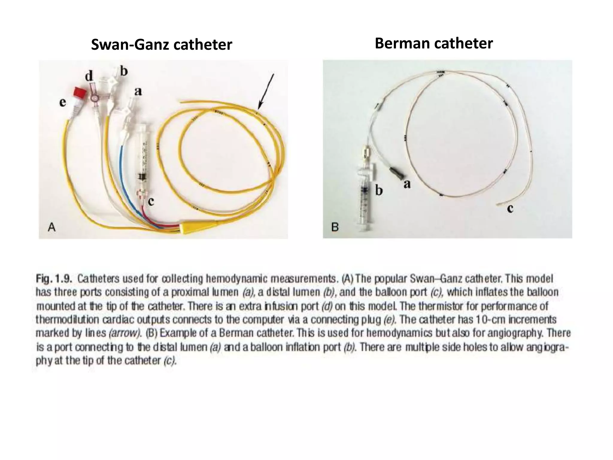
3) Different catheter types are used depending on the specific chamber being measured.
The document discusses techniques for detecting intracardiac shunts including oximetry runs, indicator dilution curves, and angiography. It provides criteria for identifying left-to-right shunts using oxygen saturation step-ups in the oximetry run. Examples are given for detecting an atrial septal defect and ventricular septal defect. The ratio of pulmonary to systemic blood flow (Qp/Qs) is discussed as a measure of shunt magnitude.
Left ventricular angiography is used to assess global and regional left ventricular function and anatomy. It involves inserting a catheter into the left ventricle and injecting contrast dye to visualize the ventricle on x-ray imaging. The procedure provides key information on mitral valve function, ventricular shape and wall motion abnormalities, and congenital defects like VSD. LV volumes and ejection fraction are calculated from the images to quantify function. Regional wall motion is graded and correlated to coronary artery territories. Characteristic appearances are seen in conditions like cardiomyopathy, mitral regurgitation, and septal defects. Potential complications include arrhythmias and endocardial injury.
The document summarizes key aspects of cardiac catheterization and hemodynamic data collection. It describes the normal cardiac cycle, pressure measurement systems, normal pressure waveforms, methods to measure cardiac output like thermodilution and Fick, how to evaluate valvular stenosis and regurgitation, determine vascular resistance and shunts. Specific details are provided on assessing aortic stenosis, mitral stenosis, right-sided valves and quantifying regurgitant fractions. Oxygen saturation analysis and Fick principles are outlined for shunt determinations.
This document discusses pressure changes that can occur during coronary angiography, specifically damping and ventricularization. Damping is defined as a significant decrease in pressure at the coronary ostium when the catheter is placed, accompanied by the disappearance of pressure waveforms, suggesting no antegrade flow. Ventricularization occurs when blood circulates within a coronary artery like a closed system, deforming the aortic pressure waveform. The document emphasizes the importance of the operator recognizing abnormal pressure changes to avoid complications, and provides solutions like catheter replacement or intracoronary nitroglycerin to address issues.
This document summarizes different devices used for closing ventricular septal defects (VSDs). It describes the common complications of VSD devices which are mostly minor, including embolization, arrhythmias, and conduction defects. Three types of Amplatzer devices are outlined - the muscular VSD device, asymmetric VSD occluder, and perimembranous VSD devices. Sizes and designs of each are provided. Results of post-myocardial infarction VSD closure show high residual leak rates. Finally, it briefly mentions some VSD devices manufactured in China including by Yatai and Lifetech, and introduces the novel NitOcclud VSD coil.
Intravascular ultrasound (IVUS) uses sound waves to visualize the inside of arteries. There are two types of IVUS systems - mechanical systems using a rotating internal cable and solid-state systems using externally mounted transducers. Both produce 360-degree images with a resolution of 100-150 μm. IVUS is used to assess plaque, vessel dimensions, stent deployment, and more. It produces cross-sectional images showing the lumen, layers of the artery wall, and plaque composition and size. Measurements include diameters, areas, plaque burden, and indices of eccentricity. IVUS helps identify vulnerable plaque and has diagnostic and interventional applications.
This document provides information about right heart catheters and angiographic catheters. It discusses the history of right heart catheters from 1929 to 1970. It then describes the diagnostic and therapeutic indications for right heart catheterization. The document outlines the parts of a catheter including the hub, body, and tip. It summarizes several general purpose catheters used for right heart catheterization including the Cournand, Goodale-Lubin, multipurpose, and Swan-Ganz balloon flotation catheters. Finally, it discusses several angiographic catheters used including the pigtail, NIH, Berman, Gensini, and Lehman catheters.
This document discusses the echocardiographic assessment of atrial septal defects (ASDs). It describes the main types of ASDs and notes that 80% are secundum defects. Echocardiography is used to identify and characterize ASDs, detect associated anomalies, diagnose complications, and guide treatment. Transthoracic echocardiography is the initial study, while transesophageal echocardiography provides better views of the atrial septum. Key measurements include ASD size, location, rim dimensions, and quantifying shunt severity with Qp/Qs. Echocardiography guides decisions about ASD device closure or surgery.
The document discusses the history and technique of transseptal puncture (TSP). It describes how TSP provides direct access to the left atrium and has become a routine skill for electrophysiologists performing procedures like atrial fibrillation ablation. The technique involves using a Brockenbrough needle and Mullins sheath inserted via the femoral or jugular vein to puncture the interatrial septum, usually at the fossa ovalis. Landmarks, equipment, steps of the procedure, challenges, and complications are reviewed in detail. The summary emphasizes the importance and increasing use of TSP as well as reviews key aspects of the technique and potential complications.
rotablation is procedure used in complex pci with heavily calcified lesion for adequate expansion of stent.if used in indicated case and well aware of contraindication is necessary for achieving good results.
Guide catheters in coronary interventionRohitWalse2
Guide catheters are essential for coronary interventions as they deliver hardware into the arteries. The document discusses the properties and types of guide catheters, highlighting how their structure provides support and torque control. It describes commonly used guide catheters like the Judkins, Amplatz and EBU catheters, noting what vessels each is best suited for. Specialty guide catheters for difficult anatomies are also reviewed. Proper guide selection and positioning are emphasized for coaxial engagement and optimal device delivery during interventions.
This document discusses fractional flow reserve (FFR), a technique used during coronary catheterization to measure pressure differences across a coronary stenosis and determine if it is causing myocardial ischemia. An FFR value below 0.75 is considered functionally significant while a value above 0.80 rules out ischemia. FFR is useful for evaluating single-vessel disease, left main stenosis, tandem lesions, diffuse disease, grafts, and ostial lesions. Limitations include inability to assess plaque morphology.
This document discusses the importance of assessing left atrial function using echocardiography. Left atrial volume and strain are superior to other echocardiographic markers for diagnosing left ventricular diastolic dysfunction. Left atrial reservoir strain in particular shows high specificity but relative low sensitivity for diagnosing heart failure with preserved ejection fraction compared to invasive exercise assessment. More research is still needed to establish robust clinical data on using left atrial function metrics for diagnosis.
This document discusses percutaneous pulmonary valve interventions. It begins by providing background on the history of pulmonary valve interventions, starting with open surgical techniques and moving to percutaneous approaches developed in the 1950s. It then discusses the first successful percutaneous pulmonary valve implantation in 2000. The document provides details on the anatomy of the pulmonary valve, causes of pulmonary valve disease, techniques for percutaneous balloon pulmonary valvuloplasty, indications and contraindications for percutaneous pulmonary valve interventions, and the evolution and indications for transcatheter pulmonary valve implantation.
Coronary artery perforation during percutaneous coronary intervention (PCI) can be classified based on its anatomical location and severity. Proximal or midvessel perforations carry a greater risk of complications while distal perforations often have a benign course. Treatment depends on the perforation type and severity, with supportive measures, prolonged balloon inflation, covered stents, or vessel occlusion techniques used for more severe cases. Emergency surgery may be needed for large perforations not responding to other treatments, though surgical outcomes in emergency settings are often disappointing.
Intravascular ultrasonography (IVUS) provides images of coronary arteries and other blood vessels. It plays a critical role in understanding coronary disease and guiding interventional cardiology procedures. IVUS uses a catheter-mounted ultrasound transducer to create images. It can assess plaque, guide stent placement, detect complications, and characterize lesion morphology. IVUS provides detailed information to evaluate patients and optimize interventional strategies.
Distal balloon occlusion devices and distal filter devices are the main types of embolic protection devices (EPDs) used during percutaneous coronary intervention (PCI). Distal balloon occlusion devices use a balloon to occlude blood flow distal to the lesion during PCI, while distal filter devices use a nitinol mesh filter to capture debris without interrupting blood flow. Major trials have shown the benefits of EPDs for saphenous vein graft interventions and for STEMI patients undergoing PCI. EPD selection depends on lesion location and vessel characteristics. EPDs are recommended for saphenous vein graft PCI but their routine use is not supported for native coronary artery PCI.
This document summarizes various devices used to close atrial septal defects (ASDs), including their designs, sizes, advantages, and disadvantages. The most commonly used device is the Amplatzer Septal Occluder, which has a double disc design and is self-expanding. Other devices discussed include the Gore HELEX, Lifetech/Cera, Figulla, Cardioseal/Starflex, and newer bioabsorbable options like the Biotrek. Complication rates of ASD device closure are generally low, below 10%, with embolization and arrhythmias being the most common issues. Larger trials have shown the Amplatzer to be very effective and easy
Contrast echocardiography uses microbubble ultrasound contrast agents to improve image quality. These microbubbles remain in the intravascular space and allow for assessment of cardiac structure, function, and perfusion. Second generation contrast agents use an inert gas encapsulated by albumin or phospholipid shells. They interact with ultrasound by reflecting at fundamental frequencies and resonating to produce harmonic frequencies. Continuous infusion provides steady contrast levels needed for perfusion assessment. Contrast echocardiography is a non-invasive technique that improves evaluation of the heart.
Percutaneous Balloon Mitral Valvuloplasty (PBMV) is a procedure to dilated the mitral valve in the setting of rheumatic mitral valve stenosis. A catheter is inserted into the femoral vein, advanced to the right atrium and across the interatrial septum. Then the mitral valve is crossed with a balloon and it is inflated to relieve the fusion of the mitral valve commissures effectively acting to increase the mitral valve area and reduce the degree of mitral stenosis. Mitral regurgitation is a potential complication and thus PBMV is contraindicated if moderate or severe regurgitation is present. The Wilkins score examines mitral valve morphology and is determined via echocardiography to assess the likelihood of using PBMV based on certain echocardiographic criteria.
The document discusses the history, anatomy, angiographic views, variations, and clinical relevance of coronary arteries. It provides a detailed overview of the typical anatomy and branches of the left main, left anterior descending, left circumflex, and right coronary arteries. It also describes common anatomical variations and anomalies seen in coronary arteries and their clinical implications. Angiographic classification methods for different coronary artery segments are presented.
This is a comprehensive description of coronay lesion assessment from routinely used angiography to advanced imaging modalities like IVUS/OCT including their functional significance by FFR
This document discusses coronary guidewires used in percutaneous coronary intervention (PCI). It begins by outlining the history of angioplasty and guidewire development. It then covers the purpose, components, classifications, and appropriate uses of guidewires. The main components include the core, tip, coils, covers, and coatings. Guidewires are classified based on flexibility, device support, and clinical usage. Complications like vessel perforation, pseudolesions, and entrapment are also discussed. Proper guidewire manipulation and strategies for difficult lesions are outlined to maximize safety and efficacy.
The document provides an overview of echocardiographic assessment of aortic valve stenosis. It describes the normal aortic valve anatomy and imaging windows used to visualize the valve. Common causes of aortic stenosis including bicuspid aortic valve and calcific stenosis are discussed. Methods for Doppler assessment of aortic stenosis including peak velocity, mean gradient, and valve area via the continuity equation are summarized. Limitations of these assessment techniques are also noted.
This document discusses hemodynamic principles and various cardiac pressures measured in the circulatory system. It begins by explaining how electrical activity leads to mechanical functions that generate pressure waves. It then discusses how to measure and interpret pressures in different parts of the heart including the aorta, pulmonary artery, right and left ventricles, and right atrium. Factors that influence pressures and common abnormalities are provided. Diagrams of normal pressure waveforms are displayed. The document concludes by defining pulmonary and systemic vascular resistances.
This document discusses various methods for quantifying intracardiac shunts in patients with congenital heart lesions. It describes invasive oximetry and indicator dilution techniques as well as noninvasive Doppler echocardiography methods. For echocardiography, it outlines techniques for quantifying left-to-right shunts using pulmonary and aortic flow measurements, as well as a simplified method using diameter ratios. It also discusses limitations and sources of error for these quantification methods.
Ppt deals shortly about various invasive monitoring modalities of cardiology for an anesthetist! This is just an overview and each topics is itself an area of deep learning! Ideal for a basic presentation for residents of anesthesiology!
1) Pressure tracing of the left ventricle involves using fluid-filled catheters connected to pressure transducers to record intracardiac pressures. Factors such as catheter size, damping, and natural frequency determine recording quality.
2) Sources of error in pressure measurements include catheter whip, end-pressure artifacts, and deterioration of frequency response. Invasive monitoring is useful for evaluating conditions like aortic stenosis and mitral stenosis when noninvasive data is discrepant or unclear.
3) Distinguishing constrictive pericarditis from restrictive cardiomyopathy involves assessing ventricular interdependence, pulmonary pressures, and left ventricular pressure tracings. Hemodynamic data aids in diagnosis and management of many conditions.
Medical Instrumentation- body parameters measurementPoornima D
This document provides information on different methods for measuring various body parameters, including blood pressure, body temperature, and respiration rate.
It describes both direct and indirect methods for measuring blood pressure, such as using a catheter or sphygmomanometer. It also discusses different categories of blood pressure readings.
For measuring body temperature, it explains the use of mercury-in-glass thermometers as well as electronic thermometers that incorporate thermistors or thermocouples.
Finally, it summarizes various techniques for monitoring respiration rate, such as the displacement method, thermistor method, impedance pneumography, measuring carbon dioxide levels, and using apnoea detectors.
This document discusses methods for measuring blood pressure, both directly and indirectly. Direct methods involve placing a sensor in the vascular system, either extravascularly using a catheter connected to a sensor, or intravascularly with a sensor at the catheter tip. Indirect methods use a sphygmomanometer with an inflatable cuff to measure blood pressure externally by detecting sounds or pulses in the artery under the cuff as pressure is released. Automated devices now replace the stethoscope with microphones or ultrasonic sensors to detect sounds and determine systolic and diastolic pressure electronically.
This document discusses various methods of basic haemodynamic monitoring including blood pressure, oxygen saturation, central venous pressure, and venous saturation monitoring. It provides details on direct and indirect blood pressure measurement, the arterial line setup, waveforms, and complications. Central venous pressure monitoring details catheter insertion sites and the procedure, normal waveforms, and complications. Venous saturation monitoring is also briefly explained.
arterial line insertion in paediatric practiceRamprasadJagtap
Invasive arterial blood pressure monitoring provides the gold standard for blood pressure measurement. It involves inserting a catheter into an artery using the Seldinger technique. The catheter is connected to a pressure transducer that converts pressure waves into electrical signals. These signals are amplified and processed to display the arterial pressure waveform on a monitor. Dynamic measures like pulse pressure variation and stroke volume variation obtained from the arterial waveform can help assess fluid responsiveness in mechanically ventilated patients. Complications of arterial lines include pain, bleeding, occlusion and infection.
Central venous pressure monitoring - Pooja Murkarpooja murkar
Hemodynamic monitoring involves continuously measuring and recording vital physiological parameters in critically ill patients using indwelling catheters and electronic monitoring equipment. It refers to measuring pressures, flows, and oxygenation within the cardiovascular system. There are invasive and non-invasive methods, with invasive methods including central venous pressure monitoring and pulmonary pressure monitoring using catheters and transducers to obtain pressure readings and waveforms. The goal is to detect life-threatening conditions early and evaluate a patient's response to treatment.
The Swan-Ganz catheter, also known as a pulmonary artery catheter, is a specialized catheter used to monitor a patient's hemodynamics. It is inserted into the internal jugular or subclavian vein and threaded through the heart into the pulmonary artery. This allows direct measurement of pressures in the right atrium, right ventricle, pulmonary artery, and indirect measurement of left-sided pressures. The catheter is useful for diagnosis and management of conditions affecting heart function or pulmonary circulation. However, randomized controlled trials found no improvement in outcomes with its use and increased risks, so the catheter's benefits must be weighed against risks for each individual patient.
This document provides an overview of arterial blood pressure monitoring. It discusses the history and development of non-invasive blood pressure measurement techniques. It then focuses on the components, principles, and technical aspects of invasive arterial blood pressure monitoring using an intra-arterial catheter connected to a transducer system. Key points covered include the components of the measuring system, optimizing the system's natural frequency and damping, and the importance of zeroing and leveling the transducer.
Cardiac catheterization is a procedure used to diagnose and treat cardiovascular conditions either diagnostically or therapeutically. It remains the gold standard for comprehensively evaluating complex heart disease through invasive physiologic assessment. It can also be used to provide minimally invasive definitive therapy for some selective cardiac defects. The procedure allows for measurement of blood flows and pressures throughout the heart and vessels, as well as calculation of systemic and pulmonary vascular resistances, to fully understand a patient's heart condition.
This document provides an overview of cardiovascular monitoring during anesthesia. It discusses the importance of monitoring heart rate, blood pressure, and other parameters to detect changes early and intervene to reduce risks. Both indirect and direct methods of measuring arterial blood pressure are described in detail, including oscillometry, Doppler, tonometry and intra-arterial cannulation. The principles of transducers, damping, natural frequency, and resonance in pressure monitoring systems are also summarized.
Types of Blood Pressure Measurement TechniquesRevathiJ10
This document discusses blood pressure measurement. It defines systolic and diastolic pressure and describes normal ranges. Both direct and indirect measurement methods are covered, with details on Korotkoff sounds, oscillometry, and rheography. Components of automated blood pressure devices and non-invasive monitoring systems are summarized. Temperature measurement using thermistors is also briefly explained.
Ventricular assist device of cardiac Cathetherizationvasanth7pv
The TandemHeart is an extracorporeal ventricular assist device that pumps blood from the left atrium to the femoral arteries. It consists of an external centrifugal pump connected to a transseptal cannula in the left atrium and outflow cannulae in the femoral arteries. It is used to provide temporary circulatory support in cardiogenic shock. Potential complications include bleeding, vascular issues, and limb ischemia which require monitoring and adjustments to cannula placement or addition of distal perfusion catheters.
This document discusses central venous pressure (CVP), how it is measured, factors that affect it, and methods of CVP monitoring. CVP reflects right atrial pressure and ventricular preload. It is normally measured via a catheter placed in the internal jugular or subclavian vein, connected to a pressure transducer. CVP monitoring provides information about cardiac function and venous return. The document outlines several methods of CVP measurement and important considerations for accurate pressure waveform analysis.
Open heart surgery involves opening the chest to operate directly on the heart or surrounding structures. Common procedures include heart valve surgery, repair of congenital defects, and coronary artery bypass grafting. Before surgery, patients undergo tests to assess cardiac function and risks. During surgery, general anesthesia is induced carefully to maintain stable hemodynamics while the heart is monitored closely using techniques like pulmonary artery catheters. Premedication aims to minimize stress on the heart during induction and intubation.
The mean arterial pressure (MAP) is the average pressure in the arteries during one cardiac cycle and is a better indicator of tissue perfusion than systolic blood pressure. MAP is calculated based on cardiac output, systemic vascular resistance, and central venous pressure. Normal MAP is between 65-110 mmHg. Pulse wave velocity measures arterial stiffness and is influenced by arterial wall thickness and diameter. As pressure waves travel further from the heart, they become steeper and narrower due to vessel properties.
CVS physiology, all details with explanation easy to recall physiology of cardiovascular system. based on Ganong's Review of Medical Physiology. all the high-yield facts are there.
Echocardiography is a key tool for diagnosing and evaluating mitral stenosis (MS). It is essential to use an integrative approach when grading MS severity by combining Doppler, 2D imaging, and measurements, rather than relying on one alone. Echocardiography plays a major role in MS by confirming diagnosis, quantifying severity, analyzing consequences, and examining valve anatomy. Mitral valve planimetry directly measures valve area and is considered the reference standard, but additional measurements like pressure gradient and half-time are also useful. Echocardiography aids clinical decision making for patients with MS.
This document discusses pulsatile versus non-pulsatile perfusion during cardiopulmonary bypass. Pulsatile perfusion is considered more physiologic as it simulates the pulsatile blood flow generated by the human heart. Pulsatile flow is associated with better organ function outcomes and increased microcirculation. It works through increasing surplus hemodynamic energy, maintaining capillary patency above the critical closing pressure, and stimulating neuroendocrine reflexes. Systems that can generate pulsatile flow include ventricular pumps, compression plate pumps, and pulsatile assist devices. However, transmitting pulsatile flow through the cardiopulmonary bypass circuit can be challenging due to pressure losses across components.
Similar to PRESSURE MEASUREMENT by Cardiac catheterisation_Dr Amol Patil.pptx (20)
This document summarizes a presentation on probiotics, prebiotics, and synbiotics. It discusses the gut microbiome and how critical illness can disrupt it. It defines probiotics, prebiotics, and synbiotics and provides examples of each. It outlines proposed mechanisms of action and potential health benefits, particularly in treating ICU patients. However, the document also notes challenges in using probiotics for critically ill patients, as a large recent trial found that the probiotic L. rhamnosus GG did not significantly reduce ventilator-associated pneumonia in ICU patients.
An 80-year-old woman presented with confusion, weakness, and nonsustained ventricular tachycardia. Her lab results showed hypokalemia (K=2.5 mmol/L), hyponatremia (Na=118 mmol/L), and she was started on IV potassium replacement. Further evaluation of her hyponatremia found she was euvolemic. Her sodium was then found to be elevated at 168 mmol/L and she received hypotonic fluids. Electrolyte imbalances can have serious consequences and require identifying the cause, urgent treatment if life-threatening, and monitoring for correction.
This document discusses early initiation of renal replacement therapy (RRT) compared to late or delayed initiation in patients with acute kidney injury (AKI). The pros of early initiation include better control of fluids, electrolytes, and acid-base balance as well as removal of toxins to prevent complications. However, early initiation can also lead to mechanical and other complications. Several studies comparing early vs late RRT are summarized, including the AKIKI trial which found no difference in 60-day mortality, and the ELAIN trial which found potential benefits of earlier initiation in preventing further organ injury. The IDEAL-ICU trial criteria for early vs delayed RRT in septic shock patients with AKI is also outlined.
This document discusses early initiation of renal replacement therapy (RRT) compared to late or delayed initiation in patients with acute kidney injury (AKI). The pros of early initiation include better control of fluids, electrolytes, and acid-base balance as well as removal of toxins to prevent complications. However, early initiation can also lead to mechanical complications and issues with biocompatibility and anticoagulation. Several studies comparing early vs late RRT are summarized, including the AKIKI trial which found no difference in 60-day mortality and more RRT-free days in the delayed group. The ELAIN study found potential benefits of earlier initiation in rapidly controlling metabolism and fluids to prevent further organ injury. The IDEAL
This document discusses anemia in pregnancy. It defines anemia as having insufficient red blood cells or hemoglobin. Anemia is common in pregnancy, affecting 18-75% of pregnant women globally. Anemia is classified as mild, moderate or severe based on hemoglobin levels. Common causes of anemia in pregnancy include iron deficiency, folic acid deficiency, vitamin B12 deficiency, and genetic disorders like sickle cell anemia. Left untreated, anemia can negatively impact both mother and baby by increasing risks of infection, hemorrhage, low birth weight, and other complications. Routine screening and treatment with iron, folic acid and other supplements can help prevent and manage anemia during pregnancy.
This document provides information on fluid therapy. It begins by classifying fluids as crystalloids or colloids based on their ability to diffuse. Crystalloids like normal saline (NS) diffuse freely while colloids do not pass as readily. NS is commonly used but can cause acidosis due to its chloride content. Ringer's lactate is more physiological with an electrolyte profile similar to plasma. Dextrose solutions like 5% dextrose provide calories but excess can cause hyperglycemia and lactate production in critical illness. The document discusses the properties, indications, and limitations of various intravenous fluids.
Current Recommendations and Changes From Previous 2016 Recommendations.pptxAshishSharma907946
- The document summarizes current recommendations (2021) and changes from previous 2016 recommendations for treating sepsis.
- Key changes include recommending a performance improvement program including sepsis screening and standard operating procedures. Screening tools are recommended against using qSOFA alone. Blood lactate measurement is suggested for suspected sepsis.
- For initial resuscitation, 30mL/kg crystalloid fluid within 3 hours is suggested over previous strong recommendation, and dynamic measures are suggested over static parameters to guide fluid resuscitation.
This document discusses drug induced liver injury (DILI), also known as hepatotoxicity. It begins by describing the physiologic functions of the liver and then defines hepatotoxicity as liver damage caused by medicinal agents, chemicals, toxins, or herbal remedies. The document outlines the classification of DILI as either intrinsic or idiosyncratic hepatotoxicity and describes various risk factors. Several specific drugs that can cause liver damage like acetaminophen, ethanol, aflatoxins, and phenytoin are discussed in more detail regarding their mechanisms of toxicity, signs and symptoms, and management approaches.
The pulmonary artery catheter can be used to obtain direct measurements of central venous pressure, right-sided cardiac pressures, pulmonary artery pressure, pulmonary artery occlusion pressure, cardiac output, and mixed venous oxygen saturation. It provides diagnostic and therapeutic benefits in critically ill patients but also carries risks of complications. The document outlines the physiologic measurements, indications, contraindications, preparation, placement technique, and interpretation of hemodynamic values and waveforms obtained from the pulmonary artery catheter.
- The document describes functional hemodynamic monitoring, including the physiology of heart-lung interactions and parameters used to predict patient response to volume like pulse pressure variation.
- It discusses cardiac output, stroke volume, preload, contractility, afterload and how they are impacted during procedures. Parameters like pulse pressure variation above 13% indicate fluid responsiveness.
- Positioning and blood loss can significantly impact hemodynamics during surgery, and continuous monitoring can help guide fluid and vasoactive drug management to support cardiac output and blood pressure.
This presentation was provided by Racquel Jemison, Ph.D., Christina MacLaughlin, Ph.D., and Paulomi Majumder. Ph.D., all of the American Chemical Society, for the second session of NISO's 2024 Training Series "DEIA in the Scholarly Landscape." Session Two: 'Expanding Pathways to Publishing Careers,' was held June 13, 2024.
Philippine Edukasyong Pantahanan at Pangkabuhayan (EPP) CurriculumMJDuyan
(𝐓𝐋𝐄 𝟏𝟎𝟎) (𝐋𝐞𝐬𝐬𝐨𝐧 𝟏)-𝐏𝐫𝐞𝐥𝐢𝐦𝐬
𝐃𝐢𝐬𝐜𝐮𝐬𝐬 𝐭𝐡𝐞 𝐄𝐏𝐏 𝐂𝐮𝐫𝐫𝐢𝐜𝐮𝐥𝐮𝐦 𝐢𝐧 𝐭𝐡𝐞 𝐏𝐡𝐢𝐥𝐢𝐩𝐩𝐢𝐧𝐞𝐬:
- Understand the goals and objectives of the Edukasyong Pantahanan at Pangkabuhayan (EPP) curriculum, recognizing its importance in fostering practical life skills and values among students. Students will also be able to identify the key components and subjects covered, such as agriculture, home economics, industrial arts, and information and communication technology.
𝐄𝐱𝐩𝐥𝐚𝐢𝐧 𝐭𝐡𝐞 𝐍𝐚𝐭𝐮𝐫𝐞 𝐚𝐧𝐝 𝐒𝐜𝐨𝐩𝐞 𝐨𝐟 𝐚𝐧 𝐄𝐧𝐭𝐫𝐞𝐩𝐫𝐞𝐧𝐞𝐮𝐫:
-Define entrepreneurship, distinguishing it from general business activities by emphasizing its focus on innovation, risk-taking, and value creation. Students will describe the characteristics and traits of successful entrepreneurs, including their roles and responsibilities, and discuss the broader economic and social impacts of entrepreneurial activities on both local and global scales.
Level 3 NCEA - NZ: A Nation In the Making 1872 - 1900 SML.pptHenry Hollis
The History of NZ 1870-1900.
Making of a Nation.
From the NZ Wars to Liberals,
Richard Seddon, George Grey,
Social Laboratory, New Zealand,
Confiscations, Kotahitanga, Kingitanga, Parliament, Suffrage, Repudiation, Economic Change, Agriculture, Gold Mining, Timber, Flax, Sheep, Dairying,
Leveraging Generative AI to Drive Nonprofit InnovationTechSoup
In this webinar, participants learned how to utilize Generative AI to streamline operations and elevate member engagement. Amazon Web Service experts provided a customer specific use cases and dived into low/no-code tools that are quick and easy to deploy through Amazon Web Service (AWS.)
The chapter Lifelines of National Economy in Class 10 Geography focuses on the various modes of transportation and communication that play a vital role in the economic development of a country. These lifelines are crucial for the movement of goods, services, and people, thereby connecting different regions and promoting economic activities.
This presentation was provided by Rebecca Benner, Ph.D., of the American Society of Anesthesiologists, for the second session of NISO's 2024 Training Series "DEIA in the Scholarly Landscape." Session Two: 'Expanding Pathways to Publishing Careers,' was held June 13, 2024.
Temple of Asclepius in Thrace. Excavation resultsKrassimira Luka
The temple and the sanctuary around were dedicated to Asklepios Zmidrenus. This name has been known since 1875 when an inscription dedicated to him was discovered in Rome. The inscription is dated in 227 AD and was left by soldiers originating from the city of Philippopolis (modern Plovdiv).
Gender and Mental Health - Counselling and Family Therapy Applications and In...PsychoTech Services
A proprietary approach developed by bringing together the best of learning theories from Psychology, design principles from the world of visualization, and pedagogical methods from over a decade of training experience, that enables you to: Learn better, faster!
LAND USE LAND COVER AND NDVI OF MIRZAPUR DISTRICT, UPRAHUL
This Dissertation explores the particular circumstances of Mirzapur, a region located in the
core of India. Mirzapur, with its varied terrains and abundant biodiversity, offers an optimal
environment for investigating the changes in vegetation cover dynamics. Our study utilizes
advanced technologies such as GIS (Geographic Information Systems) and Remote sensing to
analyze the transformations that have taken place over the course of a decade.
The complex relationship between human activities and the environment has been the focus
of extensive research and worry. As the global community grapples with swift urbanization,
population expansion, and economic progress, the effects on natural ecosystems are becoming
more evident. A crucial element of this impact is the alteration of vegetation cover, which plays a
significant role in maintaining the ecological equilibrium of our planet.Land serves as the foundation for all human activities and provides the necessary materials for
these activities. As the most crucial natural resource, its utilization by humans results in different
'Land uses,' which are determined by both human activities and the physical characteristics of the
land.
The utilization of land is impacted by human needs and environmental factors. In countries
like India, rapid population growth and the emphasis on extensive resource exploitation can lead
to significant land degradation, adversely affecting the region's land cover.
Therefore, human intervention has significantly influenced land use patterns over many
centuries, evolving its structure over time and space. In the present era, these changes have
accelerated due to factors such as agriculture and urbanization. Information regarding land use and
cover is essential for various planning and management tasks related to the Earth's surface,
providing crucial environmental data for scientific, resource management, policy purposes, and
diverse human activities.
Accurate understanding of land use and cover is imperative for the development planning
of any area. Consequently, a wide range of professionals, including earth system scientists, land
and water managers, and urban planners, are interested in obtaining data on land use and cover
changes, conversion trends, and other related patterns. The spatial dimensions of land use and
cover support policymakers and scientists in making well-informed decisions, as alterations in
these patterns indicate shifts in economic and social conditions. Monitoring such changes with the
help of Advanced technologies like Remote Sensing and Geographic Information Systems is
crucial for coordinated efforts across different administrative levels. Advanced technologies like
Remote Sensing and Geographic Information Systems
9
Changes in vegetation cover refer to variations in the distribution, composition, and overall
structure of plant communities across different temporal and spatial scales. These changes can
occur natural.
2. Overview
• Introduction
• Basic principles and physics of cardiac
catheterization pressure measurement
• Atrial pressure tracing
• PCWP tracing
• Ventricular pressure tracing
• Arterial pressure tracing
• Sources of errors and Artifact
• Pressure tracings in different clinical conditions
3. INTRODUCTION
• The beginning of invasive hemodynamics can be
traced back to the early 1700s, when Rev.
Stephen Hales measured the blood pressure of a
horse by inserting a brass rod in the femoral
artery.
• In 1844, the first invasive cardiac catheterization
was performed by Claude Bernard. Again, the
subject was a horse, and both the right and left
ventricles were entered by retrograde approach
from the jugular vein and from the carotid artery.
4. • In 1861, Chauveau and Marey-
– published their work on cardiac cycle and their
discovery that ventricular systole and apical beat are
simultaneous. In addition, they were able to measure
simultaneously left ventricular (LV) and central aortic
(Ao) pressure.
• In 1929, Werner Forssmann –
– the first right-sided heart catheterization of a human
heart on himself
– 1931, he demonstrate injecting contrast material in
the human heart.
5.
6. • Cournand and Richards-
– performed cardiac catheterization in many clinical
conditions including hypertension, circulatory
shock, and chronic lung disease.
7. • Essential components of a hemodynamic
monitoring system-
catheter,
transducer,
fluid-filled tubing to connect the catheter to the
transducer, and
physiologic recorder to display, analyze, print, and
store the hemodynamic waveforms generated.
8. • Proper equipment's for high quality catheterization
recordings
• Coronary angiography: smallest bore catheters, 5F &
even 4F
• Complex hemodynamic catheterization is optimally
performed with large-bore catheters that yield high
quality hemodynamic data. To obtain proper
hemodynamic tracings, 6F or 7F catheters may be
required.
9. • Variety of catheters used-
• The optimal catheter for hemodynamic measurements is stiff
and has a relatively large lumen opening to an end hole.
• End-hole catheter -sampling pressures within small chambers
Disadvantage-it may lead to damping or other artifact if the
end hole comes into contact with the wall of the cardiac
chamber.
• “pig-tail” catheter has multiple side holes and samples
pressure at each of these openings- adequate for sampling
pressure in a large, uniform chamber such as the aorta or left
ventricle
Disadvantage-it doesn’t have the required resolution to
discern pressure gradients within the left ventricle.
10. • The Swan–Ganz catheter is the most commonly used
catheter for measuring right-heart pressures. In addition to
the balloon at the tip for flotation, it consists of an end hole
(distal port), a side hole 30 cm from the catheter tip
(proximal port), and a thermistor for measurement of
thermodilution cardiac output.
• balloon flotation catheters
– the Berman catheter, multiple side holes near the tip and no
end hole or thermistor and is used principally for performance
of angiography, and
– the balloon-wedge catheter- contains an end hole similar to the
Swan–Ganz catheter but no thermistor for cardiac output
measurement or additional infusion or pressure monitoring
ports
12. • The use of high-fidelity manometer-tipped catheters might
also need to be considered in those instances when intricate
analysis of diastolic filling contours is required.
• Other catheters rarely used today –
– the Layman catheter and Cournand catheter consisting of
an end-hole,
– the NIH catheter that contains multiple side holes near the
tip but no end hole,
– the Goodale-Lubin catheter consisting of an end hole and
two single side holes near the tip
13. MICROMANOMETERS-
• miniaturized transducers that fit on the distal tip of
standard catheters and therefore may be used as
intracardiac manometers
• Measuring pressure directly within the vessel or
cardiac chamber with the transducer on the tip of
the catheter prevents the distortions associated with
fluid-filled systems .
• Micromanometers have a flat frequency response up
to 1 ,000 Hz, thus allowing accurate recording at high
heart rates
14. • reduce the mass and inertia of the pressure
measurement system,
• improve the frequency-response
characteristics ,
• decrease artifacts associated with
underdamping , overdamping, and catheter
whip
15. • The catheter may be subjected to gas
sterilization (ethylene oxide) along with other
catheters and instruments .
• The pressures measured with these catheters
are superior in waveform and accuracy to
those measured with standard techniques
• A disposable version is available for clinical
use
16.
17. • In fluid-filled catheter system- choose the
shortest extension tubing possible to obtain
optimal pressure contours.
• Force is transmitted through a fluid medium as a
pressure wave.
• important objective of the cardiac catheterization
procedure is to assess accurately the pressure
waves generated by various cardiac chambers.
18. the more rigid the sensing membrane, the lower the sensitivity;
the more flaccid the membrane, the higher the sensitivity
19.
20.
21.
22. • Pressure measurement systems today generally use
electrical strain gauges based on the principle of the
Wheatstone bridge.
• the strain gauge is a variable resistance transducer -
operation depends on the fact-
– when an electrical wire is stretched, its resistance
to the flow of current increases.
23. Fluid filled catheter is attached by means of a manifold to a small-
volume-displacement strain gauge type pressure transducer
24. Pressure measurement Terminology
• fundamental frequency-The number of times the
cycle occurs in 1 second
• Eg. a fundamental frequency of 2 corresponds to
a heart rate of 120 bpm.
• As a complex periodic waveform, the pressure
wave may be subjected to a type of analysis
developed by the French physicist Fourier,
whereby any complex wave form may be
considered the mathematical summation of a
series
25.
26. • The practical consequence of this analysis –
• to record pressure accurately, a system must respond
with equal amplitude for a given input throughout the
range of frequencies contained within the pressure
wave.
• If components in a particular frequency range are
either suppressed or exaggerated by the transducer
system, the recorded signal will be a grossly distorted
version of the original physiologic waveform.
• For example , the dicrotic notch of the aortic pressure
wave contains frequencies above 10 H z . If the
pressure measurement system were unable to respond
to frequencies greater than 10 Hz, the notch would be
slurred or absent.
27. • Natural Frequency and Damping
• If the sensing membrane were to be shock-
excited (like a gong) in the absence of friction,
it would oscillate for an indefinite period in
simple harmonic motion. The frequency of
this motion would be the natural frequency of
the system.
• Any means of dissipating the energy of this
oscillation, such as friction, is called damping.
28. • Natural frequency-
• Frequency of an input pressure wave at which
the ratio of output/input amplitude of an
undamaged system is maximal
29. • Damping dissipates the energy of the oscillating sensing
membrane
• optimal damping dissipates the energy gradually, thereby
maintaining the frequency response curve nearly flat
(constant output/input ratio) as it approaches the region of
the pressure measurement system's natural frequency.
30. • In practice there is always some damping, and
this has two effects:
– the amplitude of the oscillations gradually
diminishes , and
– the frequency of oscillation is reduced.
• damping helps to prevent overshoot artifacts
resulting from resonance of the system , but
at the cost of diminished frequency response.
31. frequency response and sensitivity are related reciprocally, and one can be
obtained only by sacrificing the other.
34. • Midchest level is used widely as zero Reference
• In practical terms , for measurement of left
ventricular and aortic pressure , the zero level
should be positioned approximately 5 cm below the
left sternal border at the fourth left intercostal space
(LICS)
• the right ventricle and left atrium are at different
levels in the chest than the left ventricle,
• Courtois et al. calculated that the error introduced by
use of a point 5 cm below the fourth LICS, at the left
sternal border, is approximately ± 0.8 mmHg for
chambers other than the left ventricle
35.
36. Frequency response
• Ratio of output amplitude to input amplitude over a
range of frequencies of the input pressure wave
• Frequency response of catheter system is dependent
on catheters natural frequency and amount of
damping
• The higher the natural frequency of the system, the
more accurate the pressure measurement at lower
phyliogic frequencies
42. 2. Wedge pressure
• a wedge pressure is obtained when an end-hole catheter is
positioned in a designated blood vessel with its open end-
hole facing a capillary bed, with no connecting vessels
conducting flow into or away from the designated blood
vessel between the catheter tip and the capillary bed.
• A true wedge pressure can be measured only in the absence
of flow .
• the catheter-tip pressure is equal to that on the other side of
the capillary bed.
43. • an end-hole catheter wedged in a hepatic vein may be
used to measure portal venous pressure ;
• catheter wedged in a distal pulmonary artery measures
pulmonary venous pressure;
• In the absence of cor triatriatum or obstruction to
pulmonary venous outflow, the pulmonary venous and left
atrial pressures are equal, so that pulmonary artery wedge
pressure may be used as a substitute for left atrial
pressure.
• ensure adequate wedge position by observing the pressure
waveform and by obtaining an oxygen saturation in wedge
position, which should be above 95%.
46. Atrial Pressure
• The atrial pressure waveform has three positive
deflections, the a, c, and v waves.
• The a wave is due to atrial systole and follows the P
wave on the ECG
• The height of the a wave depends on atrial contractility
and resistance to Ventricular filling.
• The X descent represents relaxation of the atrium and
downward pulling of the AV valve annulus by
Ventricular contraction.
• The X descent is interrupted by the c wave, which is a
small positive deflection caused by protrusion of the
closed AV valve annulus into the atrium.
47. • The v wave represents Ventricular systole.
• The y descent occurs after the v wave and reflects
opening of the AV valve and emptying of the atrium
into the ventricle.
• The left and right atrial pressure contours are similar,
• the left atrial pressure being generally higher than the
right atrial pressure and
• a more prominent V wave in the left atrial pressure
contour.
48.
49. PCWP
• PCWP is obtained by inflating the balloon-tipped
catheter in the distal PA position until it occludes the
PA branch.
• So it reflects pulmonary venous pressure and, thus, LA
pressure.
• The V wave is larger than the A wave, and both are
larger than mean PCWP.
• PCWP is delayed approximately 50 to 150 milliseconds
in comparison with LA pressure.
• The normal mean PCWP is 12 mmHg or less, but values
up to 15 to 18 mmHg may not lead to congestion
50. • Key points in differentiating PA pressure from PCWP-
• The V wave peaks after the T wave, whereas systolic PA
pressure peaks during T wave.
• The segment between V waves is rather horizontal or
upsloping and A waves are sometimes seen
• Where as the PA pressure tracing, the segment
between the systolic peaks is downsloping and does
not have A wave.
• In difficult cases obtain blood sample from the wedged
catheter tip and check oxygen saturation.
– The PCWP saturation = pulmonary venous saturation
(usually >95%),
– PA saturation = mixed venous saturation.
– best confirmatory method of proper wedging in rare cases
51.
52. • Conditions where PCWP tracing overestimates
the true PCWP (as well as LVEDP)
– Catheter in the upper lung zones (1 or 2)
– Catheter overwedging
– Pulmonary hypertension with hybrid PCWP-PA
tracing
– Sepsis, hypoxemia, or acute lung injury with
pulmonary venoconstriction and elevated
pulmonary interstitial
53. Atrial pressure tracing in various diseases-
• Deep X and deep Y: constrictive pericarditis
• Deep X and flat Y (diastolic flow blunting):
tamponade, but also sinus tachycardia
• Flat X (systolic flow blunting) and deep Y: severe
TR and/or RV failure
• Large V wave: severe TR and/or RV failure
• Ventricularized RA pressure: severe TR
• Large A wave: impaired RV compliance
• Equally large A and V waves: ASD
54.
55. Ventricular Pressure
• RV and LV waveforms are similar in
morphology.
• They differ mainly with respect to their
magnitudes.
• The durations of systole and isovolumic
contraction relaxation are longer and the
ejection period is shorter in the left than in
the right ventricle.
56.
57. LVEDP
• End Diastolic pressure can be measured on
the R wave of the ECG, which will coincide just
after “a” wave on LV trace.
• To be measured at the end expiration
60. • Hemodynamic findings in RV systolic or
diastolic failure-
1. -Elevated RV EDP > 8 mmHg
2. -Elevated mean RA pressure > 7 mmHg
3. -Large RA V wave
4. -Deep X and Y on RA tracing
5. -RV dip-plateau pattern
61. • There are key differences between arterial and
ventricular tracings
1. the ventricular pressure increases in diastole,
whereas arterial pressure decreases in diastole;
2. the ventricular pressure tracing in systole has a
“rectangular” shape, as opposed to the
“triangular” shape of arterial tracings;
3. ventricular pressure has an A wave in diastole;
4. arterial pressure has a dicrotic notch.
62. Aortic Pressure
• The contour of the aortic pressure is the result
of the forward wave and reflected waves .
• Arterial pressure rise slightly during isovolumic
contraction of the left ventricle due to bulging
of the semilunar valve in the aorta, before
openig called as Anacrotic notch.
63. • At the end of the isovolumic ventricular
contraction phase, with opening of the aortic
valve, the ejections phase begins and the
aortic pressure starts rising.
• The initial steep increase is followed by a peak
and then by a decline to the Dicrotic notch
• the dicrotic notch corresponds to closure of
the aortic valve and end of ejection.
64.
65. • Several aortic tracing patterns are suggestive of diseases-
1. Sharp downslope of the diastolic pressure with a widened pulse
pressure,- suggests AI. In addition, an attenuated or absent
dicrotic notch suggests AI.
2. Late peaking suggests AS (pulsus parvus and tardus).
3. Double peaking in systole suggests AI or HOCM.
4. On an appropriately damped aortic tracing, the loss of dicrotic
notch suggests severe AI or poor arterial compliance
5. Gradual reduction of the systolic pressure by more than 10
mmHg during normal quiet inspiration suggests pulsus
paradoxus, which is seen in tamponade.
6. Every-other-beat alternation in systolic pressure suggests pulsus
alternans, which reflects a severe reduction in cardiac output, as
in severe LV dysfunction or severe AS, and carries a poor
prognosis.
66.
67.
68.
69.
70. Sources of errors and Artifact
1. Deterioration in Frequency Response
2. Catheter Whip Artifact
3. End Pressure Artifact
4. Catheter Impact Artifact
5. Systolic Pressure Amplification the Periphery
6. Errors in Zero Level , Balancing, or Calibration
71. Deterioration in Frequency Response-
• Even the smallest air bubbles have a drastic
effect on pressure measurement because they
cause excessive damping and lower the
natural frequency (by serving as an added
compliance) .
• When the natural frequency of the pressure
measurement system falls,
72. • high-frequency components of the pressure
waveform may set the system in oscillation,
producing the ventricular pressure overshoot
commonly seen in early systole and diastole
• Flushing out the catheter, manifold, and
transducer dispels these small air bubbles and
restores the frequency response of the
pressure measurement system
73. The micromanometer tracing is labeled A, and the fluid -filled catheter tracing is
labeled B. Note both the early diastolic and the early ejection phase overshoots reco
rded with the fluid -filled catheter, indicating a poor frequency response, especially in
the graph on the left
74. Catheter Whip Artifact-
• Motion of the tip of the catheter within the heart
and great vessels accelerates the fluid contained
within the catheter.
• may produce superimposed waves of ± 10 mmHg.
• common in tracings from the pulmonary arteries and
are difficult to avoid.
75. End Pressure Artifact-
• Flowing blood has a kinetic energy
• when this flow suddenly comes to a halt, the
kinetic energy is converted in part into
pressure .
• End-hole catheter pointing upstream records a
pressure that is artifactually elevated by the
converted kinetic energy.
• This added pressure may range from 2 to 10
mmHg.
76. Catheter Impact Artifact-
• When a fluid-filled catheter is hit (e.g. , by valves in
the act of opening or closing or by the walls of the
ventricular chambers) , a pressure transient is
created.
• common with pigtail catheters in the left ventricular
chamber- the terminal pigtail may be hit by the
mitral valve leaflets as they open in early diastole.
77. Systolic Pressure Amplification the Periphery-
• The peripheral arterial systolic pressure may
appear to be 20 mmHg higher than the left
ventricular systolic pressure.
• More marked in Radial artery
78. • Reason-the change in waveform of arterial
pressure as it travels away from the heart is
largely a consequence of reflected waves.
• These waves, presumably reflected from the
aortic bifurcation, arterial branch points , and
small peripheral vessels , reinforce the peak
and trough of the antegrade pressure
waveform, causing amplification of the peak
systolic and pulse pressures
79.
80. • amplification in the periphery is particularly
marked in the radial artery , especially if there
is also some end pressure artifact, and may
mask and distort pressure gradients across the
aortic valve or left ventricular outflow tract.
• Use of a double-lumen catheter (e.g. , double
lumen pigtail) allows measurement of left
ventricular and central aortic pressures
simultaneously, thus avoiding this problem.
81. • Another method is the transseptal technique
with a second catheter in the central aorta
• third method is to use another pigtail catheter
inserted from the contralateral femoral artery.
• Finally, special attention to performing careful
pullback tracings may also help the operator
to avoid this particular error.
82. Errors in Zero Level , Balancing, or Calibration-
• All manometers must be zeroed at the same point,
• the zero-reference point should be changed if the
patient's position is changed during the course of the
study (e.g. , if pillows are placed to prop up the
patient) .
• Transducers requiring calibration should be
calibrated before each period of use.
83. • If possible, all transducers should be exposed
to the calibrating system simultaneously to
avoid false gradients caused by unequal
amplification of the same pressure signal.
• If the unexpected gradient persists, catheter
attachments should be switched between the
two involved manifolds. An artifactual
gradient reverses direction, whereas a true
gradient persists after this maneuver.
85. MS
• Noninvasive estimations are inconsistent &
pulmonary hypertension out of proportion to
the apparent severity of mitral valve disease
• Simultaneous PAWP and left ventricular
pressure overestimates pressure gradient
94. AS
• Discrepancy between the physical examination
and the elements of the Doppler echocardiogram
• Optimal technique: record simultaneously
obtained left ventricular and ascending aortic
pressures
• Mean gradient differences not the peak to peak
gradient(peak left ventricular pressure doesn’t
occur simultaneously with the peak aortic
pressue)
95.
96. • Pullback traces with a single catheter from the
left ventricle to the aorta can be helpful, but
only if pt is in sinus rhythm with regular rate.
• Carabello sign:
Critical AS
Catheter across the valve itself will cause
further outflow obstruction
This sign occurs in valve areas 0.6cm2 or less
& when 7F or 8F catheters are used to cross
the valve
97.
98.
99. Don’t use femoral arterial pressure:
1. Peripheral amplification of arterial wave decrease the
gradient falsely
2. PAD- increase gradient falsely
100. Dynamic LVOT obstruction
• Dynamic LVOT obstruction:
• Spike and dome pattern with an initial rapid
upstroke
• Late peaking left ventricular pressure.
101.
102.
103.
104.
105. • If there is a gradient 50mm Hg at rest,
provocative maneuvers such as the valsalva
maneuver or induction of a premature
ventricular contraction should be performed
• However, if a gradient is not provoked with
these maneuvers, infusion of isoproterenol is
helpful because direct stimulation of beta1
and beta2 receptors simulates exercise and
may uncover labile outflow tract gradient.
112. • Features shared by constrictive pericarditis,
restrictive cardiomyopathy, and decompensated
ventricular failure
1. Elevated right- and left-sided filling pressures
(elevated RA pressure and PCWP)
2. Ventricular dip-plateau pattern
3. Deep atrial X and Y descents with an atrial M
morphology
113.
114.
115.
116.
117.
118.
119. Which chambers are represented by this simultaneous recording
? What diagnosis is suggested?
120. • LV pressure and PCWP tracings are represented in Figure.
• V wave is gigantic, over 2 times the mean PCWP, suggesting severe
MR.
• The large V wave makes PCWP simulate PA pressure tracing.
However, as
• opposed to PA pressure: 1. V wave peaks after the end of T wave
• 2. The interval between V waves is horizontal (blue bars)
• 3. V wave peak almost intersects with the descent of LV pressure
• The patient has atrial fibrillation, hence A waves are not seen. Only
• V waves and Y descents are seen. This patient has severe acute MR
related
• to papillary muscle fibrosis and dysfunction related to infarction.
121.
122. • There is a very large diastolic pressure gradient of
~25 mmHg between the RA and the RV,
suggesting tricuspid stenosis.
• RA A wave is severely increased, a typical finding
in tricuspid stenosis. The RV systolic pressure is
severely reduced (~10 mmHg), lower than the
mean RA pressure, and the RV diastolic pressure
has lost its A wave as a result of the reduced RV
filling.
• This patient is found to have an RA myxoma that
is causing severe tricuspid obstruction.