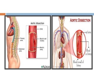
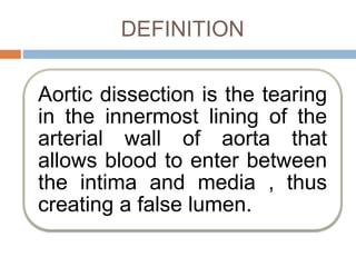
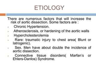
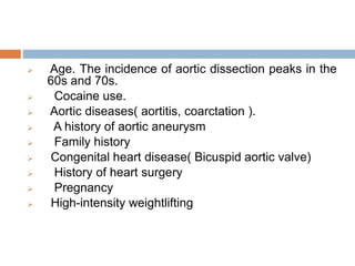
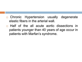
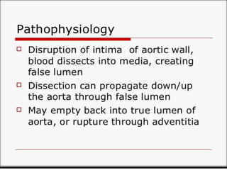
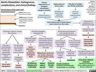
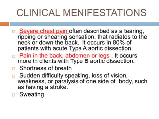
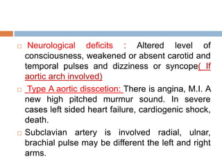
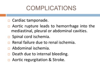
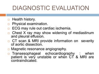
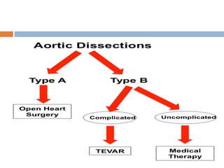
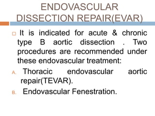
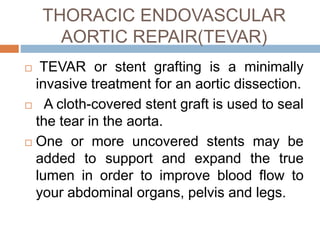
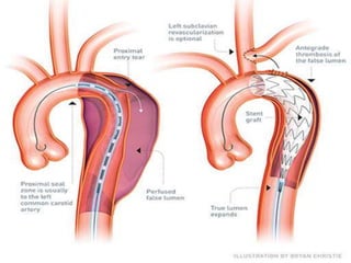
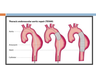
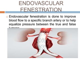
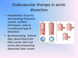
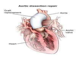
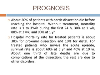
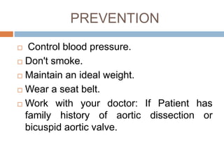
INTRODUCTION
 Aortic dissection is a serious condition in which there
is a tear in the wall of aorta.
 Aortic dissection is the surging of blood through a tear
in the aortic intima with separation of the intima and
media and creation of a false lumen (channel).
 During an aortic dissection the inner layer of aorta
tears, letting blood in where it usually doesn’t go.
 This causes the inner and middle layers to separate,
or dissect.
 If the blood bursts through the outer wall of aorta, it’s
life-threatening and needs immediate repair.
 It occurs when blood enters the medial layer
of the aortic wall through a tear or penetrating
ulcer in the intima and tracks along the media,
forming a second blood-filled channel within
the wall.
DEFINITION
Aortic dissection is the tearing
in the innermost lining of the
arterial wall of aorta that
allows blood to enter between
the intima and media , thus
creating a false lumen.
INCIDENCE
 Aortic dissection affect men 2 to 5 times
more often than women.
 Occurs most frequently in the sixth and
seventh decades of the life.
CLASSIFICATION
DeBakey classification system.
Stanford system.
Based on symptom onset.
DEBAKEY CLASSIFICATION
SYSTEM
 type I: involves ascending and
descending aorta (= Stanford A)
 type II: involves ascending aorta only
(= Stanford A)
 type III: involves descending aorta
only, commencing after the origin of
the left subclavian artery (= Stanford
B)
STANFORD SYSTEM
 TYPE A: A affects ascending aorta and arch
 accounts for ~60% of aortic dissections & start before left
subclavian artery.
 surgical management
 may result in:
 coronary artery occlusion
 aortic incompetence
 rupture into pericardial sac with resulting cardiac tamponade
 TYPE B: B affects descending aorta.
 accounts for ~40% of aortic dissections
 dissection commences distal to the left subclavian artery
 medical management with blood pressure control
BASED ON SYMPTOM ONSET
Acute(First 14 days).
Subacute( 14-90 days).
Chronic( greater than 90
days)
 Approximately two third of dissections involve
the ascending aorta and are acute in onset.
 Chronic dissections are almost type B.
ETIOLOGY
There are numerous factors that will increase the
risk of aortic dissection. Some factors are :
 Chronic Hypertension.
 Atherosclerosis, or hardening of the aortic walls
 Hypercholesterolemia
 Rare: traumatic injury to chest area( Blunt or
Iatrogenic).
 Sex. Men have about double the incidence of
aortic dissection.
 Connective tissue disorders( Marfan’s or
Ehlers-Danlos) Syndrome.
 Age. The incidence of aortic dissection peaks in the
60s and 70s.
 Cocaine use.
 Aortic diseases( aortitis, coarctation ).
 A history of aortic aneurysm
 Family history
 Congenital heart disease( Bicuspid aortic valve)
 History of heart surgery
 Pregnancy
 High-intensity weightlifting
 Chronic Hypertension usually degenerate
elastic fibers in the arterial wall.
 Half of the all acute aortic dissections in
patients younger than 40 years of age occur in
patients with Marfan’s syndrome.
PATHOPHSIOLOGY
PATHOPHYSIOLOGY
CLINICAL MENIFESTATIONS
 Severe chest pain often described as a tearing,
ripping or shearing sensation, that radiates to the
neck or down the back. It occurs in 80% of
patients with acute Type A aortic dissection.
 Pain in the back, abdomen or legs . It occurs
more in clients with Type B aortic dissection.
 Shortness of breath
 Sudden difficulty speaking, loss of vision,
weakness, or paralysis of one side of body, such
as having a stroke.
 Sweating
 Neurological deficits : Altered level of
consciousness, weakened or absent carotid and
temporal pulses and dizziness or syncope( If
aortic arch involved)
 Type A aortic disscetion: There is angina, M.I. A
new high pitched murmur sound. In severe
cases left sided heart failure, cardiogenic shock,
death.
 Subclavian artery is involved radial, ulnar,
brachial pulse may be different the left and right
arms.
COMPLICATIONS
 Cardiac tamponade.
 Aortic rupture leads to hemorrhage into the
mediastinal, pleural or abdominal cavities.
 Spinal cord ischemia.
 Renal failure due to renal ischemia.
 Abdominal ischemia.
 Death due to internal bleeding.
 Aortic regurgitation & Stroke.
DIAGNOSTIC EVALUATION
 Health history.
 Physical examination.
 ECG may rule out cardiac ischemia.
 Chest X ray may show widening of mediastinum
and pleural effusion.
 CT scan & MRI provide information on severity
of aortic dissection.
 Magnetic resonance angiography.
 Transesophageal echocardiography : when
patient is very unstable or when CT & MRI are
contraindicated.
MANAGEMENT
. An aortic dissection is a medical emergency
requiring immediate treatment. Therapy may
include surgery or medications, depending on the
area of the aorta involved.
The initial goals of therapy without complications are
heart rate and BP control
General measures includes:
1) Bed rest.
2) ICU hospitalization.
3) Blood transfusion (if needed)
PHARAMACOLOGICAL
MANAGEMENT
 I.V. Beta blocker(esmolol) to lower Blood
pressure & heart rate.
 Calcium channel blockers(diltiazem) can
be used to lower heart rate when beta
blockers are contraindicated.
 Morphine reduces pain through
decreasing sympathetic nervous system
stimulation.
CONSERVATIVE THERAPY
 The patient with acute or chronic Type
B aortic dissection without
complication can be treated through:
1) Pain relief.
2) BP and Heart rate control.
3) CVD risk factor modification
4) Close surveillance imaging with CT or
MRI.
SURGICAL MANAGEMENT
ENDOVASCULAR
DISSECTION REPAIR(EVAR)
 It is indicated for acute & chronic
type B aortic dissection . Two
procedures are recommended under
these endovascular treatment:
A. Thoracic endovascular aortic
repair(TEVAR).
B. Endovascular Fenestration.
THORACIC ENDOVASCULAR
AORTIC REPAIR(TEVAR)
 TEVAR or stent grafting is a minimally
invasive treatment for an aortic dissection.
 A cloth-covered stent graft is used to seal
the tear in the aorta.
 One or more uncovered stents may be
added to support and expand the true
lumen in order to improve blood flow to
your abdominal organs, pelvis and legs.
 Complications are Major or minor stroke (up
to 5–7% of cases); insufficient blood supply to
the spinal cord, which can lead to leg paralysis
(up to 3%), and extension of the dissection (up
to 3%).
ENDOVASCULAR
FENESTRATION
 Endovascular fenestration is done to improve
blood flow to a specific branch artery or to help
equalize pressure between the true and false
lumens.
OPEN SURGICAL REPAIR
 Indications of open surgery are:
I. Acute type A aortic dissection.
II. Chronic dissection with connective
tissue disorder.
III. A descending thoracic aortic diameter
greater than 5.5 cm.
 Once surgeon has found the tear, he or
she will use man-made (synthetic) grafts
to replace the damaged parts of the aorta.
If r aortic valve is damaged, surgeon may
also put in a replacement valve.
 When surgeon has made all of the repairs,
he or she will remove the heart bypass
machine and close incision.
 Surgeon will make a cut (incision) in
chest or belly (abdomen). The exact spot
will depend on where dissection is
located.
 A heart bypass machine will take over
pumping blood around heart and lungs.
NURSING DIAGNOSIS
 Ineffective tissue perfusion related to
compromised arterial blood
flow secondary to blood extravasation via
aortic dissection.
NURSING INTERVENTION
 Continuously monitor arterial BP during acute
phase to evaluate the patient’s response to
therapy.
 Monitor hourly urine output because a drop in
output may indicate renal artery dissection or a
decrease in arterial blood flow.
 Continuously monitor ECG for dysrythmia
formation, ST segment or T-wave changes,
suggesting coronary sequelae or a decrease in
arterial blood flow.
 Assess neurologic status to evaluate the
course of dissection. Confusion or changes in
sensation and motor strength may indicate
compromised cerebral blood flow (CBF).
 Auscultate for changes in heart sound
and signs and symptoms of heart failure,
which may indicate that the dissection involves
the aortic valve.
 Compare BP and pulses in both arms and legs
to determine differences.
PROGNOSIS
 About 20% of patients with aortic dissection die before
reaching the hospital. Without treatment, mortality
rate is 1 to 3%/h during the first 24 h, 30% at 1 wk,
80% at 2 wk, and 90% at 1 yr.
 Hospital mortality rate for treated patients is about
30% for proximal dissection and 10% for distal. For
treated patients who survive the acute episode,
survival rate is about 60% at 5 yr and 40% at 10 yr.
About one third of late deaths are due to
complications of the dissection; the rest are due to
other disorders.
PREVENTION
 Control blood pressure.
 Don't smoke.
 Maintain an ideal weight.
 Wear a seat belt.
 Work with your doctor: If Patient has
family history of aortic dissection or
bicuspid aortic valve.
Aortic dissection Nikhil

Aortic dissection Nikhil

  • 2.
    INTRODUCTION  Aortic dissectionis a serious condition in which there is a tear in the wall of aorta.  Aortic dissection is the surging of blood through a tear in the aortic intima with separation of the intima and media and creation of a false lumen (channel).  During an aortic dissection the inner layer of aorta tears, letting blood in where it usually doesn’t go.  This causes the inner and middle layers to separate, or dissect.  If the blood bursts through the outer wall of aorta, it’s life-threatening and needs immediate repair.
  • 3.
     It occurswhen blood enters the medial layer of the aortic wall through a tear or penetrating ulcer in the intima and tracks along the media, forming a second blood-filled channel within the wall.
  • 5.
    DEFINITION Aortic dissection isthe tearing in the innermost lining of the arterial wall of aorta that allows blood to enter between the intima and media , thus creating a false lumen.
  • 6.
    INCIDENCE  Aortic dissectionaffect men 2 to 5 times more often than women.  Occurs most frequently in the sixth and seventh decades of the life.
  • 7.
  • 8.
    DEBAKEY CLASSIFICATION SYSTEM  typeI: involves ascending and descending aorta (= Stanford A)  type II: involves ascending aorta only (= Stanford A)  type III: involves descending aorta only, commencing after the origin of the left subclavian artery (= Stanford B)
  • 9.
    STANFORD SYSTEM  TYPEA: A affects ascending aorta and arch  accounts for ~60% of aortic dissections & start before left subclavian artery.  surgical management  may result in:  coronary artery occlusion  aortic incompetence  rupture into pericardial sac with resulting cardiac tamponade  TYPE B: B affects descending aorta.  accounts for ~40% of aortic dissections  dissection commences distal to the left subclavian artery  medical management with blood pressure control
  • 13.
    BASED ON SYMPTOMONSET Acute(First 14 days). Subacute( 14-90 days). Chronic( greater than 90 days)
  • 14.
     Approximately twothird of dissections involve the ascending aorta and are acute in onset.  Chronic dissections are almost type B.
  • 15.
    ETIOLOGY There are numerousfactors that will increase the risk of aortic dissection. Some factors are :  Chronic Hypertension.  Atherosclerosis, or hardening of the aortic walls  Hypercholesterolemia  Rare: traumatic injury to chest area( Blunt or Iatrogenic).  Sex. Men have about double the incidence of aortic dissection.  Connective tissue disorders( Marfan’s or Ehlers-Danlos) Syndrome.
  • 16.
     Age. Theincidence of aortic dissection peaks in the 60s and 70s.  Cocaine use.  Aortic diseases( aortitis, coarctation ).  A history of aortic aneurysm  Family history  Congenital heart disease( Bicuspid aortic valve)  History of heart surgery  Pregnancy  High-intensity weightlifting
  • 17.
     Chronic Hypertensionusually degenerate elastic fibers in the arterial wall.  Half of the all acute aortic dissections in patients younger than 40 years of age occur in patients with Marfan’s syndrome.
  • 18.
  • 19.
  • 20.
    CLINICAL MENIFESTATIONS  Severechest pain often described as a tearing, ripping or shearing sensation, that radiates to the neck or down the back. It occurs in 80% of patients with acute Type A aortic dissection.  Pain in the back, abdomen or legs . It occurs more in clients with Type B aortic dissection.  Shortness of breath  Sudden difficulty speaking, loss of vision, weakness, or paralysis of one side of body, such as having a stroke.  Sweating
  • 21.
     Neurological deficits: Altered level of consciousness, weakened or absent carotid and temporal pulses and dizziness or syncope( If aortic arch involved)  Type A aortic disscetion: There is angina, M.I. A new high pitched murmur sound. In severe cases left sided heart failure, cardiogenic shock, death.  Subclavian artery is involved radial, ulnar, brachial pulse may be different the left and right arms.
  • 22.
    COMPLICATIONS  Cardiac tamponade. Aortic rupture leads to hemorrhage into the mediastinal, pleural or abdominal cavities.  Spinal cord ischemia.  Renal failure due to renal ischemia.  Abdominal ischemia.  Death due to internal bleeding.  Aortic regurgitation & Stroke.
  • 23.
    DIAGNOSTIC EVALUATION  Healthhistory.  Physical examination.  ECG may rule out cardiac ischemia.  Chest X ray may show widening of mediastinum and pleural effusion.  CT scan & MRI provide information on severity of aortic dissection.  Magnetic resonance angiography.  Transesophageal echocardiography : when patient is very unstable or when CT & MRI are contraindicated.
  • 24.
    MANAGEMENT . An aorticdissection is a medical emergency requiring immediate treatment. Therapy may include surgery or medications, depending on the area of the aorta involved. The initial goals of therapy without complications are heart rate and BP control General measures includes: 1) Bed rest. 2) ICU hospitalization. 3) Blood transfusion (if needed)
  • 25.
    PHARAMACOLOGICAL MANAGEMENT  I.V. Betablocker(esmolol) to lower Blood pressure & heart rate.  Calcium channel blockers(diltiazem) can be used to lower heart rate when beta blockers are contraindicated.  Morphine reduces pain through decreasing sympathetic nervous system stimulation.
  • 26.
    CONSERVATIVE THERAPY  Thepatient with acute or chronic Type B aortic dissection without complication can be treated through: 1) Pain relief. 2) BP and Heart rate control. 3) CVD risk factor modification 4) Close surveillance imaging with CT or MRI.
  • 27.
  • 29.
    ENDOVASCULAR DISSECTION REPAIR(EVAR)  Itis indicated for acute & chronic type B aortic dissection . Two procedures are recommended under these endovascular treatment: A. Thoracic endovascular aortic repair(TEVAR). B. Endovascular Fenestration.
  • 30.
    THORACIC ENDOVASCULAR AORTIC REPAIR(TEVAR) TEVAR or stent grafting is a minimally invasive treatment for an aortic dissection.  A cloth-covered stent graft is used to seal the tear in the aorta.  One or more uncovered stents may be added to support and expand the true lumen in order to improve blood flow to your abdominal organs, pelvis and legs.
  • 31.
     Complications areMajor or minor stroke (up to 5–7% of cases); insufficient blood supply to the spinal cord, which can lead to leg paralysis (up to 3%), and extension of the dissection (up to 3%).
  • 34.
    ENDOVASCULAR FENESTRATION  Endovascular fenestrationis done to improve blood flow to a specific branch artery or to help equalize pressure between the true and false lumens.
  • 36.
    OPEN SURGICAL REPAIR Indications of open surgery are: I. Acute type A aortic dissection. II. Chronic dissection with connective tissue disorder. III. A descending thoracic aortic diameter greater than 5.5 cm.
  • 38.
     Once surgeonhas found the tear, he or she will use man-made (synthetic) grafts to replace the damaged parts of the aorta. If r aortic valve is damaged, surgeon may also put in a replacement valve.  When surgeon has made all of the repairs, he or she will remove the heart bypass machine and close incision.
  • 39.
     Surgeon willmake a cut (incision) in chest or belly (abdomen). The exact spot will depend on where dissection is located.  A heart bypass machine will take over pumping blood around heart and lungs.
  • 41.
    NURSING DIAGNOSIS  Ineffectivetissue perfusion related to compromised arterial blood flow secondary to blood extravasation via aortic dissection.
  • 42.
    NURSING INTERVENTION  Continuouslymonitor arterial BP during acute phase to evaluate the patient’s response to therapy.  Monitor hourly urine output because a drop in output may indicate renal artery dissection or a decrease in arterial blood flow.  Continuously monitor ECG for dysrythmia formation, ST segment or T-wave changes, suggesting coronary sequelae or a decrease in arterial blood flow.
  • 43.
     Assess neurologicstatus to evaluate the course of dissection. Confusion or changes in sensation and motor strength may indicate compromised cerebral blood flow (CBF).  Auscultate for changes in heart sound and signs and symptoms of heart failure, which may indicate that the dissection involves the aortic valve.  Compare BP and pulses in both arms and legs to determine differences.
  • 44.
    PROGNOSIS  About 20%of patients with aortic dissection die before reaching the hospital. Without treatment, mortality rate is 1 to 3%/h during the first 24 h, 30% at 1 wk, 80% at 2 wk, and 90% at 1 yr.  Hospital mortality rate for treated patients is about 30% for proximal dissection and 10% for distal. For treated patients who survive the acute episode, survival rate is about 60% at 5 yr and 40% at 10 yr. About one third of late deaths are due to complications of the dissection; the rest are due to other disorders.
  • 45.
    PREVENTION  Control bloodpressure.  Don't smoke.  Maintain an ideal weight.  Wear a seat belt.  Work with your doctor: If Patient has family history of aortic dissection or bicuspid aortic valve.