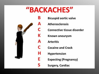
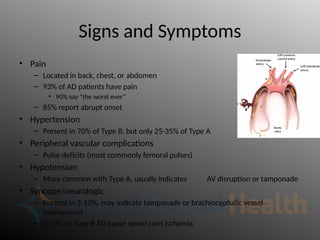
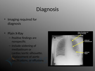
Aortic Dissection and Acute
Aortic Syndrome
Brant Clatterbuck
• Nothing to disclose
The problem
• Most common catastrophic event affecting the
aorta
– Incidence ranging from 2.9 – 3.5 per 100,000
person years
• Without treatment, majority of patients die
within 3 months
Anatomy
• Aorta is largest artery in the body
– Aortic Root: includes AV, starts at
annulus ends at sinotubual junction.
– Ascending Aorta: sinotubual
junction to takeoff of the Inominate
Artery (brachiocephalic artery).
– Aortic Arch: ends at the Subclavian
Artery. Curves anterior to posterior
– Descending Aorta: from subclavian
to diaphragm
– Abdominal aorta: from diaphragm
until division into iliacs
Anatomy – Layers
• Endothelium
• Intima
– Smooth surface for laminar
flow
• Media
– Muscles and elastic fibers
• Allows for stretch and
contraction
• Adventitia
– Surrounding connective
tissue
– Vasa vasorum originate here
Pathophysiology
• Aortic Dissection (AD) occurs when a defect in the
intimal layer of the aorta permits blood to create a false
channel within the aortic wall
– Typically occurs between media and adventitia
• Typically transverse and longitudinal
– Originates
• ascending aorta in 65%
• descending aorta in 25%
• arch and abdominal aorta in 10%
• Aorta is divided into true and false lumens, separated by
a dissection flap
• Blood courses antegrade or retrograde through the
newly created space
Pathophysiology
• Penetrating atherosclerotic ulcer and intramural hematoma
are also considered to be variants of aortic dissection.
– These lesions may eventually extend into the media and evolve
into a true dissection
Pathophysiology
• Fenestrations (connections
between the true and false
lumens) occur within the
intimal flap downstream
– usually at branch vessel
ostia
• These serve as sites where
blood flows back from the
false lumen into the true
lumen
– This flow allows the false
lumen to persist, rather than
thrombose
Malperfusion Syndromes
• Systemic effects of AD
largely depend on
involved branches
• Aortic branch
compromise is
present in 31% of
cases
Aortic Branch Vessel Compromise
• Dynamic obstruction: the
compressed true lumen cannot
provide enough volume due to low
flow or intimal flap prolapse into the
branch vessel
– Most common, 80%
– Severity based on circumference of AD,
cardiac output, BP, HR, and peripheral
resistance at the branch vessel
• Static Obstruction: dissection
extends into the branch vessel,
narrowing the lumen
– False lumen is highly thrombogenic, so
thrombosis beyond the compromised
area can occur
Anatomical Classification
• 2 schemes of classification based on intimal tear location
– Origin of the entry tear is known to be key predictor of early outcomes
• Debakey also considered extent of dissection in its classification
• Debakey
– Type I: Dissection originates in the ascending aorta, extends through the aortic
arch, and continues into the descending aorta and/or abdominal aorta for a
varying distance
– Type II: Dissection originates in and is confined to the ascending aorta
– Type IIIa: Dissection originates in the descending aorta and is limited to same.
– Type IIIb: Dissection involves descending and variable extents of the abdominal
aorta.
• Stanford
– Type A: originates in the ascending aorta
– Type B: originates in the descending aorta distal to the origin of the left
subclavian
Anatomical Classification
Temporal Classification
• Acute
– Traditionally a dissection is considered acute for the first
2 weeks after onset of symptoms
• Based on autopsy studies, 74% on patient deaths occur within
the first 2 weeks
• Subacute
– 2 weeks to 3 months
• Dissection flap remains pliable
• Chronic
– > 90 days, remodeling is taking place (for better or worse)
Risk Factors - Traditional
• HTN
– Especially if uncontrolled
• Male sex
– 4:1 predilection over females
• Age 60-80
– Avg age range for Type A is 50-60
– Avg age range for Type B is 60-70
• Family History
• Connective Tissue disease
– Marfan syndrome accounts for 50% of AD in patients < 40 years old
• Pregnancy
– Uncommon overall (5.5 per million pregnant women)
– 4x increased risk when compared to similar non-pregnant women
• Associated with pre-eclampsia
Risk Factors – Less Common
• Cardiovascular conditions
– Bicuspid aortic valve with or without aortic root
dilatation
– Congenital aortic diseases (coarctation, aortic arch
hypoplasia, etc…)
• Turner’s syndrome
• Coccaine/Crack use
• Thoracic Aortic aneurysm
• Trauma/Iatrogenic
Signs and Symptoms
• Pain
– Located in back, chest, or abdomen
– 93% of AD patients have pain
• 90% say “the worst ever”
– 85% report abrupt onset
• Hypertension
– Present in 70% of Type B, but only 25-35% of Type A
• Peripheral vascular complications
– Pulse deficits (most commonly femoral pulses)
• Hypotension
– More common with Type A, usually indicates AV disruption or tamponade
• Syncope/neurologic
– Present in 5-10%, may indicate tamponade or brachiocephalic vessel
involvement
– 2-10% of Type B AD cause spinal cord ischemia
Diagnosis
• Imaging required for
diagnosis
• Plain X-Ray
– Positive findings are
nonspecific
– Include widening of
mediastinum or
cardiac/aortic silhouette,
displacement of aortic
calcifications, or effusions
Diagnosis - CT
• CT with contrast is the mainstay of
imaging diagnosis
– Fast
– 90% sensitive, up to 100% specific
– Excellent anatomic data
• ability to localize the entry tear and
fenestrations
• assessment of branch vessel patency
• Detect extravasation of contrast
material consistent with rupture
– Effect of iodinated contrast on renal
function is main drawback
Diagnosis - Echocardiography
• Transthoracic Echo
– Widely variable
sensitivity/specificity
– Blind spot behind trachea and left
mainstem bronchus
• Transesophageal Echo
– Sensitivity as high as 98%
– May give useful surgical anatomical
information
– Often done in peri-operative period
Management – If suspected
• Obtain EKG, look for signs of ACS
– Extension of type A dissection to coronary ostia can cause coronary
ischemia
• Right coronary most commonly affected
• Labs:
– D-dimer. If <500 ng/dL is less likely to be aortic dissection
– CBC, basic electrolytes, LDH, coagulation parameters
– Cardiac markers
– Type and crossmatch
• This could become very important very soon
• Chest radiograph
– Widened mediastinum and/or unexplained pleural effusion are consistent
with dissection, particularly if unilateral.
• Proceed with vascular imaging as patient stability allows
Management – Once Diagnosed
• Two large bore IV’s. Continuous monitoring. Consider early arterial line
• Control heart rate and blood pressure.
– Maintain heart rate <60 BPM and systolic blood pressure between 100 and 120
mmHg.
• Esmolol - Loading dose plus drip
• Labetalol – 20 mg bolus followed q10 min boluses, or start drip
• Verapamil, diltiazem, or nicardipine if beta blockers not tolerated
• Once heart rate is consistently <60 BPM, give vasodilator therapy.
– For SBP > 120, start nitroprousside drip or nicardipine drip
– DO NOT use vasodilators without first controlling the heart rate
• Give IV opioids for analgesia (eg, fentanyl).
• Place Foley catheter for assessment of urine output and kidney
perfusion.
• Surgical Consultation
Surgical Consultation
• Obtain immediate surgical consultation (CT surgery, vascular surgery) as
soon as the diagnosis is strongly suspected
– Especially if ascending aorta is involved
• Type A aortic dissection is a cardiac surgical emergency
– Transesophageal echocardiography should be routinely performed in the
operating room to assess AV function, tamponade, etc
• Type B aortic dissection WITH evidence of malperfusion is treated with
urgent aortic stent-grafting or surgery
• Type B aortic dissection without evidence of malperfusion is admitted to
the ICU for medical management of hemodynamics and serial aortic
imaging
• If appropriate surgical services are not available, transfer without delay
Surgical Repair – Type A
• Surgical treatment
involves:
– Excision of the intimal
tear
– Obliteration of entry
into the false lumen
proximally
– Reconstitution of the
aorta with
interposition of a
synthetic vascular
graft
– Repair or replacement
of the aortic valve
Surgical Repair – Type A
• Endovascular repair has not been utilized
widely, still being studied
Management – Type B
• Type B dissection is generally managed medically
initially, with surgical intervention reserved for those
who develop complications related to the dissection
(eg, dissection extension, malperfusion)
– Early intervention to prevent malperfusion becoming more
common
– For uncomplicated Type B, endovascular intervention vs best
medical mgmt had equivocal 2-year survival (INSTEAD trial)
• Endovascular treatment had much more favorable aortic
remodeling (91% vs 14%) when compared to medical, but clinically
little difference was seen at 5 years (INSTEAD-XL trial)
Surgical Repair – Type B
• Indications for repair
– Evidence of end-organ malperfusion (see 'End-organ malperfusion' below)
– Refractory pain in spite of adequate medical treatment (subacute or
chronic)
– A rapidly expanding false lumen seen in first months after diagnosis
– Impending or frank rupture (acute or chronic)
– Aneurysmal dilation in a chronic type B dissection
• Open repair generally met with high mortality
– Reserved for those with anatomically unfavorable aorta or threatened or
actual rupture of proximal intima with no identifiable proximal landing zone
– Mortality ranges 6 – 70%
– Spinal cord ischemia in 32% of successful cases
Surgical Repair – Type B
• Endovascular repair was initially
used for complicated type B
dissections with malperfusion or
(impending) rupture
– promotion of aortic remodeling has
promise for all patients with type B
dissections
• Goals of TEVAR
– Cover proximal entry tear
– Obliterate false lumen 
thrombosis
– Restore flow to visceral vessels
Surgical Repair – Type B
• Fenestrated grafts allow for selected
angioplasty/stenting of involved branch vessels
– Promotes false lumen obliteration and reverses
malperfusion
– Technically challenging, not available everywhere
Aortic Dissection and Acute Aortic Syndrome

Aortic Dissection and Acute Aortic Syndrome

  • 1.
    Aortic Dissection andAcute Aortic Syndrome Brant Clatterbuck
  • 2.
  • 3.
    The problem • Mostcommon catastrophic event affecting the aorta – Incidence ranging from 2.9 – 3.5 per 100,000 person years • Without treatment, majority of patients die within 3 months
  • 4.
    Anatomy • Aorta islargest artery in the body – Aortic Root: includes AV, starts at annulus ends at sinotubual junction. – Ascending Aorta: sinotubual junction to takeoff of the Inominate Artery (brachiocephalic artery). – Aortic Arch: ends at the Subclavian Artery. Curves anterior to posterior – Descending Aorta: from subclavian to diaphragm – Abdominal aorta: from diaphragm until division into iliacs
  • 5.
    Anatomy – Layers •Endothelium • Intima – Smooth surface for laminar flow • Media – Muscles and elastic fibers • Allows for stretch and contraction • Adventitia – Surrounding connective tissue – Vasa vasorum originate here
  • 6.
    Pathophysiology • Aortic Dissection(AD) occurs when a defect in the intimal layer of the aorta permits blood to create a false channel within the aortic wall – Typically occurs between media and adventitia • Typically transverse and longitudinal – Originates • ascending aorta in 65% • descending aorta in 25% • arch and abdominal aorta in 10% • Aorta is divided into true and false lumens, separated by a dissection flap • Blood courses antegrade or retrograde through the newly created space
  • 7.
    Pathophysiology • Penetrating atheroscleroticulcer and intramural hematoma are also considered to be variants of aortic dissection. – These lesions may eventually extend into the media and evolve into a true dissection
  • 8.
    Pathophysiology • Fenestrations (connections betweenthe true and false lumens) occur within the intimal flap downstream – usually at branch vessel ostia • These serve as sites where blood flows back from the false lumen into the true lumen – This flow allows the false lumen to persist, rather than thrombose
  • 9.
    Malperfusion Syndromes • Systemiceffects of AD largely depend on involved branches • Aortic branch compromise is present in 31% of cases
  • 10.
    Aortic Branch VesselCompromise • Dynamic obstruction: the compressed true lumen cannot provide enough volume due to low flow or intimal flap prolapse into the branch vessel – Most common, 80% – Severity based on circumference of AD, cardiac output, BP, HR, and peripheral resistance at the branch vessel • Static Obstruction: dissection extends into the branch vessel, narrowing the lumen – False lumen is highly thrombogenic, so thrombosis beyond the compromised area can occur
  • 11.
    Anatomical Classification • 2schemes of classification based on intimal tear location – Origin of the entry tear is known to be key predictor of early outcomes • Debakey also considered extent of dissection in its classification • Debakey – Type I: Dissection originates in the ascending aorta, extends through the aortic arch, and continues into the descending aorta and/or abdominal aorta for a varying distance – Type II: Dissection originates in and is confined to the ascending aorta – Type IIIa: Dissection originates in the descending aorta and is limited to same. – Type IIIb: Dissection involves descending and variable extents of the abdominal aorta. • Stanford – Type A: originates in the ascending aorta – Type B: originates in the descending aorta distal to the origin of the left subclavian
  • 12.
  • 13.
    Temporal Classification • Acute –Traditionally a dissection is considered acute for the first 2 weeks after onset of symptoms • Based on autopsy studies, 74% on patient deaths occur within the first 2 weeks • Subacute – 2 weeks to 3 months • Dissection flap remains pliable • Chronic – > 90 days, remodeling is taking place (for better or worse)
  • 14.
    Risk Factors -Traditional • HTN – Especially if uncontrolled • Male sex – 4:1 predilection over females • Age 60-80 – Avg age range for Type A is 50-60 – Avg age range for Type B is 60-70 • Family History • Connective Tissue disease – Marfan syndrome accounts for 50% of AD in patients < 40 years old • Pregnancy – Uncommon overall (5.5 per million pregnant women) – 4x increased risk when compared to similar non-pregnant women • Associated with pre-eclampsia
  • 15.
    Risk Factors –Less Common • Cardiovascular conditions – Bicuspid aortic valve with or without aortic root dilatation – Congenital aortic diseases (coarctation, aortic arch hypoplasia, etc…) • Turner’s syndrome • Coccaine/Crack use • Thoracic Aortic aneurysm • Trauma/Iatrogenic
  • 17.
    Signs and Symptoms •Pain – Located in back, chest, or abdomen – 93% of AD patients have pain • 90% say “the worst ever” – 85% report abrupt onset • Hypertension – Present in 70% of Type B, but only 25-35% of Type A • Peripheral vascular complications – Pulse deficits (most commonly femoral pulses) • Hypotension – More common with Type A, usually indicates AV disruption or tamponade • Syncope/neurologic – Present in 5-10%, may indicate tamponade or brachiocephalic vessel involvement – 2-10% of Type B AD cause spinal cord ischemia
  • 18.
    Diagnosis • Imaging requiredfor diagnosis • Plain X-Ray – Positive findings are nonspecific – Include widening of mediastinum or cardiac/aortic silhouette, displacement of aortic calcifications, or effusions
  • 19.
    Diagnosis - CT •CT with contrast is the mainstay of imaging diagnosis – Fast – 90% sensitive, up to 100% specific – Excellent anatomic data • ability to localize the entry tear and fenestrations • assessment of branch vessel patency • Detect extravasation of contrast material consistent with rupture – Effect of iodinated contrast on renal function is main drawback
  • 20.
    Diagnosis - Echocardiography •Transthoracic Echo – Widely variable sensitivity/specificity – Blind spot behind trachea and left mainstem bronchus • Transesophageal Echo – Sensitivity as high as 98% – May give useful surgical anatomical information – Often done in peri-operative period
  • 22.
    Management – Ifsuspected • Obtain EKG, look for signs of ACS – Extension of type A dissection to coronary ostia can cause coronary ischemia • Right coronary most commonly affected • Labs: – D-dimer. If <500 ng/dL is less likely to be aortic dissection – CBC, basic electrolytes, LDH, coagulation parameters – Cardiac markers – Type and crossmatch • This could become very important very soon • Chest radiograph – Widened mediastinum and/or unexplained pleural effusion are consistent with dissection, particularly if unilateral. • Proceed with vascular imaging as patient stability allows
  • 23.
    Management – OnceDiagnosed • Two large bore IV’s. Continuous monitoring. Consider early arterial line • Control heart rate and blood pressure. – Maintain heart rate <60 BPM and systolic blood pressure between 100 and 120 mmHg. • Esmolol - Loading dose plus drip • Labetalol – 20 mg bolus followed q10 min boluses, or start drip • Verapamil, diltiazem, or nicardipine if beta blockers not tolerated • Once heart rate is consistently <60 BPM, give vasodilator therapy. – For SBP > 120, start nitroprousside drip or nicardipine drip – DO NOT use vasodilators without first controlling the heart rate • Give IV opioids for analgesia (eg, fentanyl). • Place Foley catheter for assessment of urine output and kidney perfusion. • Surgical Consultation
  • 24.
    Surgical Consultation • Obtainimmediate surgical consultation (CT surgery, vascular surgery) as soon as the diagnosis is strongly suspected – Especially if ascending aorta is involved • Type A aortic dissection is a cardiac surgical emergency – Transesophageal echocardiography should be routinely performed in the operating room to assess AV function, tamponade, etc • Type B aortic dissection WITH evidence of malperfusion is treated with urgent aortic stent-grafting or surgery • Type B aortic dissection without evidence of malperfusion is admitted to the ICU for medical management of hemodynamics and serial aortic imaging • If appropriate surgical services are not available, transfer without delay
  • 25.
    Surgical Repair –Type A • Surgical treatment involves: – Excision of the intimal tear – Obliteration of entry into the false lumen proximally – Reconstitution of the aorta with interposition of a synthetic vascular graft – Repair or replacement of the aortic valve
  • 26.
    Surgical Repair –Type A • Endovascular repair has not been utilized widely, still being studied
  • 27.
    Management – TypeB • Type B dissection is generally managed medically initially, with surgical intervention reserved for those who develop complications related to the dissection (eg, dissection extension, malperfusion) – Early intervention to prevent malperfusion becoming more common – For uncomplicated Type B, endovascular intervention vs best medical mgmt had equivocal 2-year survival (INSTEAD trial) • Endovascular treatment had much more favorable aortic remodeling (91% vs 14%) when compared to medical, but clinically little difference was seen at 5 years (INSTEAD-XL trial)
  • 28.
    Surgical Repair –Type B • Indications for repair – Evidence of end-organ malperfusion (see 'End-organ malperfusion' below) – Refractory pain in spite of adequate medical treatment (subacute or chronic) – A rapidly expanding false lumen seen in first months after diagnosis – Impending or frank rupture (acute or chronic) – Aneurysmal dilation in a chronic type B dissection • Open repair generally met with high mortality – Reserved for those with anatomically unfavorable aorta or threatened or actual rupture of proximal intima with no identifiable proximal landing zone – Mortality ranges 6 – 70% – Spinal cord ischemia in 32% of successful cases
  • 29.
    Surgical Repair –Type B • Endovascular repair was initially used for complicated type B dissections with malperfusion or (impending) rupture – promotion of aortic remodeling has promise for all patients with type B dissections • Goals of TEVAR – Cover proximal entry tear – Obliterate false lumen  thrombosis – Restore flow to visceral vessels
  • 30.
    Surgical Repair –Type B • Fenestrated grafts allow for selected angioplasty/stenting of involved branch vessels – Promotes false lumen obliteration and reverses malperfusion – Technically challenging, not available everywhere

Editor's Notes

  • #4 Aortic Root: begins at the aortic annulus and incorporates the aortic sinuses, aortic valve, and ends at the sinotubual junction. Ascending Aorta: starts at the sinotubual junction and ends at the takeoff of the Inominate Artery (brachiocephalic artery). Aortic Arch: continues after the Inominate Artery and ends at the Subclavian Artery.  This portion of the aorta curves (arch) from the anterior portion of the chest cavity towards the back. Descending Aorta: begins after the takeoff of the Subclavian Artery and courses down the thorax through the diaphragm. Abdominal aorta: the Thoracic (Descending) Aorta becomes the Abdominal Aorta once it passes through the diaphragm.  The Abdominal Aorta ends in the pelvis when it divides in
  • #10 Branch vessel compromise occurs via 1 mechanisms
  • #11 Debakey system uses entry tear and extent of dissection, Stanford uses entry tear alone Classic Stanford A is confined to thoracic ascending aorta, and really anything that occurs distal to the innominate artery is more or less a type b. However, anything occurring proximal to the left subclavian will be a thoracic surgery problem, since repair will require cardiopulmonary arrest and bypass
  • #12 Because the origin of the entry tear is the key predictor of early outcomes, most patients are now stratified into Stanford type A or B at the time of presentation in order to direct initial therapy
  • #13 Both anatomic and temporal classifications are important for choosing treatment plan
  • #19 Sensitivity drops off when imaging the proximal aorta, TTE is better in this case
  • #21 TEE of proximal dissection
  • #26 A) Complex aortic aneurysm extending between aortic arch and thoracic aorta region, (B) Open surgical repair, (C) Endovascular repair, (D) Frozen elephant trunk hybrid repair. BA—Brachiocephalic artery, LCCA—Left common carotid artery, LSA—Left subclavian artery.
  • #31 Angiography pictures of TEVAR