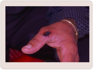
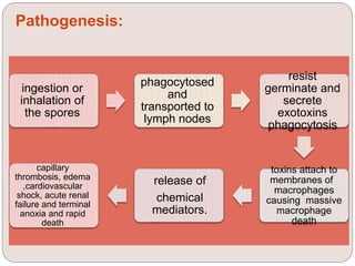
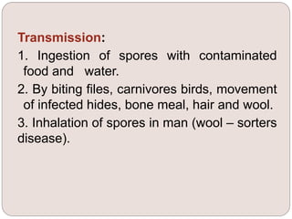
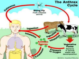
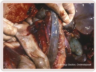
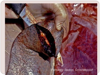
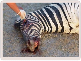
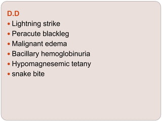
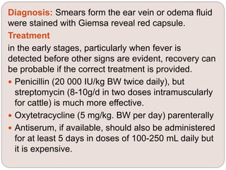
Lecturer Muna Tawfeeq
AL-Muthanna university
Epidemiology & infectious
diseases
anthrax
ANTHRAX
 synonym: Splenic fever , charban
disease
 Etiology: It is an acute septicemic disease
caused by Bacillus anthracis, gram
positive capsulated, spore forming rod-
shaped aerobic organism
Epidemiology:
Global occurrence and often occurs as outbreaks.
Spores survive in soil for many years and disease
is enzootic in certain areas. Animals become
infected while grazing on grass contaminated with
spores. When the pathogen is returned to the soil
in animal excrement or carcasses ,it can sporulate
and become a long-term reservoir of infection for
the animal population.
Source of the infection
B. anthracis can infect animals directly from
the soil or from fodder grown on infected
soil, from contaminated bone meal or
protein concentrates, or from infected
excreta, blood, or other discharges from
infected animals.
Transmission:
1. Ingestion of spores with contaminated
food and water.
2. By biting files, carnivores birds, movement
of infected hides, bone meal, hair and wool.
3. Inhalation of spores in man (wool – sorters
disease).
Risk factors:
Host risk factors
The disease occurs in all vertebrates but is most
common in cattle and sheep and less frequent in
goats and horses. Humans occupy an intermediate
position between this group and the relatively
resistant pigs, dogs, and cats.
Environment risk factors
Outbreaks originating from a soil-borne infection
always occur after a major climate change, for
example heavy rain after a prolonged drought. close
grazing of tough, scratchy feed in dry times, which
results in abrasions of the oral mucosa, and confined
grazing on heavily contaminated areas around water
Pathogen risk factors
When material containing anthrax bacilli is exposed to the air,
spores are formed that protract the infectivity of the
environment for very long periods. The spores are resistant to
most external influences including the salting of hides, normal
environmental temperatures and standard disinfectants.
Economic importance
heavy losses may still occur in individual herds in spite of
vaccination of animals in enzootic area Loss occurs due to
mortality but also from withholding of milk in infected dairy
herds and for a period following vaccination.
Disease forms:
 Cutaneous anthrax, caused by spores entering
the skin through small cuts and abrasions. The
most common and least dangerous of all forms
appear as black eschar.
 pulmonary anthrax (wool sorter’s disease)
associated with the inhalation of airborne spores,
either from animal products or from contaminated
soil.
 gastrointestinal form of infection is another rare
Pathogenesis:
ingestion or
inhalation of
the spores
phagocytosed
and
transported to
lymph nodes
resist
germinate and
secrete
exotoxins
phagocytosis
toxins attach to
membranes of
macrophages
causing massive
macrophage
death
release of
chemical
mediators.
capillary
thrombosis, edema
,cardiovascular
shock, acute renal
failure and terminal
anoxia and rapid
death
Clinical signs:
 Incubation period: 1 to 14 days.
 Acute cases: high temperature (42°C). The
respiration rapid and deep, the mucosa congested
and hemorrhagic, and the heart rate much
increased.
 Pregnant cows may abort. In milking cows the
yield is very much reduced and the milk may be
blood stained or deep yellow in color.
 Local edema of the tongue and edematous lesions
in the region of the throat, sternum, perineum, and
flanks may occur.
 Dark colored blood oozes from the natural orifices
just before death.
Clinical Pathology
In the living animal the organism may be detected in a
stained smear of peripheral blood or local edema fluid
by microscopic examination, of a clearly defined
metachromatic capsule on square-ended bacilli (often in
chains) in a blood smear stained with aged polychrome
methylene blue.
Necropsy Findings:
1. Carcass readily undergoes purification with much gas
formation.
2. The blood is dark colored and fail to coagulate.
3. Hemorrhages on the muscular subcutaneous and
serous tissues.
4. In horses, dog and pigs extensive gelatinous odema
over abdomen, thorax, limbs and external genetalia.
5. Spleenomegaly in cattle, which is absent in sheep,
horse, dog and swine.
D.D
 Lightning strike
 Peracute blackleg
 Malignant edema
 Bacillary hemoglobinuria
 Hypomagnesemic tetany
 snake bite
Diagnosis: Smears form the ear vein or odema fluid
were stained with Giemsa reveal red capsule.
Treatment
in the early stages, particularly when fever is
detected before other signs are evident, recovery can
be probable if the correct treatment is provided.
 Penicillin (20 000 IU/kg BW twice daily), but
streptomycin (8-10g/d in two doses intramuscularly
for cattle) is much more effective.
 Oxytetracycline (5 mg/kg. BW per day) parenterally
 Antiserum, if available, should also be administered
for at least 5 days in doses of 100-250 mL daily but
it is expensive.
Control:
anthrax is a reportable disease when an
outbreak occurs, the placing of the farm in
quarantine, destruction of discharges and
dead body, and vaccination of survivors, are
part of the animal disease control program
and indirectly reduce human exposure.
prevention of movement of milk and meat
from the farm during the quarantine period
should prevent entry of the infection into the
human food chain.
Disposal of infected material and Infected
carcasses should not be opened but
Thank you

Anthrax

  • 1.
    Lecturer Muna Tawfeeq AL-Muthannauniversity Epidemiology & infectious diseases anthrax
  • 2.
    ANTHRAX  synonym: Splenicfever , charban disease  Etiology: It is an acute septicemic disease caused by Bacillus anthracis, gram positive capsulated, spore forming rod- shaped aerobic organism
  • 3.
    Epidemiology: Global occurrence andoften occurs as outbreaks. Spores survive in soil for many years and disease is enzootic in certain areas. Animals become infected while grazing on grass contaminated with spores. When the pathogen is returned to the soil in animal excrement or carcasses ,it can sporulate and become a long-term reservoir of infection for the animal population.
  • 4.
    Source of theinfection B. anthracis can infect animals directly from the soil or from fodder grown on infected soil, from contaminated bone meal or protein concentrates, or from infected excreta, blood, or other discharges from infected animals.
  • 5.
    Transmission: 1. Ingestion ofspores with contaminated food and water. 2. By biting files, carnivores birds, movement of infected hides, bone meal, hair and wool. 3. Inhalation of spores in man (wool – sorters disease).
  • 7.
    Risk factors: Host riskfactors The disease occurs in all vertebrates but is most common in cattle and sheep and less frequent in goats and horses. Humans occupy an intermediate position between this group and the relatively resistant pigs, dogs, and cats. Environment risk factors Outbreaks originating from a soil-borne infection always occur after a major climate change, for example heavy rain after a prolonged drought. close grazing of tough, scratchy feed in dry times, which results in abrasions of the oral mucosa, and confined grazing on heavily contaminated areas around water
  • 8.
    Pathogen risk factors Whenmaterial containing anthrax bacilli is exposed to the air, spores are formed that protract the infectivity of the environment for very long periods. The spores are resistant to most external influences including the salting of hides, normal environmental temperatures and standard disinfectants. Economic importance heavy losses may still occur in individual herds in spite of vaccination of animals in enzootic area Loss occurs due to mortality but also from withholding of milk in infected dairy herds and for a period following vaccination.
  • 9.
    Disease forms:  Cutaneousanthrax, caused by spores entering the skin through small cuts and abrasions. The most common and least dangerous of all forms appear as black eschar.  pulmonary anthrax (wool sorter’s disease) associated with the inhalation of airborne spores, either from animal products or from contaminated soil.  gastrointestinal form of infection is another rare
  • 11.
    Pathogenesis: ingestion or inhalation of thespores phagocytosed and transported to lymph nodes resist germinate and secrete exotoxins phagocytosis toxins attach to membranes of macrophages causing massive macrophage death release of chemical mediators. capillary thrombosis, edema ,cardiovascular shock, acute renal failure and terminal anoxia and rapid death
  • 12.
    Clinical signs:  Incubationperiod: 1 to 14 days.  Acute cases: high temperature (42°C). The respiration rapid and deep, the mucosa congested and hemorrhagic, and the heart rate much increased.  Pregnant cows may abort. In milking cows the yield is very much reduced and the milk may be blood stained or deep yellow in color.  Local edema of the tongue and edematous lesions in the region of the throat, sternum, perineum, and flanks may occur.  Dark colored blood oozes from the natural orifices just before death.
  • 15.
    Clinical Pathology In theliving animal the organism may be detected in a stained smear of peripheral blood or local edema fluid by microscopic examination, of a clearly defined metachromatic capsule on square-ended bacilli (often in chains) in a blood smear stained with aged polychrome methylene blue.
  • 16.
    Necropsy Findings: 1. Carcassreadily undergoes purification with much gas formation. 2. The blood is dark colored and fail to coagulate. 3. Hemorrhages on the muscular subcutaneous and serous tissues. 4. In horses, dog and pigs extensive gelatinous odema over abdomen, thorax, limbs and external genetalia. 5. Spleenomegaly in cattle, which is absent in sheep, horse, dog and swine.
  • 20.
    D.D  Lightning strike Peracute blackleg  Malignant edema  Bacillary hemoglobinuria  Hypomagnesemic tetany  snake bite
  • 21.
    Diagnosis: Smears formthe ear vein or odema fluid were stained with Giemsa reveal red capsule. Treatment in the early stages, particularly when fever is detected before other signs are evident, recovery can be probable if the correct treatment is provided.  Penicillin (20 000 IU/kg BW twice daily), but streptomycin (8-10g/d in two doses intramuscularly for cattle) is much more effective.  Oxytetracycline (5 mg/kg. BW per day) parenterally  Antiserum, if available, should also be administered for at least 5 days in doses of 100-250 mL daily but it is expensive.
  • 22.
    Control: anthrax is areportable disease when an outbreak occurs, the placing of the farm in quarantine, destruction of discharges and dead body, and vaccination of survivors, are part of the animal disease control program and indirectly reduce human exposure. prevention of movement of milk and meat from the farm during the quarantine period should prevent entry of the infection into the human food chain. Disposal of infected material and Infected carcasses should not be opened but
  • 23.