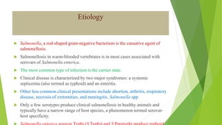
This document discusses Salmonellosis, caused by the Salmonella bacterium. It affects various warm-blooded animals and has two main clinical presentations: enteritis, characterized by diarrhea and enterocolitis; and septicemia, a systemic infection most common in young animals. The bacterium is transmitted orally through contaminated food, water or feces. Control relies on reducing exposure through biosecurity, sanitation and antibiotic treatment of clinical cases.
local names, definition, etiology,epidemiology lifecycle, pathogenesis, clinical findings, necropsy finding, diagnosis,treatment, control and prevention
West Nile fever is an infection by the West Nile virus, which is typically spread by mosquitoes. It causes disease in humans, horses, and several species of birds
Largest viruses that infect vertebrates
Can be seen under light microscope
Poxvirus diseases are characterized by skin lesions – localized or generalized
Important diseases caused by poxviruses are-
Smallpox
Monkeypox
Cowpox
Tanapox
Molluscum contagiosum
etiology, local names, definition, transmission, source of infection, epidemiology, pathogenesis, clinical signs, diagnosis, differential diagnosis, treatment prevention and control
Colibacillosis refers to any localized or systemic infection caused entirely or partly by avian pathogenic Escherichia coli (APEC), It manifests in diverse ways, including as acute fatal septicemia, subacute pericarditis, peritonitis, and cellulitis.
It is frequently associated with immunosuppressive diseases such as Infectious Bursal Disease Virus (Gumboro Disease) in chickens or Haemorrhagic Enteritis in turkeys, or in young birds that are immunologically immature.
local names, definition, etiology,epidemiology lifecycle, pathogenesis, clinical findings, necropsy finding, diagnosis,treatment, control and prevention
Salmonella & Vibrio Cholerae an overview.pdfAlanShwan2
medical Bacteriology
Salmonella & Vibrio
Salmonella infection (salmonellosis) is a common bacterial disease that affects the intestinal tract. Salmonella bacteria typically live in animal and human intestines and are shed through stool (feces). Humans become infected most frequently through contaminated water or food.
Some people with salmonella infection have no symptoms. Most people develop diarrhea, fever and stomach (abdominal) cramps within 8 to 72 hours after exposure. Most healthy people recover within a few days to a week without specific treatment.
Cholera is a bacterial disease usually spread through contaminated water. Cholera causes severe diarrhea and dehydration. Left untreated, cholera can be fatal within hours, even in previously healthy people.
Modern sewage and water treatment have virtually eliminated cholera in industrialized countries. But cholera still exists in Africa, Southeast Asia and Haiti. The risk of a cholera epidemic is highest when poverty, war or natural disasters force people to live in crowded conditions without adequate sanitation.
Guinea pigs are generally hardy, healthy animals but are susceptible to certain problems and diseases. Here are the common bacterial infections of guinea pigs.
prof . dr. ihsan edan alsaimary
department of microbiology - college of medicine - university of basrah - basrah -IRAQ
ihsanalsaimary@gmail.com
00964 7801410838
Explore natural remedies for syphilis treatment in Singapore. Discover alternative therapies, herbal remedies, and lifestyle changes that may complement conventional treatments. Learn about holistic approaches to managing syphilis symptoms and supporting overall health.
Anti ulcer drugs and their Advance pharmacology ||
Anti-ulcer drugs are medications used to prevent and treat ulcers in the stomach and upper part of the small intestine (duodenal ulcers). These ulcers are often caused by an imbalance between stomach acid and the mucosal lining, which protects the stomach lining.
||Scope: Overview of various classes of anti-ulcer drugs, their mechanisms of action, indications, side effects, and clinical considerations.
Pulmonary Thromboembolism - etilogy, types, medical- Surgical and nursing man...VarunMahajani
Disruption of blood supply to lung alveoli due to blockage of one or more pulmonary blood vessels is called as Pulmonary thromboembolism. In this presentation we will discuss its causes, types and its management in depth.
MANAGEMENT OF ATRIOVENTRICULAR CONDUCTION BLOCK.pdfJim Jacob Roy
Cardiac conduction defects can occur due to various causes.
Atrioventricular conduction blocks ( AV blocks ) are classified into 3 types.
This document describes the acute management of AV block.
Factory Supply Best Quality Pmk Oil CAS 28578–16–7 PMK Powder in Stockrebeccabio
Factory Supply Best Quality Pmk Oil CAS 28578–16–7 PMK Powder in Stock
Telegram: bmksupplier
signal: +85264872720
threema: TUD4A6YC
You can contact me on Telegram or Threema
Communicate promptly and reply
Free of customs clearance, Double Clearance 100% pass delivery to USA, Canada, Spain, Germany, Netherland, Poland, Italy, Sweden, UK, Czech Republic, Australia, Mexico, Russia, Ukraine, Kazakhstan.Door to door service
Hot Selling Organic intermediates
Title: Sense of Smell
Presenter: Dr. Faiza, Assistant Professor of Physiology
Qualifications:
MBBS (Best Graduate, AIMC Lahore)
FCPS Physiology
ICMT, CHPE, DHPE (STMU)
MPH (GC University, Faisalabad)
MBA (Virtual University of Pakistan)
Learning Objectives:
Describe the primary categories of smells and the concept of odor blindness.
Explain the structure and location of the olfactory membrane and mucosa, including the types and roles of cells involved in olfaction.
Describe the pathway and mechanisms of olfactory signal transmission from the olfactory receptors to the brain.
Illustrate the biochemical cascade triggered by odorant binding to olfactory receptors, including the role of G-proteins and second messengers in generating an action potential.
Identify different types of olfactory disorders such as anosmia, hyposmia, hyperosmia, and dysosmia, including their potential causes.
Key Topics:
Olfactory Genes:
3% of the human genome accounts for olfactory genes.
400 genes for odorant receptors.
Olfactory Membrane:
Located in the superior part of the nasal cavity.
Medially: Folds downward along the superior septum.
Laterally: Folds over the superior turbinate and upper surface of the middle turbinate.
Total surface area: 5-10 square centimeters.
Olfactory Mucosa:
Olfactory Cells: Bipolar nerve cells derived from the CNS (100 million), with 4-25 olfactory cilia per cell.
Sustentacular Cells: Produce mucus and maintain ionic and molecular environment.
Basal Cells: Replace worn-out olfactory cells with an average lifespan of 1-2 months.
Bowman’s Gland: Secretes mucus.
Stimulation of Olfactory Cells:
Odorant dissolves in mucus and attaches to receptors on olfactory cilia.
Involves a cascade effect through G-proteins and second messengers, leading to depolarization and action potential generation in the olfactory nerve.
Quality of a Good Odorant:
Small (3-20 Carbon atoms), volatile, water-soluble, and lipid-soluble.
Facilitated by odorant-binding proteins in mucus.
Membrane Potential and Action Potential:
Resting membrane potential: -55mV.
Action potential frequency in the olfactory nerve increases with odorant strength.
Adaptation Towards the Sense of Smell:
Rapid adaptation within the first second, with further slow adaptation.
Psychological adaptation greater than receptor adaptation, involving feedback inhibition from the central nervous system.
Primary Sensations of Smell:
Camphoraceous, Musky, Floral, Pepperminty, Ethereal, Pungent, Putrid.
Odor Detection Threshold:
Examples: Hydrogen sulfide (0.0005 ppm), Methyl-mercaptan (0.002 ppm).
Some toxic substances are odorless at lethal concentrations.
Characteristics of Smell:
Odor blindness for single substances due to lack of appropriate receptor protein.
Behavioral and emotional influences of smell.
Transmission of Olfactory Signals:
From olfactory cells to glomeruli in the olfactory bulb, involving lateral inhibition.
Primitive, less old, and new olfactory systems with different path
- Video recording of this lecture in English language: https://youtu.be/lK81BzxMqdo
- Video recording of this lecture in Arabic language: https://youtu.be/Ve4P0COk9OI
- Link to download the book free: https://nephrotube.blogspot.com/p/nephrotube-nephrology-books.html
- Link to NephroTube website: www.NephroTube.com
- Link to NephroTube social media accounts: https://nephrotube.blogspot.com/p/join-nephrotube-on-social-media.html
Title: Sense of Taste
Presenter: Dr. Faiza, Assistant Professor of Physiology
Qualifications:
MBBS (Best Graduate, AIMC Lahore)
FCPS Physiology
ICMT, CHPE, DHPE (STMU)
MPH (GC University, Faisalabad)
MBA (Virtual University of Pakistan)
Learning Objectives:
Describe the structure and function of taste buds.
Describe the relationship between the taste threshold and taste index of common substances.
Explain the chemical basis and signal transduction of taste perception for each type of primary taste sensation.
Recognize different abnormalities of taste perception and their causes.
Key Topics:
Significance of Taste Sensation:
Differentiation between pleasant and harmful food
Influence on behavior
Selection of food based on metabolic needs
Receptors of Taste:
Taste buds on the tongue
Influence of sense of smell, texture of food, and pain stimulation (e.g., by pepper)
Primary and Secondary Taste Sensations:
Primary taste sensations: Sweet, Sour, Salty, Bitter, Umami
Chemical basis and signal transduction mechanisms for each taste
Taste Threshold and Index:
Taste threshold values for Sweet (sucrose), Salty (NaCl), Sour (HCl), and Bitter (Quinine)
Taste index relationship: Inversely proportional to taste threshold
Taste Blindness:
Inability to taste certain substances, particularly thiourea compounds
Example: Phenylthiocarbamide
Structure and Function of Taste Buds:
Composition: Epithelial cells, Sustentacular/Supporting cells, Taste cells, Basal cells
Features: Taste pores, Taste hairs/microvilli, and Taste nerve fibers
Location of Taste Buds:
Found in papillae of the tongue (Fungiform, Circumvallate, Foliate)
Also present on the palate, tonsillar pillars, epiglottis, and proximal esophagus
Mechanism of Taste Stimulation:
Interaction of taste substances with receptors on microvilli
Signal transduction pathways for Umami, Sweet, Bitter, Sour, and Salty tastes
Taste Sensitivity and Adaptation:
Decrease in sensitivity with age
Rapid adaptation of taste sensation
Role of Saliva in Taste:
Dissolution of tastants to reach receptors
Washing away the stimulus
Taste Preferences and Aversions:
Mechanisms behind taste preference and aversion
Influence of receptors and neural pathways
Impact of Sensory Nerve Damage:
Degeneration of taste buds if the sensory nerve fiber is cut
Abnormalities of Taste Detection:
Conditions: Ageusia, Hypogeusia, Dysgeusia (parageusia)
Causes: Nerve damage, neurological disorders, infections, poor oral hygiene, adverse drug effects, deficiencies, aging, tobacco use, altered neurotransmitter levels
Neurotransmitters and Taste Threshold:
Effects of serotonin (5-HT) and norepinephrine (NE) on taste sensitivity
Supertasters:
25% of the population with heightened sensitivity to taste, especially bitterness
Increased number of fungiform papillae
Ozempic: Preoperative Management of Patients on GLP-1 Receptor Agonists Saeid Safari
Preoperative Management of Patients on GLP-1 Receptor Agonists like Ozempic and Semiglutide
ASA GUIDELINE
NYSORA Guideline
2 Case Reports of Gastric Ultrasound
Triangles of Neck and Clinical Correlation by Dr. RIG.pptx
Salmonellosis-1.pptx
1. Salmonellosis
Dr. Bipin Kumar
Assistant Professor
Department of Veterinary Medicine
Bihar Veterinary College, Patna
(Bihar Animal Sciences University, Patna)
2. Etiology
Salmonella, a rod-shaped gram-negative bacterium is the causative agent of
salmonellosis.
Salmonellosis in warm-blooded vertebrates is in most cases associated with
serovars of Salmonella enterica.
The most common type of infection is the carrier state.
Clinical disease is characterized by two major syndromes: a systemic
septicemia (also termed as typhoid) and an enteritis.
Other less common clinical presentations include abortion, arthritis, respiratory
disease, necrosis of extremities, and meningitis. Salmonella spp.
Only a few serotypes produce clinical salmonellosis in healthy animals and
typically have a narrow range of host species, a phenomenon termed serovar-
host specificity.
3. Young calves, piglets, lambs, and foals may develop both the enteritis and
septicemic form.
cattle—S Typhimurium, S Dublin, and S Newport;
sheep and goats—S Typhimurium, S Dublin, S Abortusovis, S Anatum,
and S Montevideo;
pigs—S Typhimurium and S Choleraesuis;
horses—S Typhimurium, S Anatum, S Newport, S Enteritidis,
and Salmonella serovar IIIa 18:z4:z23; and
poultry—S Enteritidis, S Typhimurium, S Gallinarum, and S Pullorum.
4. Transmission
Human:
People are often infected when they eat contaminated foods of animal origin such as meat or
eggs.
They can also be infected by ingesting organisms in animal feces, either directly or in
contaminated food or water.
Directly transmitted human infections are most often acquired from the feces of reptiles, chicks
and ducklings. Livestock, dogs, cats, adult poultry and cage birds can also be involved.
Animals:
Salmonella spp. are mainly transmitted by the fecal-oral route.
Vertical transmission occurs in birds, with contamination of the vitelline membrane, albumen and
the yolk of eggs.
Salmonella spp. can also be transmitted in utero in mammals.
Animals may also become infected from contaminated feed (including pastures), drinking water,
or close contact with infected animal (including humans).
5. Mechanisms of Pathogenicity
Bacterial products involved in virulence:
Salmonellae owe their pathogenicity largely to their ability to invade tissue and to survive within
macrophages.
The Vi antigen is a capsule that affords salmonellae some protection from phagocytosis.
Once phagocytosed, Salmonella inhibits generation of oxidative free radicals and
intraphagosomal killing.
Additionally, salmonellae have endotoxic lipopolysaccharide, which is responsible for septic
shock in patients with bacteriemia.
Salmonellae that cause enteritis produce at least two enterotoxins that are responsible for many of
the clinical signs of enteritis.
The first of these is a small (25-30kD) protein that binds to GM1 gangliosides and cause
hypersecretion of fluids and electrolytes by elevating levels of c-AMP.
It appears that both protein kinase C and prostaglandin E2 are involved in this process.
The second enterotoxin is larger (about 100 kD) and is unrelated in structure and mechanism of
activity to the first enterotoxin
Salmonella strains that produce enterotoxins have been reported to invade the intestinal wall more
6. The Salmonella infection cycle.
• Intestinal infection with salmonellae can follow one of two infection cycle.
One cycle causes enteritis, other causes typhoid
a. Enteritis
Most serotypes cause enteritis, an infection that is limited to the terminal ileum.
The salmonellae invade the intestinal wall and produce enterotoxins that cause nausea, vomiting and
diarrhea.
b. Enteric fever (Typhoid): • Two serotypes Typhi and Paratyphi can cause typhoid.
• The salmonella invade the wall of the terminal ileum and than spread to the intestinal lymphatics,
where they are phagocytosed by PMNs and macrophages.
• Salmonella phagocytosed by PMNs(polymorphonuclear leukocytes/HYPOCHLORITE AND Ph
basic) are killed, but those phagocytosed by macrophages survive and multiply within phagocytic
vacuoles.
• Wandering macrophages that contain salmonellae act as “taxi/cabs” that deliver salmonellae to
various reticuloendothelial tissues.
• Infected macrophages are eventually destroyed and salmonellae released from lysed macrophages
cause septicemia.
7. Clinical manifestation
Salmonellae often localize in the mucosae of the ileum, caecum and colon, and in the mesenteric
lymph nodes of infected animals.
Although most organisms are cleared from the tissues by host defense mechanisms, subclinical
infection may persist with shedding of small numbers of salmonellae in the faeces.
Latent infections, in which salmonellae are present in the gall bladder but are not excreted, also
occur.
Clinical disease may develop from subclinical and latent infections if affected animals are
stressed.
Other factors which determine the clinical outcome of infection include the number of
salmonellae ingested, the virulence of the infecting serotype or strain and the susceptibility of the
host.
Host susceptibility may be related to immunological status, genetic make- up or age.
Young and debilitated or aged animaIs are particularly susceptible and may develop the
septicemic form of the disease.
In most animal species, both enteric and septicemic forms of salmonellosis are recorded.
8. Enteric Salmonellosis
Enterocolitis caused by salmonella organisms can affect most species of farm
animals, irrespective of age.
Acute disease is characterized by fever, depression, anorexia and profuse foul-
smelling diarrhoea often containing blood, mucus and epithelial casts.
Dehydration and weight loss follow and pregnant animals may abort.
Severely affected young animals become recumbent and may die within a few
days of acquiring infection.
On farms with endemic salmonellosis, the milder clinical signs often observed
may be attributed to the influence of acquired immunity.
Chronic enterocolitis can follow acute salmonellosis in pigs, cattle and horses.
Intermittent fever, soft faeces and gradual weight loss, leading to emaciation,
9. Septicaemic Salmonellosis
The septicaemic form can occur in all age groups but is most common in calves, in neonatal foals and in
pigs less than four months of age.
Onset of clinical disease is sudden with high fever, depression and recumbency.
If treatment is delayed, many young animals with septicaemic salmonellosis die within 48 hours.
Surviving animals can develop persistent diarrhoea, arthritis, meningitis or pneumonia.
In pigs with septicaemic Salmonella Choleraesuis infection, there is a characteristic bluish discolouration
of the ears and snout.
Intercurrent viral infections often predispose to severe clinical forms of the disease.
The close clinical and pathological relationships which have been recognized in animals infected with
Salmonella Choleraesuis (hog- cholera bacillus) and classical swine fever virus, either jointly or
separately, exemplify both the importance of intercurrent infections and the difficulty of clinically
distinguishing the diseases caused by these agents.
10. Salmonellosis in poultry
Salmonella Pullorum, Salmonella Gallinarum and Salmonella Enteritidis can infect the ovaries of
hens and be transmitted through eggs.
Pullorum disease or bacillary white diarrhoea (Salmonella Pullorum):
Infects young chicks and turkey poults up to 2 to 3 weeks of age.
The mortality rate is high and affected birds huddle under a heat source and are anorexic,
depressed and have whitish faecal pasting around their vents.
Characteristic lesions includes whitish nodes throughout the lungs and focal necrosis of liver and
spleen.
Fowl typhoid (Salmonella Gallinarum):
Can produce lesions in young chicks and poults similar to those of pullorum disease, but it is also
a serious concern in growing and adult poultry
However, in countries where fowl typhoid is endemic, a septicaemic disease of adult birds occurs,
often resulting in sudden deaths.
Characteristic findings include an enlarged, friable, bile-stained liver and enlarged spleen.
Paratyphoid is a name given to infections of poultry by non-host-adapted salmonellae such as
Salmonella Enteritidis and Salmonella Typhimurium. These infections are often subclinical in
laying birds.
11. Diagnosis
A history of previous outbreaks of the disease may suggest salmonellosis.
At postmortem, enterocolitis with blood-stained luminal contents and enlarged mesenteric lymph
nodes are commonly observed.
Specimens for submission should include faeces and blood from live animals.
Intestinal contents and samples from tissue lesions should be submitted from dead animals and
abomasal contents from aborted foetuses.
Isolation of salmonellae from blood or parenchymatous organs is deemed to be confirmatory for
septicaemic sallmonellosis.
Serological tests such as ELISA and agglutination techniques are of greatest value when used on a
herd or flock basis.
A rising antibody titre using paired serum samples is indicative of active infection.
DNA probes can be used to screen large numbers of faecal samples for salmonellae (Maddox and
Fales, 1991).
12. Treatment
Antibiotic therapy should be based on results of susceptibility testing because
R-plasmids coding for multiple resistance are comparatively common in
salmonellae.
Oral antimicrobial therapy should be used judiciously for treating enteric
salmonellosis because it may disturb the normal intestinal flora, extend the
duration of salmonella excretion and increase the probability of drug resistance
developing.
In the septicaemic form of the disease, intravenous antibiotic therapy must be
used.
Fluid and electrolyte replacement therapy is required to counteract dehydration
and shock.
13. Control
Control is based on reducing the risk of exposure to infection.
Measures for excluding infection from a herd or flock free of
salmonellosis:
A closed-herd policy should be implemented when feasible.
Animals should be purchased from reliable sources and isolated until
negative for salmonellae on three consecutive samplings.
Steps should be taken to prevent contamination of foodstuffs and water
- In this context, rodent control is important.
Protective clothing and footwear should be worn by personnel entering
hatcheries and minimal disease pig units.
Measures for reducing environmental contamination:
Effective routine cleaning and disinfection of buildings and equipment is
14. Strategies for enhancing resistance and reducing the likelihood of clinical disease:
Vaccination procedures are used in cattle, sheep, poultry and pigs.
Modified live vaccines which stimulate humoral immunity and cell- mediated
immunity are preferabIe to bacterins.
Measures for controlIing an outbreak of salmonellosis:
Detection and elimination of the source of infection is essential.
Clinically affected animals should be isolated.
Movement of animals, vehicles and humans should be curtailed.
Foot baths containing suitable disinfectant, such as 3% iodophor, should be
placed at strategic locations to limit spread of salmonellae.
Careful disposal of contaminated carcases and bedding is mandatory.
A 3% concentration of sodium hypochlorite or iodophors is suitable for clean
surfaces.
Phenolic disinfectants are suitable for buildings with residual organic matter.