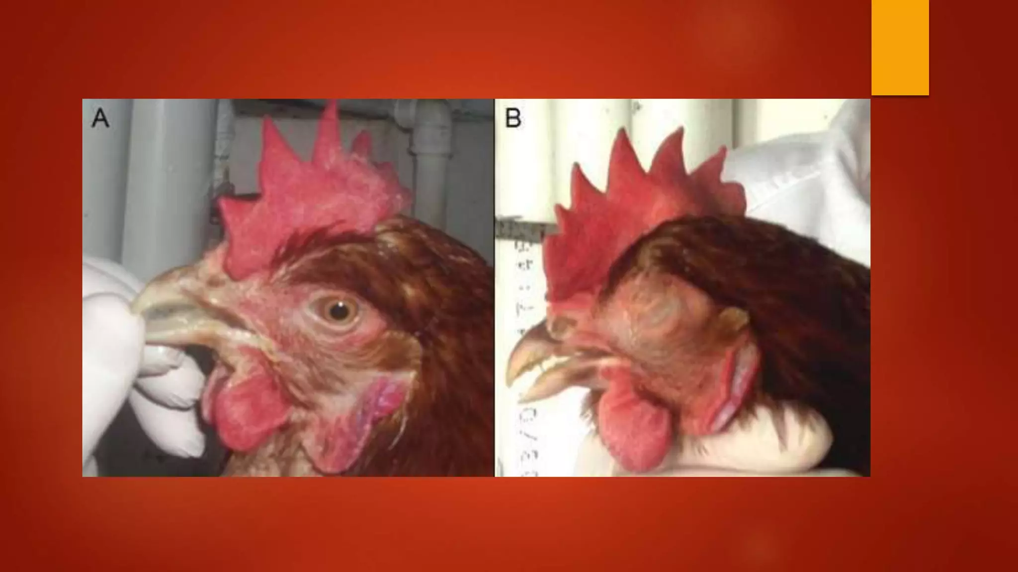
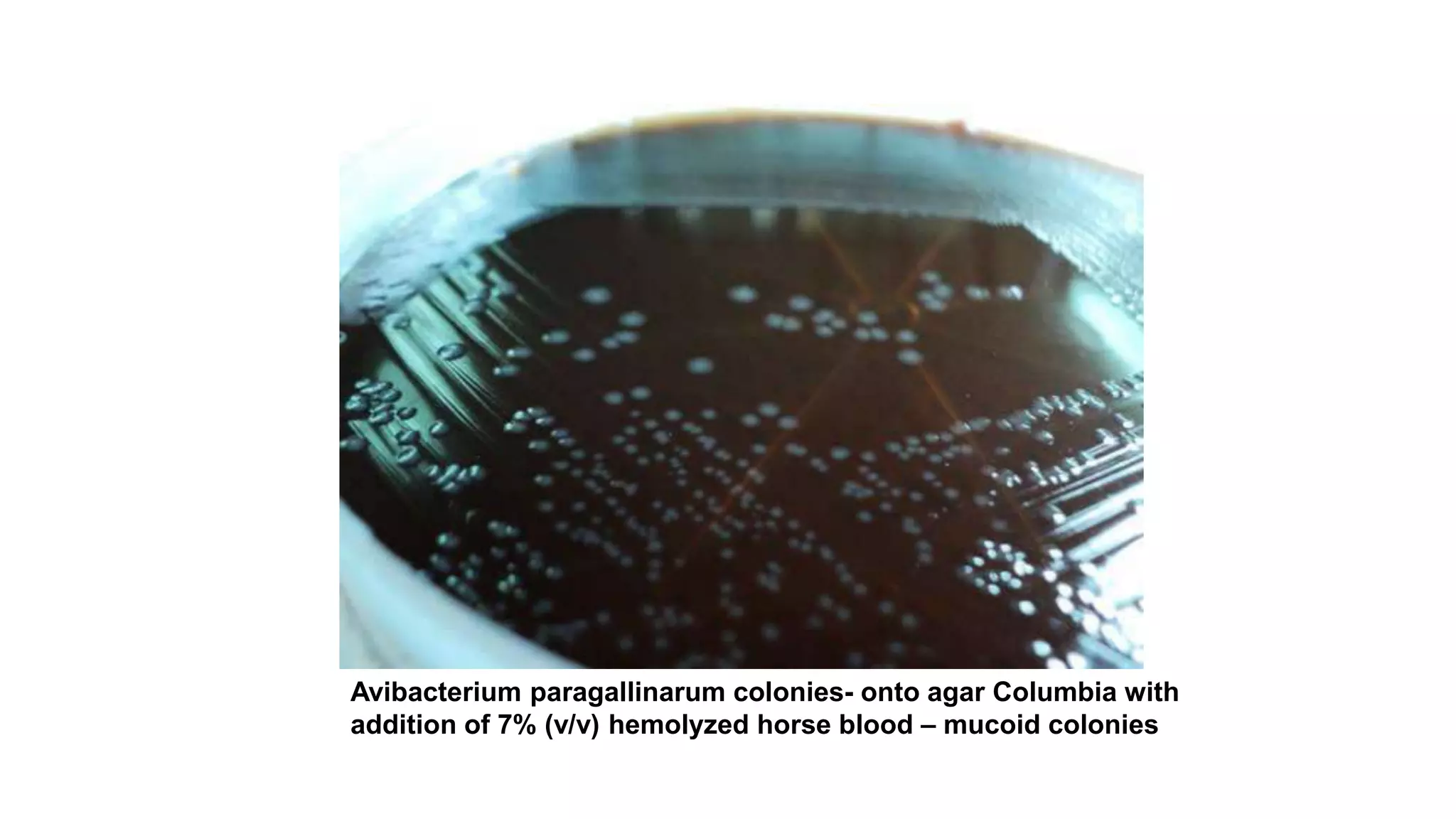
INFECTIOUS CORYZA
FOWL CORYZA, ROUP, COLD, SWOLLEN HEAD DISEASE, WATTLE DISEASE
BY RANJINI MANUEL
INTRODUCTION
 Highly contagious
 Acute disease of the upper respiratory tract of chickens – starts as acute
and progresses as chronic
 Worldwide distribution
 Economic losses due to increased culling rate and reduction in egg
production
 Limited to chickens and has no public health significance
 Disease is characterized by nasal discharge, facial swelling, sneezing,
labored breathing and fetid odor of the exudates
HISTORY
 clinical syndrome was first diagnosed in 1931 by De Blieck.
 Since the disease proved to be infectious and primarily affected nasal
passages, the name "infectious coryza" was adopted (Blackall, 1989)
HISTORY
 1930’S – 1960’S – causative agent – Haemophilus gallinarum
 It required X (haemin ) and V factor (nicotinamide adenine dinucleotide
NAD) for the growth in vitro
 1960- 1980’s – disease producing agent required only V factor – known as
Haemophilus paragallinarum
 Later named to Avibacterium (Blackall et al., 2005)
 V factor independent H. paragallinarum was also prevalent
ETIOLOGY
 Avibacterium paragallinarum
 can be V factor dependent or independent
 Gram negative
 Non-motile
 Non spore forming
 Capsulated rod forming bacterium
 Microaerophilic and high humidity – promote the growth
PROPERTIES
 Ability to reduce nitrate to nitrite
 Ferment D- glucose without gas formation
 Inability to grow in MacConkey agar
 Indole negative, catalase positive, oxidase negative
 Do not hydrolyse urea and gelatin
 Organism is delicate difficult to survive outside the host- not more than
48hrs, 18-24 degrees
 There are two serotyping schemes - page serotyping and kume serotyping
 Page serotyping scheme – serogroup A, B, C
 Kume serotyping scheme- serogroup A-1, A-2, A-3, A-4, B-1, C-1, C-2, C-3
and C-4 (Blackall et al., 1990).
EPIDEMIOLOGY
 World wide distribution
 Argentina, India, Morocco, South Africa, U.S and Thailand – common
 Prevalence of serotypes:
 Serogroups A and C have been reported in Japan, Australia Indonesia,and
Malaysia
 In Germany, serogroups A and B have been identified
 Serogroup A has been reported in China (Chen et al.,1993), while serogroup
C has been found in Taiwan(Lin et al., 1995).
 serovars A and C have been identified in India (Tongaonkar et al.,2003).
INDIA
 In Karnool district of India, infectious coryza has been reported as the
second most important bacterial disease associated with mortality after
Salmonellosis
(Shrinivasa et al., 1989)
 It has been reported as an epidemic in Madhya Pradesh.
 In India it is most common in areas of high altitude, Bihar plateau but may
occur elsewhere in cold damp weather. (Rajurkar et al.,2009)
HOST
 Limited to chickens
 Cultured from pheasents, guinea fowls and quails
 All age and sex susceptible but older are more severely affected
TRANSMISSION
 Clinically affected and carrier birds
 Direct and indirect contact
 Contaminated drinking water, feed, bedding material etc
PATHOGENESIS
Organism adhere to cilia of
URT
Capsule and
haemagglutinin -
colonization
toxins
clinical signs
If complicated – moves to
LRT
CLINICAL SIGNS
 Incubation period – 1-3 days
 Clinical signs within 1-7 days
 If not complicated resolve within
 Mild – 10days
 Severe- 3 weeks
 Seromucoid nasal and ocular discharge
 Facial edema
 Severe – marked conjunctivitis with
cosed eyes
 Swollen wattles
 Laboured breathing
 Low egg production (10-40 %)
A) Mild conjunctivitis.
B) Conjunctivitis with partial closure of the eye and swelling of
periorbital area and paranasal sinuses.
C) & D) Conjunctivitis, eyelids closed but unbounded and obvious
swelling of periorbital area and paranasal sinuses
LESIONS
 Catarrhal fibrinopurulent inflammation of the nasal passages
 Conjunctivitis
 Subcutenous edema of face and wattle
 Loss of cilia and microvilli, cell edema, degeneration and desquamation of
mucosal and glandular epithelium
DIAGNOSIS
 Clinical signs
 Culture and identification
 Specimen- swab from sinuses,
exudates, conjunctiva
Swabbing the inside of the infraorbital sinus
 Blood agar is used for culture
 After 24-48hrs – 37 degrees atm 5% carbon dioxide- presence of
transluscent colonies
 PCR
 Agglutination
 FAT
 ELISA
Avibacterium paragallinarum colonies- onto agar Columbia with
addition of 7% (v/v) hemolyzed horse blood – mucoid colonies
CONTROL
 DEPOPULATION
 SANITATION
 GOOD BIOSECURITY
DRUG THERAPY
 Antibiotic susceptibility pattern using six different antibiotics were carried
out. All the isolates were sensitive to colistin, co-trimoxazole and cefalexin
and resistant to tetracycline, ceftriaxone-tazobactam and enrofloxacin
(N Sarika et al.,2018)
vaccination
 Currently inactivated bivalent (serovars A-1 and C-1) or trivalent (A-1, B-1
and C-2) bacterins are used according to the incidence of serovars in
different geographical regions
 Vaccination – inactivated sero grp specific
first dose – 12- 16 weeks
Second dose 3-4 weeks apart

Infectious coryza/ fowl coryza

  • 1.
    INFECTIOUS CORYZA FOWL CORYZA,ROUP, COLD, SWOLLEN HEAD DISEASE, WATTLE DISEASE BY RANJINI MANUEL
  • 2.
    INTRODUCTION  Highly contagious Acute disease of the upper respiratory tract of chickens – starts as acute and progresses as chronic  Worldwide distribution  Economic losses due to increased culling rate and reduction in egg production  Limited to chickens and has no public health significance  Disease is characterized by nasal discharge, facial swelling, sneezing, labored breathing and fetid odor of the exudates
  • 3.
    HISTORY  clinical syndromewas first diagnosed in 1931 by De Blieck.  Since the disease proved to be infectious and primarily affected nasal passages, the name "infectious coryza" was adopted (Blackall, 1989)
  • 4.
    HISTORY  1930’S –1960’S – causative agent – Haemophilus gallinarum  It required X (haemin ) and V factor (nicotinamide adenine dinucleotide NAD) for the growth in vitro  1960- 1980’s – disease producing agent required only V factor – known as Haemophilus paragallinarum  Later named to Avibacterium (Blackall et al., 2005)  V factor independent H. paragallinarum was also prevalent
  • 5.
    ETIOLOGY  Avibacterium paragallinarum can be V factor dependent or independent  Gram negative  Non-motile  Non spore forming  Capsulated rod forming bacterium  Microaerophilic and high humidity – promote the growth
  • 6.
    PROPERTIES  Ability toreduce nitrate to nitrite  Ferment D- glucose without gas formation  Inability to grow in MacConkey agar  Indole negative, catalase positive, oxidase negative  Do not hydrolyse urea and gelatin  Organism is delicate difficult to survive outside the host- not more than 48hrs, 18-24 degrees
  • 7.
     There aretwo serotyping schemes - page serotyping and kume serotyping  Page serotyping scheme – serogroup A, B, C  Kume serotyping scheme- serogroup A-1, A-2, A-3, A-4, B-1, C-1, C-2, C-3 and C-4 (Blackall et al., 1990).
  • 8.
    EPIDEMIOLOGY  World widedistribution  Argentina, India, Morocco, South Africa, U.S and Thailand – common  Prevalence of serotypes:  Serogroups A and C have been reported in Japan, Australia Indonesia,and Malaysia  In Germany, serogroups A and B have been identified  Serogroup A has been reported in China (Chen et al.,1993), while serogroup C has been found in Taiwan(Lin et al., 1995).  serovars A and C have been identified in India (Tongaonkar et al.,2003).
  • 9.
    INDIA  In Karnooldistrict of India, infectious coryza has been reported as the second most important bacterial disease associated with mortality after Salmonellosis (Shrinivasa et al., 1989)  It has been reported as an epidemic in Madhya Pradesh.  In India it is most common in areas of high altitude, Bihar plateau but may occur elsewhere in cold damp weather. (Rajurkar et al.,2009)
  • 10.
    HOST  Limited tochickens  Cultured from pheasents, guinea fowls and quails  All age and sex susceptible but older are more severely affected
  • 11.
    TRANSMISSION  Clinically affectedand carrier birds  Direct and indirect contact  Contaminated drinking water, feed, bedding material etc
  • 12.
    PATHOGENESIS Organism adhere tocilia of URT Capsule and haemagglutinin - colonization toxins clinical signs If complicated – moves to LRT
  • 13.
    CLINICAL SIGNS  Incubationperiod – 1-3 days  Clinical signs within 1-7 days  If not complicated resolve within  Mild – 10days  Severe- 3 weeks  Seromucoid nasal and ocular discharge  Facial edema  Severe – marked conjunctivitis with cosed eyes  Swollen wattles  Laboured breathing  Low egg production (10-40 %)
  • 14.
    A) Mild conjunctivitis. B)Conjunctivitis with partial closure of the eye and swelling of periorbital area and paranasal sinuses. C) & D) Conjunctivitis, eyelids closed but unbounded and obvious swelling of periorbital area and paranasal sinuses
  • 16.
    LESIONS  Catarrhal fibrinopurulentinflammation of the nasal passages  Conjunctivitis  Subcutenous edema of face and wattle  Loss of cilia and microvilli, cell edema, degeneration and desquamation of mucosal and glandular epithelium
  • 17.
    DIAGNOSIS  Clinical signs Culture and identification  Specimen- swab from sinuses, exudates, conjunctiva Swabbing the inside of the infraorbital sinus
  • 18.
     Blood agaris used for culture  After 24-48hrs – 37 degrees atm 5% carbon dioxide- presence of transluscent colonies  PCR  Agglutination  FAT  ELISA
  • 19.
    Avibacterium paragallinarum colonies-onto agar Columbia with addition of 7% (v/v) hemolyzed horse blood – mucoid colonies
  • 20.
  • 21.
    DRUG THERAPY  Antibioticsusceptibility pattern using six different antibiotics were carried out. All the isolates were sensitive to colistin, co-trimoxazole and cefalexin and resistant to tetracycline, ceftriaxone-tazobactam and enrofloxacin (N Sarika et al.,2018)
  • 22.
    vaccination  Currently inactivatedbivalent (serovars A-1 and C-1) or trivalent (A-1, B-1 and C-2) bacterins are used according to the incidence of serovars in different geographical regions  Vaccination – inactivated sero grp specific first dose – 12- 16 weeks Second dose 3-4 weeks apart