Download to read offline
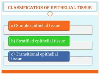
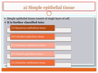
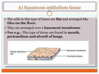
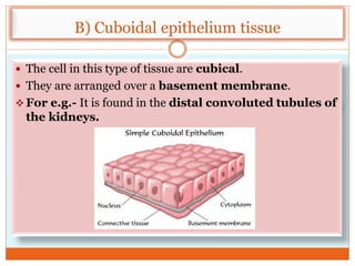


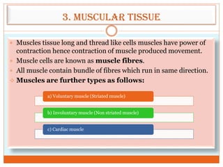


The document provides a comprehensive overview of animal tissues, detailing their classification into epithelial, connective, muscular, and nervous tissues. It explains the structure and function of each tissue type, highlighting specific subcategories, such as simple and stratified epithelial tissues, different connective tissues, and types of muscular and nervous tissues. The document emphasizes the roles that these tissues play in supporting, protecting, and facilitating various functions within multicellular organisms.