Download to read offline
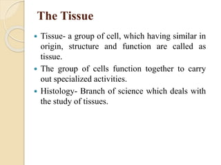
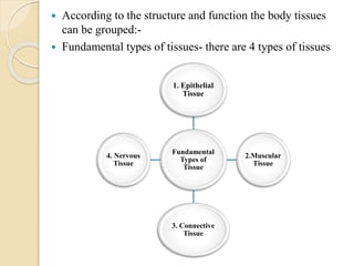
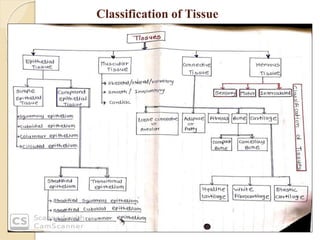
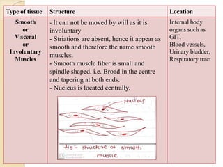
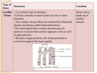
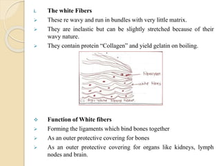
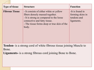
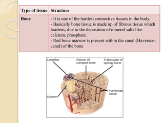
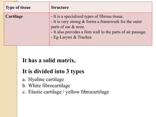
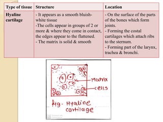
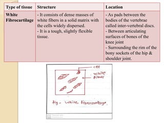
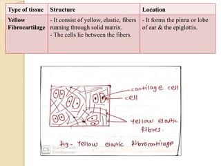
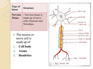


The document provides a comprehensive overview of tissue types in the human body, categorizing them into four fundamental types: epithelial, muscular, connective, and nervous tissue. Each type is further described with subcategories, functions, and examples of locations in the body. It also covers the components of nervous tissue and the significance of body fluids in metabolic processes.


