Download as PDF, PPTX
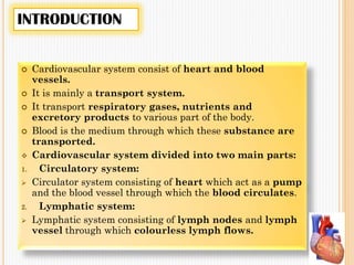
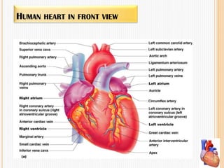
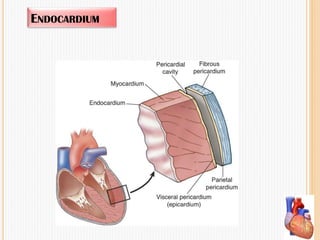
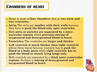
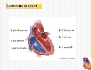
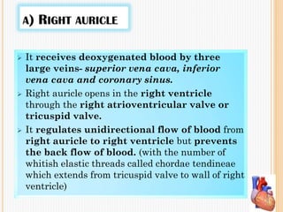
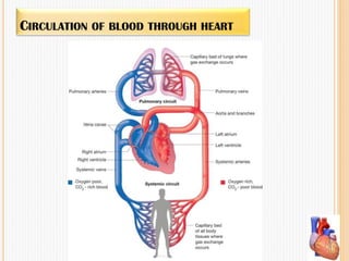

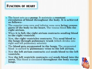


The cardiovascular system comprises the heart and blood vessels, functioning primarily as a transport system for gases, nutrients, and waste. The heart, a muscular organ, has four chambers (two atria and two ventricles) and is surrounded by three tissue layers, ensuring unidirectional blood flow through valves while facilitating both pulmonary and systemic circulation. Key concepts include the cardiac cycle, heart sounds, ECG recordings, and potential disorders affecting heart rate and blood pressure.