Downloaded 11 times


![Brain
It is the main controlling centre of our body. It is the
major part of the CNS.
It is present in a cavity called skull.
It is surrounded by bony box-cranium.
It is covered with membranes called meninges.
The fluid present in vacant spaces and between membranes
is called CSF (Cerebrospinal Fluid). [which act as cushion]
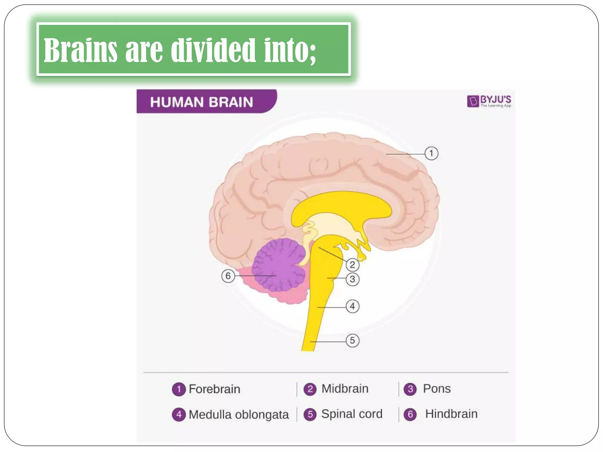
It is divided into:
1. Fore brain
2. Mid brain
3. Hind brain](https://image.slidesharecdn.com/controlandcoordinationinhumans-200727190558/75/Control-and-coordination-in-humans-22-2048.jpg)


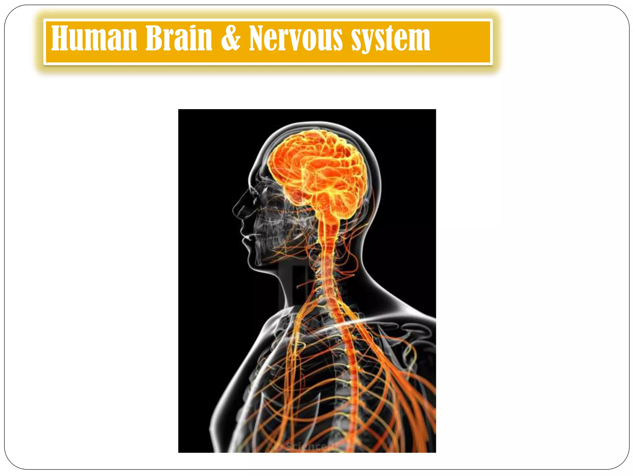
The document discusses the human brain and nervous system, detailing the structures and functions of neurons, including types of nerves and how impulses are conducted through synapses. It emphasizes the importance of the central nervous system, including the brain's different parts and their roles in control and coordination, as well as the concept of reflex actions. Additionally, it touches on the spinal cord's structure and function, alongside the method of recording brain activity using EEG.