Download as PDF, PPTX
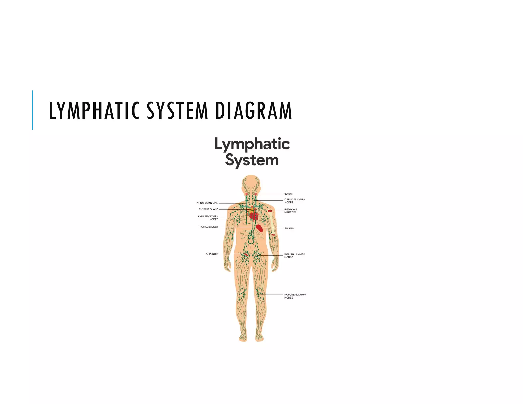


The document provides a detailed overview of the lymphatic system, including its components such as lymph, lymph vessels, and lymph nodes, as well as their functions in filtering infection and returning excess fluid to circulation. It describes the structure and functions of specific organs within the lymphatic system, including the spleen, thymus, and tonsils, highlighting their roles in immune response and blood cell production. Additionally, it covers the terminology related to lymph ducts and the significance of Peyer's patches in intestinal immunity.


![Lymphatic system ppt[1].pptx .](https://cdn.slidesharecdn.com/ss_thumbnails/lymphaticsystemppt1-250112055350-023a9ad6-thumbnail.jpg?width=640&height=640&fit=bounds)


