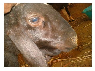
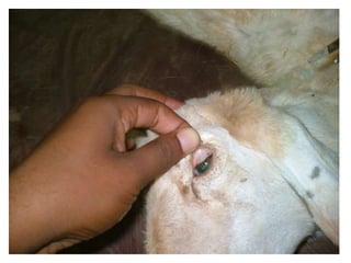
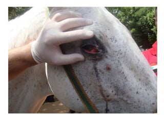
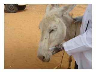
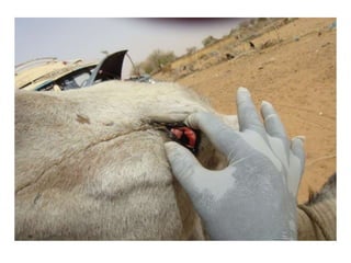
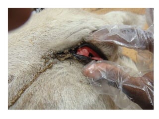
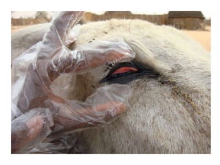
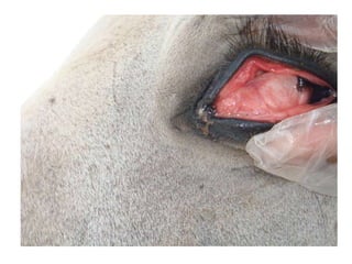
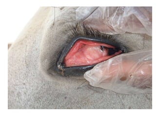
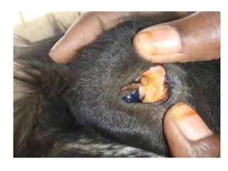
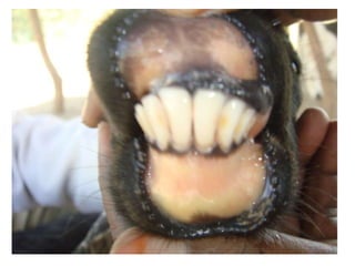
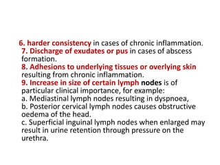
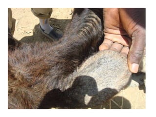
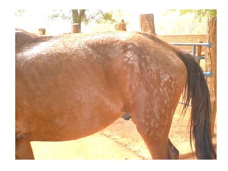
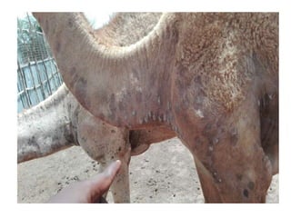
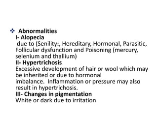
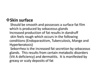
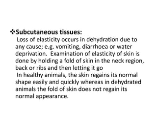
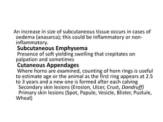
2nd , Mar , 2016
SHAFI’I. A. M. SHEIKH
It is important physical and chemical process
and gas exchange (inspiration and expiration).
It is voluntary and non-voluntary process,
based on movement of different reparatory
muscles(diaphragm, intercostal, sternal and
abdominal) and controlled by respiratory
center in medulla oblongata
Respiratory system consists of …. And lung
consists of bronchi, bronchioles and aceni (air
sacs)
The exchange of O2 and CO2 is done by simple
diffusion which depends on partial pressure of
membrane that separates aceni and blood
Respiratory rate, rhythm, type and quality
should always be noted when assessing the
animal’s health
In rest position we can observe respiration by
putting hands on flank region or accurately by
auscultation
Rate is the number of respiratory cycles
(Inspiration Expiration pause) in one minute. It
will increase (Polypnoea) or decrease
(Oligopnoea)
Polypnoea is due to physiological (fear,
excitation, irritation, effort, heat, humidity) or
pathological (fever, anaemia, res. Dis) causes
Oligopnea is due to lesions of CNS,
hydrocephalus, uremia and Res. Passages
stenos is
Hyperpnoea is the increased pulmonary
ventilation (increased respiratory rate with or
without increase in the amplitude or width of
the movement
Types of respiration:
1- costo-abdominal: this type of respiration is
seen in equines
2- abdominal respiration: this type of respiration
is evident in cattle and small ruminants
3-costal respirations This type of respiration is
evident in dogs and cats and birds
Changes in respiration:
Wholly costal is due to: diaphragmatic problems,
bloat and lung and pleurisy problems
Wholly abdominal is due to: Pleuritis
Double expiration is due to: Chronic alveolar
emphysema
Respiratory rhythm
It is regular or irregular res. Cycles
Expiration is long in pulmonary emphysema
Pause is canceled in sick animals and long in
toxaemia, and heart and kineys diseases
Examination of Mucous Membranes
Has great clinical importance as it reflects the
general condition of the animal as well as certain
disease conditions (e.g. jaundice in case of liver
diseases).
The visible mucous membranes are oral, nasal,
conjunctival and vaginal.
Changes in color may result from disturbance in
oxidation process of the blood, disturbances in
blood circulation or of certain other disease
conditions.
 Noticeable changes in mucous membranes
include changes in color, presence of exudates
and swelling.
1- Changes in color:
The normal color of the visible mucous
membranes varies between species. In
general, different shades of pink (light pink,
pink or rosy red) are considered normal. The
following changes occur in various diseases
1. Pale mucous membranes
a. Moist pale :
due to anaemia
b- dry pale:
due to shock
2. Congested mucous membranes
Mucous membrane is redish in color due to fever
3. Yellowish or icteric mucous membranes
Icterus of jaundice occurs due to increase of
blood bilirubin concentration (blood parasites,
leptospirosis, hepatitis)
4. Brick red mucous membranes
Due to heavy metals intoxication
5. Cyanosed mucous membranes
Bluish discoloration of visible mucous
membranes resulting from presence of
reduced haemoglobin in blood capillaries.
2- Swelling of mucous membranes
Inflammation of mucous membranes results in its
swelling; in which case the mucous membranes
may also be hot and tender (i.e. showing cardinal
signs of inflammation).
Marked swelling of conjunctival mucous
membranes is characteristic of equine
influenza. A slight degree of swelling is noticed in
contagious pleuropneumonia of horse and cattle
plague, anthrax and fowl diphtheria
3- Presence of exudate
Escape of serous fluid and fibrinogen from
blood vessels, which also contains
desquamated epithelial cells, leukocytes and
erythrocytes
Examination of Superficial Lymph
Nodes
 Lymph Nodes to be examined:
1- Sub-maxillary LN
2- Pharyngearl LN: a- para-phayngeal (Sub-
parotid) b- Retropharyngeal (Supra-pharyngal)
3- Cervical LN (Anterior, Meddile, Posterior)
4- Pre-scapular LN 5- Popliteal LN 6- Supra-
mammary
The examination includes:
Size, tenderness, consistency, lobulation,
temperature of overlying skin, discharge if
present and adhesions.
A normal lymph node varies in size depending on
animal species and age. It is commonly larger in
younger animals; of firm consistency. Small
lymph nodes may appear to be smooth;
lobulation can be palpated in large lymph
nodes. Normally, lymph nodes should be freely
movable under the skin or surrounding tissues
Abnormalities:
1. Swelling is caused by:
a- Acute local inflammation b- Systemic infectious
diseases c- Chronic inflammation d- Infection of
the lymph node itself e- Lymphomas.
2. Tenderness: Acute infection and inflammation
causes pain on palpation.
3. Loss of lobulation in cases of acute infection.
4. Hotness due to acute inflammation.
5. Abnormal consistency, fluctuation in cases of
suppuration
6. harder consistency in cases of chronic inflammation.
7. Discharge of exudates or pus in cases of abscess
formation.
8. Adhesions to underlying tissues or overlying skin
resulting from chronic inflammation.
9. Increase in size of certain lymph nodes is of
particular clinical importance, for example:
a. Mediastinal lymph nodes resulting in dyspnoea,
b. Posterior cervical lymph nodes causes obstructive
oedema of the head.
c. Superficial inguinal lymph nodes when enlarged may
result in urine retention through pressure on the
urethra.
Examination of Skin and Associated
Structures
it is considered the mirror of health
Bad management and inappropriate diet are
quickly reflected on the skin condition
Aspects of examination of skin include: condition
of hair, surface of skin, subcutaneous tissue
elasticity and examination of horny structures
A shiny coat and well-pigmented hide is an
indicator of optimum conditions of management,
feeding and productivity
Hair
We examine consistency, hair color and detachment
some animal breeds have naturally smooth or naturally
curly hair. Loss of luster and dryness reflects bad
management and/or nutritional deficiency
Hair color is also breed-specific; in an animal with a
natural dark hair color, deficient diet (qualitative or
quantitative) may result in paleness of hair; this is
termed ?hunger hair?. Easily detached hair is also a
sign of nutritional or trace element deficiency
Presence of parasites such as lice, fleas, mites or ticks
result in Alopecia (areas of hair loss) and itching
 Abnormalities
I- Alopecia
due to (Senility:, Hereditary, Hormonal, Parasitic,
Follicular dysfunction and Poisoning (mercury,
selenium and thallium)
II- Hypertrichosis
Excessive development of hair or wool which may
be inherited or due to hormonal
imbalance. Inflammation or pressure may also
result in hypertrichosis.
III- Changes in pigmentation
White or dark due to irritation
Skin surface
Should be smooth and possesses a surface fat film
which is produced by sebaceous glands
Increased production of fat results in dandruff
skin feels rough which occurs in the following
conditions (Endoparasitism, Tuberculosis, Mange and
Hyperkeratosis)
Seborrhea is the increased fat secretion by sebaceous
glands. This results from certain metabolic disorders
(Vit A deficiency) and dermatitis. It is manifested by
greasy or scaly deposits of fat
Subcutaneous tissues:
Loss of elasticity occurs in dehydration due to
any cause; e.g. vomiting, diarrhoea or water
deprivation. Examination of elasticity of skin is
done by holding a fold of skin in the neck region,
back or ribs and then letting it go
In healthy animals, the skin regains its normal
shape easily and quickly whereas in dehydrated
animals the fold of skin does not regain its
normal appearance.
An increase in size of subcutaneous tissue occurs in cases of
oedema (anasarca); this could be inflammatory or non-
inflammatory.
Subcutaneous Emphysema
Presence of soft yielding swelling that crepitates on
palpation and sometimes
Cutaneous Appendages
Where horns are examined, counting of horn rings is useful
to estimate age or the animal as the first ring appears at 2.5
to 3 years and a new one is formed after each calving
Secondary skin lesions (Erosion, Ulcer, Crust, Dandruff)
Primary skin lesions (Spot, Papule, Vesicle, Blister, Pustule,
Wheal)
Examination of Musculo-Skeletal
System
The primary function of the musculskeletal
system is to support the body in various ways
to insure normal location and posture
The components of the musculoskeletal
system are muscles, tendons, bones and joints
Muscles
Superficial muscles are examined by inspection
and palpation. The main clinical affections of
muscles are:
1- Atrophy 2- hypertrophy 3- Increased tone of
musculature a. Continuous (tonic) as in
tetanus b. Tremors
Animal parameters lecture

Animal parameters lecture

  • 1.
    2nd , Mar, 2016 SHAFI’I. A. M. SHEIKH
  • 2.
    It is importantphysical and chemical process and gas exchange (inspiration and expiration). It is voluntary and non-voluntary process, based on movement of different reparatory muscles(diaphragm, intercostal, sternal and abdominal) and controlled by respiratory center in medulla oblongata
  • 3.
    Respiratory system consistsof …. And lung consists of bronchi, bronchioles and aceni (air sacs) The exchange of O2 and CO2 is done by simple diffusion which depends on partial pressure of membrane that separates aceni and blood
  • 4.
    Respiratory rate, rhythm,type and quality should always be noted when assessing the animal’s health In rest position we can observe respiration by putting hands on flank region or accurately by auscultation Rate is the number of respiratory cycles (Inspiration Expiration pause) in one minute. It will increase (Polypnoea) or decrease (Oligopnoea)
  • 5.
    Polypnoea is dueto physiological (fear, excitation, irritation, effort, heat, humidity) or pathological (fever, anaemia, res. Dis) causes Oligopnea is due to lesions of CNS, hydrocephalus, uremia and Res. Passages stenos is Hyperpnoea is the increased pulmonary ventilation (increased respiratory rate with or without increase in the amplitude or width of the movement
  • 6.
    Types of respiration: 1-costo-abdominal: this type of respiration is seen in equines 2- abdominal respiration: this type of respiration is evident in cattle and small ruminants 3-costal respirations This type of respiration is evident in dogs and cats and birds Changes in respiration: Wholly costal is due to: diaphragmatic problems, bloat and lung and pleurisy problems
  • 7.
    Wholly abdominal isdue to: Pleuritis Double expiration is due to: Chronic alveolar emphysema Respiratory rhythm It is regular or irregular res. Cycles Expiration is long in pulmonary emphysema Pause is canceled in sick animals and long in toxaemia, and heart and kineys diseases
  • 8.
    Examination of MucousMembranes Has great clinical importance as it reflects the general condition of the animal as well as certain disease conditions (e.g. jaundice in case of liver diseases). The visible mucous membranes are oral, nasal, conjunctival and vaginal. Changes in color may result from disturbance in oxidation process of the blood, disturbances in blood circulation or of certain other disease conditions.
  • 9.
     Noticeable changesin mucous membranes include changes in color, presence of exudates and swelling. 1- Changes in color: The normal color of the visible mucous membranes varies between species. In general, different shades of pink (light pink, pink or rosy red) are considered normal. The following changes occur in various diseases
  • 10.
    1. Pale mucousmembranes a. Moist pale : due to anaemia b- dry pale: due to shock 2. Congested mucous membranes Mucous membrane is redish in color due to fever 3. Yellowish or icteric mucous membranes Icterus of jaundice occurs due to increase of blood bilirubin concentration (blood parasites, leptospirosis, hepatitis)
  • 11.
    4. Brick redmucous membranes Due to heavy metals intoxication 5. Cyanosed mucous membranes Bluish discoloration of visible mucous membranes resulting from presence of reduced haemoglobin in blood capillaries.
  • 12.
    2- Swelling ofmucous membranes Inflammation of mucous membranes results in its swelling; in which case the mucous membranes may also be hot and tender (i.e. showing cardinal signs of inflammation). Marked swelling of conjunctival mucous membranes is characteristic of equine influenza. A slight degree of swelling is noticed in contagious pleuropneumonia of horse and cattle plague, anthrax and fowl diphtheria
  • 13.
    3- Presence ofexudate Escape of serous fluid and fibrinogen from blood vessels, which also contains desquamated epithelial cells, leukocytes and erythrocytes
  • 27.
    Examination of SuperficialLymph Nodes  Lymph Nodes to be examined: 1- Sub-maxillary LN 2- Pharyngearl LN: a- para-phayngeal (Sub- parotid) b- Retropharyngeal (Supra-pharyngal) 3- Cervical LN (Anterior, Meddile, Posterior) 4- Pre-scapular LN 5- Popliteal LN 6- Supra- mammary
  • 31.
    The examination includes: Size,tenderness, consistency, lobulation, temperature of overlying skin, discharge if present and adhesions. A normal lymph node varies in size depending on animal species and age. It is commonly larger in younger animals; of firm consistency. Small lymph nodes may appear to be smooth; lobulation can be palpated in large lymph nodes. Normally, lymph nodes should be freely movable under the skin or surrounding tissues
  • 32.
    Abnormalities: 1. Swelling iscaused by: a- Acute local inflammation b- Systemic infectious diseases c- Chronic inflammation d- Infection of the lymph node itself e- Lymphomas. 2. Tenderness: Acute infection and inflammation causes pain on palpation. 3. Loss of lobulation in cases of acute infection. 4. Hotness due to acute inflammation. 5. Abnormal consistency, fluctuation in cases of suppuration
  • 35.
    6. harder consistencyin cases of chronic inflammation. 7. Discharge of exudates or pus in cases of abscess formation. 8. Adhesions to underlying tissues or overlying skin resulting from chronic inflammation. 9. Increase in size of certain lymph nodes is of particular clinical importance, for example: a. Mediastinal lymph nodes resulting in dyspnoea, b. Posterior cervical lymph nodes causes obstructive oedema of the head. c. Superficial inguinal lymph nodes when enlarged may result in urine retention through pressure on the urethra.
  • 37.
    Examination of Skinand Associated Structures it is considered the mirror of health Bad management and inappropriate diet are quickly reflected on the skin condition Aspects of examination of skin include: condition of hair, surface of skin, subcutaneous tissue elasticity and examination of horny structures A shiny coat and well-pigmented hide is an indicator of optimum conditions of management, feeding and productivity
  • 38.
    Hair We examine consistency,hair color and detachment some animal breeds have naturally smooth or naturally curly hair. Loss of luster and dryness reflects bad management and/or nutritional deficiency Hair color is also breed-specific; in an animal with a natural dark hair color, deficient diet (qualitative or quantitative) may result in paleness of hair; this is termed ?hunger hair?. Easily detached hair is also a sign of nutritional or trace element deficiency Presence of parasites such as lice, fleas, mites or ticks result in Alopecia (areas of hair loss) and itching
  • 45.
     Abnormalities I- Alopecia dueto (Senility:, Hereditary, Hormonal, Parasitic, Follicular dysfunction and Poisoning (mercury, selenium and thallium) II- Hypertrichosis Excessive development of hair or wool which may be inherited or due to hormonal imbalance. Inflammation or pressure may also result in hypertrichosis. III- Changes in pigmentation White or dark due to irritation
  • 46.
    Skin surface Should besmooth and possesses a surface fat film which is produced by sebaceous glands Increased production of fat results in dandruff skin feels rough which occurs in the following conditions (Endoparasitism, Tuberculosis, Mange and Hyperkeratosis) Seborrhea is the increased fat secretion by sebaceous glands. This results from certain metabolic disorders (Vit A deficiency) and dermatitis. It is manifested by greasy or scaly deposits of fat
  • 47.
    Subcutaneous tissues: Loss ofelasticity occurs in dehydration due to any cause; e.g. vomiting, diarrhoea or water deprivation. Examination of elasticity of skin is done by holding a fold of skin in the neck region, back or ribs and then letting it go In healthy animals, the skin regains its normal shape easily and quickly whereas in dehydrated animals the fold of skin does not regain its normal appearance.
  • 48.
    An increase insize of subcutaneous tissue occurs in cases of oedema (anasarca); this could be inflammatory or non- inflammatory. Subcutaneous Emphysema Presence of soft yielding swelling that crepitates on palpation and sometimes Cutaneous Appendages Where horns are examined, counting of horn rings is useful to estimate age or the animal as the first ring appears at 2.5 to 3 years and a new one is formed after each calving Secondary skin lesions (Erosion, Ulcer, Crust, Dandruff) Primary skin lesions (Spot, Papule, Vesicle, Blister, Pustule, Wheal)
  • 49.
    Examination of Musculo-Skeletal System Theprimary function of the musculskeletal system is to support the body in various ways to insure normal location and posture The components of the musculoskeletal system are muscles, tendons, bones and joints
  • 50.
    Muscles Superficial muscles areexamined by inspection and palpation. The main clinical affections of muscles are: 1- Atrophy 2- hypertrophy 3- Increased tone of musculature a. Continuous (tonic) as in tetanus b. Tremors