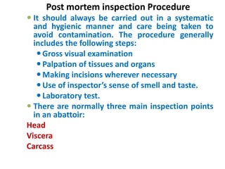
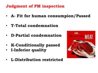
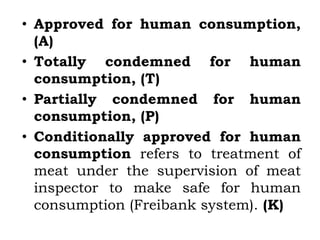
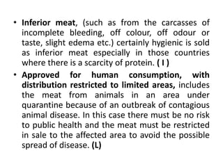
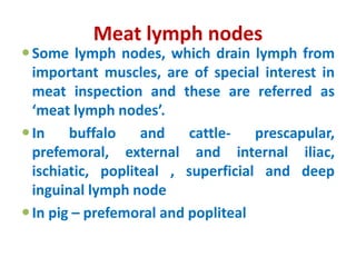
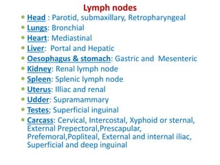
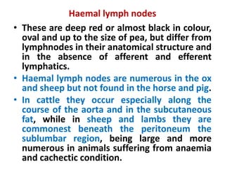
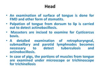
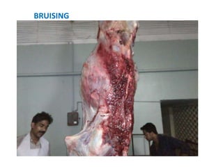
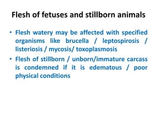
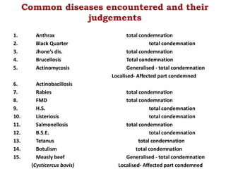
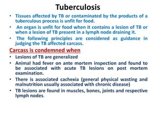
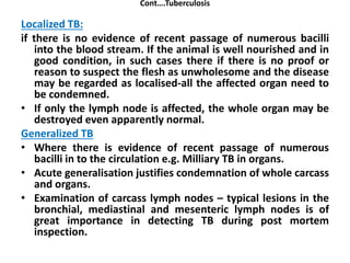
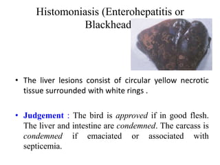
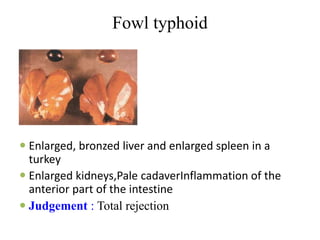
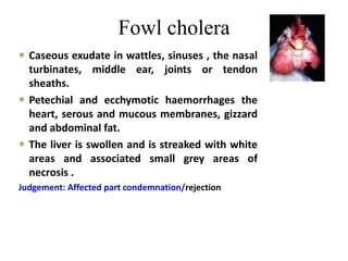
Dr. Sethulekshmi C
Assistant Professor, Dept. of
Veterinary Public Health, CVASM
POST-MORTEM INSPECTION
 Post-Mortem Inspection is the systematic
examination of dressed carcasses, their organs
including blood in a hygienic manner immediately
after slaughter in the presence of adequate light by a
meat inspector with the objective of providing
wholesome meat to the consumers
 Many diseases and abnormal conditions not
detectable on ante-mortem necessitate a careful
postmortem inspection.
 Conducted by a qualified veterinarian and may
be assisted by a lay inspector for recording.
 A meat inspector can efficiently examine 75
cattle or 250 calves or 200 pigs or 400 sheep per
day (8 hours) in an abattoir
Objectives
 To detect and eliminate abnormalities
from the carcass and organs including
contamination, thus ensuring that only meat
fit for human consumption is passed for food
 To check the efficiency of slaughter and
carcass dressing
 To diagnose disease conditions for control
purposes
 To ensure that carcasses, parts or organs
marked unfit for human consumption are
properly disposed off or destroyed
Facilities required
 Inspection points should have sufficient and well
distributed light of 540 lux or 50 foot candles
 Adequate hand washing units with supply of hot
and cold running water, liquid soap and towels.
 Meat inspection knives.
 Sterilizers for complete immersion of knives, saws,
cleavers facilities etc.
 Stamping ink—A solution containing 1 to 2% of
fuchsine in acetic acid is used as stamping ink for
carcasses.
Methyl violet 35 g, Cane sugar 450 g, Ethyl alcohol
1363 ml and water 1636 ml. Sugar is first dissolved
in water and methyl violet is added at the end.
General Principles
 Identification of carcass and the offal of the
corresponding animal.
 As a thumb rule, viscera and head should remain
identifiable with the carcass until the inspection is
completed.
 Viewing, palpation, incision and olfactory
techniques.
 Classify the lesions into acute or chronic
 Establish whether the condition is localized or
generalized
 Co-ordinate all observations of Ante mortem and
post mortem findings to make a final decision
 Submit samples to laboratory for diagnostic
support
Post mortem inspection Procedure
 It should always be carried out in a systematic
and hygienic manner and care being taken to
avoid contamination. The procedure generally
includes the following steps:
Gross visual examination
Palpation of tissues and organs
Making incisions wherever necessary
Use of inspector’s sense of smell and taste.
Laboratory test.
 There are normally three main inspection points
in an abattoir:
Head
Viscera
Carcass
Judgment of PM inspection
Generally four option may come
• A- Fit for human consumpion/Passed
• T-Total condemnation
• D-Partial condemnation
• K-Conditionally passed
• I-Inferior quality
• L-Distribution restricted
• Approved for human consumption,
(A)
• Totally condemned for human
consumption, (T)
• Partially condemned for human
consumption, (P)
• Conditionally approved for human
consumption refers to treatment of
meat under the supervision of meat
inspector to make safe for human
consumption (Freibank system). (K)
• Inferior meat, (such as from the carcasses of
incomplete bleeding, off colour, off odour or
taste, slight edema etc.) certainly hygienic is sold
as inferior meat especially in those countries
where there is a scarcity of protein. ( I )
• Approved for human consumption, with
distribution restricted to limited areas, includes
the meat from animals in an area under
quarantine because of an outbreak of contagious
animal disease. In this case there must be no risk
to public health and the meat must be restricted
in sale to the affected area to avoid the possible
spread of disease. (L)
Meat lymph nodes
Some lymph nodes, which drain lymph from
important muscles, are of special interest in
meat inspection and these are referred as
‘meat lymph nodes’.
In buffalo and cattle- prescapular,
prefemoral, external and internal iliac,
ischiatic, popliteal , superficial and deep
inguinal lymph node
In pig – prefemoral and popliteal
Lymph nodes
 Head : Parotid, submaxillary, Retropharyngeal
 Lungs: Bronchial
 Heart: Mediastinal
 Liver: Portal and Hepatic
 Oesophagus & stomach: Gastric and Mesenteric
 Kidney: Renal lymph node
 Spleen: Splenic lymph node
 Uterus: Illiac and renal
 Udder: Supramammary
 Testes; Superficial inguinal
 Carcass: Cervical, Intercostal, Xyphoid or sternal,
External Prepectoral,Prescapular,
Prefemoral,Popliteal, External and internal iliac,
Superficial and deep inguinal
Haemal lymph nodes
• These are deep red or almost black in colour,
oval and up to the size of pea, but differ from
lymphnodes in their anatomical structure and
in the absence of afferent and efferent
lymphatics.
• Haemal lymph nodes are numerous in the ox
and sheep but not found in the horse and pig.
• In cattle they occur especially along the
course of the aorta and in the subcutaneous
fat, while in sheep and lambs they are
commonest beneath the peritoneum the
sublumbar region, being large and more
numerous in animals suffering from anaemia
and cachectic condition.
Head
• An examination of surface of tongue is done for
FMD and other form of stomatits.
• Palpation of tongue from dorsum to lip is carried
out to detect actinobacillosis.
• Masseters are incised to examine for Cysticercus
bovis.
• A detailed examination of retropharyngeal,
submaxillary and parotid lymphnodes becomes
necessary to detect tuberculosis and
actinobacillosis.
• In case of pigs, the portions of muscles from tongue
are examined under microscope or trichinoscope
for trichinellosis
Viscera
• All viscera should be inspected as they are
removed from the carcass.
• Every organ and associated lymphnodes is
examined.
• In case of any abnormal condition, the organs are
incised not to contaminate other organs or
carcass parts
Lungs
 Visual inspection followed by palpation is carried out
for evidence of pleurisy, pneumonia, tuberculosis,
fascioliasis and hydatid cysts.
 Bronchial and mediastinal lymphnodes should be
incised to detect tuberculosis
Heart
• Pericardium is opened and looked for tuberculosis
pericarditis.
• Heart is incised to detect petechial haemorrhages or
cysts.
Liver
The surface and substance of liver is
examined for fatty changes, actinobacillosis,
and abscesses, telangiectasis, parasitic
infections like cysticercus bovis, fascioliasis,
hydatid cysts or larval stage of
oesophagostomum.
Kidney
Renal lymphnodes and adrenal glands should
be examined before the removal of kidneys
Spleen
• The surface and substance of spleen is
inspected for anthrax, tuberculosis or
presence of artifacts.
Stomach and intestine
• The serous surface of these organs could
indicate tuberculosis or actinobacillosis while
anterior part of reticulum could show
penetration of a foreign body.
• Mesenteric Iliac lymph node is incised as a
routine to look for tuberculosis.
Other viscera
• Urinary bladder: The outer and inner surfaces
of urinary bladder are observed for diseased
condition.
• Uterus and ovaries: These are opened for
examination of septic conditions.
• Udder: This is examined for septic mastitis or
abscesses. Supra mammary lymph nodes are
incised for evidence of abscesses or
tuberculosis.
• Testicles: The outer surface, substance and
superficial inguinal lymphnodes are inspected.
Postmortem Inspection of Sheep and Goat
Carcasses
The cutting of head, heart, pulmonary lymph
nodes and body lymph nodes should be
carried out only in cases of suspicion.
Carcass is examined for satisfactory bleeding,
liver for fascioliasis and lungs for parasitic
infections like nematodes or hydatid cysts etc.
Postmortem Inspection of Swine
Carcasses
• Skin lesions serve as important diagnostic
feature of erysipelas, swine fever and urticaria,
feet for abscess formation, udder for mastitis or
actinomycosis and tail for necrosis.
• In places where Cysticercus cellulosae is
common, the external masseter muscles and
root of tongue are incised.
• If trichinosis is known to occur in a particular
area or suspected, muscle sampling should be
done for appropriate examination.
• Visceral organs should be examined for
pneumonia, pericarditis, peritonitis and also
submaxillary lymph nodes for tuberculosis.
Carcass
 1. State of nutrition Poorness/ Emaciation
 2. Evidence of bruising or discolouration
 Anthracosis
 Bile Staining
 Carotene Pigmentation
 DFD
 Jaundice or icterus
 Melanosis
 PSE
 3. Local or general edema
 4. Efficiency of bleeding
 5. Swelling deformities or other abnormalities of
bones or musculature.
 6. Age and sex of the animal from which the
carcass was derived.
 7. Any abnormal odour (Diet,Drugs
administered,Sexual odour,Acetonemia,Bone
taint,Abscess,Putrefaction)
 8. The condition of the pleura and peritoneum
 9. Any other evidence of abnormality
 10. Carcass lymph nodes
Emaciation
It is a pathological condition, which develops
during the course of an illness, which, either
lowers the food intake or increases the
metabolic rate of the animal.
May be due to disease like JD, TB, muscular
tissue atrophies, and parasite or simply due
to prolonged starvation.
Characterized by
Reduced size of organ, loss of body fat, jelly
like consistency.
Carcass do not set well
Muscle pits on pressure
BRUISING
Odema
• Hydrothorax (Thoracic cavity)
• Ascites (Abdominal cavity) ------- Less serious
• Anasarca (subcutaneous / connective tissue)---
More serious
• Detain the carcass for 12 hrs.
• Anasarca if accompanied by emaciation needs total
condemnation
Abscesses
• Injection, wound, jowl abscess or organ
specific lesions.
• Local condemnation
Icterus / jaundice
• It is a symptom of presence of bilirubin (orange / yellow)
and biliverdin (green) bile pigments in the circulating blood.
It can be a obstructive jaundice or hemolytic jaundice
Yellow coloration of fatty tissues, serous, connective tissue,
nerve trunks, endothelial linings of arteries such as iliac,
brachial & femoral, cortex of kidney, sclerotic membrane of
eyes, liver, tendons and cartilaginous extremities of long
bones.
Carcass may render repugnant odour.
• Test –Remington Fowrie test - for presence of abnormal
odour & bile pigments.
• Distinguish jaundice with carotene in cattle or xanthophyll in
sheep (sheep are unable to oxidize xanthophylls in its food
• Carcass may be detained for 24 hrs to note if this color fades
due to action of the muscular enzymes..
SEPTICAEMIA / VIRAEMIA/TOXEMIA
 Serious condition of Toxemia, fever and the presence of
large number of pathogenic bacteria in blood stream.
 Characterized by high fever
 Petechial haemorrhages in myocardium, liver, kidney,
enlarged edematous lymph nodes, cloudy swelling of
liver, kidney, blood stained serous exudates in thoracic
cavity abdominal cavity.
 Meat become alkaline, soft, dark in color, sweetish
repugnant odour.
 Total condemnation for two reasons
 Poor keeping quality
 Pathogenic organism or toxin.
Anaemia
• Pale colour of the meat, watery blood
• Totally condemned if associated with poor
condition or haematoma
• Conditional approval in less severe cases.
Flesh of fetuses and stillborn animals
• Flesh watery may be affected with specified
organisms like brucella / leptospirosis /
listeriosis / mycosis/ toxoplasmosis
• Flesh of stillborn / unborn/immature carcass
is condemned if it is edematous / poor
physical conditions
Diseases judgements
1. Anthrax total condemnation
2. Black Quarter total condemnation
3. Jhone’s dis. total condemnation
4. Brucellosis Total condemnation
5. Actinomycosis Generalised - total condemnation
Localised- Affected part condemned
6. Actinobacillosis
7. Rabies total condemnation
8. FMD total condemnation
9. H.S. total condemnation
10. Listeriosis total condemnation
11. Salmonellosis total condemnation
12. B.S.E. total condemnation
13. Tetanus total condemnation
14. Botulism total condemnation
15. Measly beef Generalised - total condemnation
(Cysticercus bovis) Localised- Affected part condemned
Tuberculosis
• Tissues affected by TB or contaminated by the products of a
tuberculous process is unfit for food.
• An organ is unfit for food when it contains a lesion of TB or
when a lesion of TB present in a lymph node draining it.
• The following principles are considered as guidance in
judging the TB affected carcass.
Carcass is condemned when
• Lesions of TB are generalized
• Animal had fever on ante mortem inspection and found to
be associated with acute TB lesions on post mortem
examination.
• There is associated cachexia (general physical wasting and
malnutrition usually associated with chronic disease)
• TB lesions are found in muscles, bones, joints and respective
lymph nodes.
Cont….Tuberculosis
Localized TB:
if there is no evidence of recent passage of numerous bacilli
into the blood stream. If the animal is well nourished and in
good condition, in such cases there if there is no proof or
reason to suspect the flesh as unwholesome and the disease
may be regarded as localised-all the affected organ need to
be condemned.
• If only the lymph node is affected, the whole organ may be
destroyed even apparently normal.
Generalized TB
• Where there is evidence of recent passage of numerous
bacilli in to the circulation e.g. Milliary TB in organs.
• Acute generalisation justifies condemnation of whole carcass
and organs.
• Examination of carcass lymph nodes – typical lesions in the
bronchial, mediastinal and mesenteric lymph nodes is of
great importance in detecting TB during post mortem
inspection.
Brucellosis
• Total condemnation or heat treatment may be
appropriate, depending on local incidence and
economics.
• Confirmation: Cultural examination of blood or
lesion material (placenta, fetus, milk, and semen).
Anthrax
 The entire carcass including blood, hide, fat and all
intestinal organs must be destroyed along with the
contaminated animals and meat.
 Zoonotic importance
 The type of anthrax in man usually depends on the
mode of infection. It can be a cutaneous, pulmonary
or intestinal disease. Cutaneous form is common
and results form the handling of infected material.
 The knives, saws and other instruments should be boiled in
water for half an hour and thoroughly cleansed in a hot 5%
solution of NaOH or 10% formaldehyde or ethylene oxide in
plastic bags.
 The clothing must be disinfected with 10% formaldehyde.
 The abattoir premises, pen, trucks, cars and other areas are
disinfected with 10% NaOH or 5% formaldehyde.
 Hands / arms of the workers are washed with liquid soap
followed by hot water and immersed for about one minute in
an organic iodine solution or 1 ppm HgCl2, followed by
potable water rinse.
Swine erysipelas
Total condemnation of carcass in septicemic
form while in urticarial form affected tissues
are rejected and carcass is passed for food.
Pox disease
• Total condemnation of the carcass in acute
stage (poor bleeding) while in recovered
cases passed for food.
Foot and Mouth disease
• Total condemnation of the diseased carcass or subject to
heat treatment.
Cysticercus bovis
 Total condemnation of the carcass in cases of generalized
condition due to its zoonotic importance. In localized cases
affected tissue is removed and carcass is passed for food.
 Cooking temperature of above 60 ˚ C and salting for 21 days
kills the cyst.
 In offal stored at 6.5˚ C for not less than 3 weeks or - 10 ˚ C
for 2 weeks destroys the cyst
Cysticercus cellulosae
Total condemnation of carcass in generalized
cases
Hydatidosis
Carcass is passed for food after removal of
affected organs.
Trichinosis
Total condemnation and proper disposal of
the carcass is required due to its zoonotic
importance.
Sarcocyst
• In earlier times,the condemnation of the specific muscle or
tissue or carcass in generalized cases is only on the aesthetic
grounds.
• Three main species
• Sarcocystis cruzi
• S.Hominis…….microscopic……PATHOGENIC TO HUMANS
(Humans act as definite host)
• S.hirsuta….macroscopic…NON PATHOGENIC
Newcastle disease
• Heamorrhagic conjunctiva
– The mucosal lining of the proventriculus
haemorrhage
– Junction between the oesophagus and
proventriculus
– Bursal heamorrhage
• Judgement : Total rejection.
Mareks disease
• Sciatic nerve enlargement, nerves are 2–3 times their
normal size
• Judgement : Total rejection.
Coccidiosis
• Birds affected with severe coccidiosis associated
with emaciation or anaemia are condemned.
Otherwise, the carcass is approved, after
the condemnation of affected tissue.
Avian influenza
• Head and Neck edema,
• Comb and wattle Swollen and cyanotic .
• Hemorrhages and edema around the heart
• Judgement : Total rejection and condemnation.
Histomoniasis (Enterohepatitis or
Blackhead)
• The liver lesions consist of circular yellow necrotic
tissue surrounded with white rings .
• Judgement : The bird is approved if in good flesh.
The liver and intestine are condemned. The carcass is
condemned if emaciated or associated with
septicemia.
Fowl typhoid
 Enlarged, bronzed liver and enlarged spleen in a
turkey
 Enlarged kidneys,Pale cadaverInflammation of the
anterior part of the intestine
 Judgement : Total rejection
Fowl cholera
 Caseous exudate in wattles, sinuses , the nasal
turbinates, middle ear, joints or tendon
sheaths.
 Petechial and ecchymotic haemorrhages the
heart, serous and mucous membranes, gizzard
and abdominal fat.
 The liver is swollen and is streaked with white
areas and associated small grey areas of
necrosis .
Judgement: Affected part condemnation/rejection
Rendering
• Rendering has historically been defined as separation of fat
from animal tissues by the application of heat
• Rendering of animal tissues involves conversion of carcasses
into three end products—
carcass meal (proteinaceous solids)
amelted fat
water
Using
• Mechanical processes (e.g., grinding, mixing, pressing,
centrifuging)
• Thermal processes (e.g., cooking, evaporating, and
drying), and sometimes chemical processes (e.g., solvent
extraction)..
Raw materials for rendering
• Packinghouse by-products, such as organ fats, offal,
bones and blood
• Boning house material which consists of bones and
meat trimmings
• Meat market trimmings including adipose and inter-
muscular fats, bone, cartilage and meat trimmings
• Restaurant greases: Recovered cooking oils (these
are processed and stored separately)
• Fallen animals
Wet rendering(Autoclave)
• The pre-ground raw materials are cooked a 140 oC in a closed
digester (autoclave) under a pressure of 40-60 psi by direct
injection of steam on to the raw materials for 3-4 hours
• pressure is slowly released and the liquid and the solid phases are
allowed to separate
• The fat, which floats on top, is drawn off and can be polished
further in disk centrifuge to remove residual water and fine
particles
• Residual solid material (Cracklings or Greaves- residual
meat fibers/ solid material left behind after all fat have
been extracted) is removed and pressed to remove
additional water (stick water) before being dried and
milled
Comparison of different rendering systems
Wet rendering
Adv:
Wet rendering produces good quality fat
Disadv:
 Requires long cooking times (3-4 hours)
 It is labour intensive
 Significant losses (up to 20% loss in stick
water) because soluble and suspended
materials are lost
 Energy intensive
Dry batch rendering
• The digester or cooker of this process is a double-jacketed
vessel
• No steam or hot water is added to the material, whereas the
steam is allowed into the outer jacket
• The raw material is first ground to less than 2.5 cm and batch
fed to the cooker
• Steam filled agitator improves heat transfer
• The end point temperature is often 120-140o C and the
resident time extends up to 3 hours
• .The cooker contents are then discharged into a percolating
tank to remove free drain fat
• The solid material is pressed (continuous operation) or
centrifuged (batch process) to remove additional fat
• Then the solid materials are ground in a mill to make meal
Dry batch rendering
Adv:
• Little material is lost
• Heat recovery is possible from the vent steam
• Cooking, sterilization and drying can be carried out in
the same vessel
• Being a batch process, separate cookers can be set for
different raw materials
Disadv
• Produces darker tallow compared to WR or LTR
• High temperature used produces fines, which can pass
into the fat, degrading its quality
• Meal contains higher fat(10-16%) compared to LTR( 3-
8%)
• To produce good quality tallow, raw material must be
cut and washed, resulting in loss of fat and addition of water
Continuous low temperature rendering or
mechanical dewatering systems
• Uses either a dry or wet(steam injection) cooker
• Raw material minced, passed to low temp cooker called as
coagulator or pre-heater or melting section
• Material heated to 60-90 ˚C in short time (10-30 min)
• Cells break, liberate tallow
• Liquid tallow pressed out in a continuous screw press along
with water( equal in volume to that in raw material, or
higher if live steam is injected)
• Solids sent to a cooker/drier
• The liquid mixture sent to an evaporator or a centrifuge.
Evaporaor operates a low pressure, hence at temp much
less than 100 ˚ C
• Water, fat and solids removed
Continuous LTR
Adv:
• Raw material need not be washed
• The colour of paunch contents do not get
fixed on to tallow
• Fat in the meal is less (3-8% )compared to
high temp systems(Batch HTR- 10-16%)
Post mortem inspection in animals

Post mortem inspection in animals

  • 1.
    Dr. Sethulekshmi C AssistantProfessor, Dept. of Veterinary Public Health, CVASM
  • 2.
    POST-MORTEM INSPECTION  Post-MortemInspection is the systematic examination of dressed carcasses, their organs including blood in a hygienic manner immediately after slaughter in the presence of adequate light by a meat inspector with the objective of providing wholesome meat to the consumers  Many diseases and abnormal conditions not detectable on ante-mortem necessitate a careful postmortem inspection.  Conducted by a qualified veterinarian and may be assisted by a lay inspector for recording.  A meat inspector can efficiently examine 75 cattle or 250 calves or 200 pigs or 400 sheep per day (8 hours) in an abattoir
  • 3.
    Objectives  To detectand eliminate abnormalities from the carcass and organs including contamination, thus ensuring that only meat fit for human consumption is passed for food  To check the efficiency of slaughter and carcass dressing  To diagnose disease conditions for control purposes  To ensure that carcasses, parts or organs marked unfit for human consumption are properly disposed off or destroyed
  • 4.
    Facilities required  Inspectionpoints should have sufficient and well distributed light of 540 lux or 50 foot candles  Adequate hand washing units with supply of hot and cold running water, liquid soap and towels.  Meat inspection knives.  Sterilizers for complete immersion of knives, saws, cleavers facilities etc.  Stamping ink—A solution containing 1 to 2% of fuchsine in acetic acid is used as stamping ink for carcasses. Methyl violet 35 g, Cane sugar 450 g, Ethyl alcohol 1363 ml and water 1636 ml. Sugar is first dissolved in water and methyl violet is added at the end.
  • 5.
    General Principles  Identificationof carcass and the offal of the corresponding animal.  As a thumb rule, viscera and head should remain identifiable with the carcass until the inspection is completed.  Viewing, palpation, incision and olfactory techniques.  Classify the lesions into acute or chronic  Establish whether the condition is localized or generalized  Co-ordinate all observations of Ante mortem and post mortem findings to make a final decision  Submit samples to laboratory for diagnostic support
  • 6.
    Post mortem inspectionProcedure  It should always be carried out in a systematic and hygienic manner and care being taken to avoid contamination. The procedure generally includes the following steps: Gross visual examination Palpation of tissues and organs Making incisions wherever necessary Use of inspector’s sense of smell and taste. Laboratory test.  There are normally three main inspection points in an abattoir: Head Viscera Carcass
  • 7.
    Judgment of PMinspection Generally four option may come • A- Fit for human consumpion/Passed • T-Total condemnation • D-Partial condemnation • K-Conditionally passed • I-Inferior quality • L-Distribution restricted
  • 8.
    • Approved forhuman consumption, (A) • Totally condemned for human consumption, (T) • Partially condemned for human consumption, (P) • Conditionally approved for human consumption refers to treatment of meat under the supervision of meat inspector to make safe for human consumption (Freibank system). (K)
  • 9.
    • Inferior meat,(such as from the carcasses of incomplete bleeding, off colour, off odour or taste, slight edema etc.) certainly hygienic is sold as inferior meat especially in those countries where there is a scarcity of protein. ( I ) • Approved for human consumption, with distribution restricted to limited areas, includes the meat from animals in an area under quarantine because of an outbreak of contagious animal disease. In this case there must be no risk to public health and the meat must be restricted in sale to the affected area to avoid the possible spread of disease. (L)
  • 10.
    Meat lymph nodes Somelymph nodes, which drain lymph from important muscles, are of special interest in meat inspection and these are referred as ‘meat lymph nodes’. In buffalo and cattle- prescapular, prefemoral, external and internal iliac, ischiatic, popliteal , superficial and deep inguinal lymph node In pig – prefemoral and popliteal
  • 11.
    Lymph nodes  Head: Parotid, submaxillary, Retropharyngeal  Lungs: Bronchial  Heart: Mediastinal  Liver: Portal and Hepatic  Oesophagus & stomach: Gastric and Mesenteric  Kidney: Renal lymph node  Spleen: Splenic lymph node  Uterus: Illiac and renal  Udder: Supramammary  Testes; Superficial inguinal  Carcass: Cervical, Intercostal, Xyphoid or sternal, External Prepectoral,Prescapular, Prefemoral,Popliteal, External and internal iliac, Superficial and deep inguinal
  • 12.
    Haemal lymph nodes •These are deep red or almost black in colour, oval and up to the size of pea, but differ from lymphnodes in their anatomical structure and in the absence of afferent and efferent lymphatics. • Haemal lymph nodes are numerous in the ox and sheep but not found in the horse and pig. • In cattle they occur especially along the course of the aorta and in the subcutaneous fat, while in sheep and lambs they are commonest beneath the peritoneum the sublumbar region, being large and more numerous in animals suffering from anaemia and cachectic condition.
  • 13.
    Head • An examinationof surface of tongue is done for FMD and other form of stomatits. • Palpation of tongue from dorsum to lip is carried out to detect actinobacillosis. • Masseters are incised to examine for Cysticercus bovis. • A detailed examination of retropharyngeal, submaxillary and parotid lymphnodes becomes necessary to detect tuberculosis and actinobacillosis. • In case of pigs, the portions of muscles from tongue are examined under microscope or trichinoscope for trichinellosis
  • 14.
    Viscera • All viscerashould be inspected as they are removed from the carcass. • Every organ and associated lymphnodes is examined. • In case of any abnormal condition, the organs are incised not to contaminate other organs or carcass parts
  • 15.
    Lungs  Visual inspectionfollowed by palpation is carried out for evidence of pleurisy, pneumonia, tuberculosis, fascioliasis and hydatid cysts.  Bronchial and mediastinal lymphnodes should be incised to detect tuberculosis
  • 16.
    Heart • Pericardium isopened and looked for tuberculosis pericarditis. • Heart is incised to detect petechial haemorrhages or cysts.
  • 17.
    Liver The surface andsubstance of liver is examined for fatty changes, actinobacillosis, and abscesses, telangiectasis, parasitic infections like cysticercus bovis, fascioliasis, hydatid cysts or larval stage of oesophagostomum.
  • 19.
    Kidney Renal lymphnodes andadrenal glands should be examined before the removal of kidneys
  • 20.
    Spleen • The surfaceand substance of spleen is inspected for anthrax, tuberculosis or presence of artifacts.
  • 21.
    Stomach and intestine •The serous surface of these organs could indicate tuberculosis or actinobacillosis while anterior part of reticulum could show penetration of a foreign body. • Mesenteric Iliac lymph node is incised as a routine to look for tuberculosis.
  • 22.
    Other viscera • Urinarybladder: The outer and inner surfaces of urinary bladder are observed for diseased condition. • Uterus and ovaries: These are opened for examination of septic conditions. • Udder: This is examined for septic mastitis or abscesses. Supra mammary lymph nodes are incised for evidence of abscesses or tuberculosis. • Testicles: The outer surface, substance and superficial inguinal lymphnodes are inspected.
  • 23.
    Postmortem Inspection ofSheep and Goat Carcasses The cutting of head, heart, pulmonary lymph nodes and body lymph nodes should be carried out only in cases of suspicion. Carcass is examined for satisfactory bleeding, liver for fascioliasis and lungs for parasitic infections like nematodes or hydatid cysts etc.
  • 25.
    Postmortem Inspection ofSwine Carcasses • Skin lesions serve as important diagnostic feature of erysipelas, swine fever and urticaria, feet for abscess formation, udder for mastitis or actinomycosis and tail for necrosis. • In places where Cysticercus cellulosae is common, the external masseter muscles and root of tongue are incised. • If trichinosis is known to occur in a particular area or suspected, muscle sampling should be done for appropriate examination. • Visceral organs should be examined for pneumonia, pericarditis, peritonitis and also submaxillary lymph nodes for tuberculosis.
  • 26.
    Carcass  1. Stateof nutrition Poorness/ Emaciation  2. Evidence of bruising or discolouration  Anthracosis  Bile Staining  Carotene Pigmentation  DFD  Jaundice or icterus  Melanosis  PSE  3. Local or general edema  4. Efficiency of bleeding  5. Swelling deformities or other abnormalities of bones or musculature.  6. Age and sex of the animal from which the carcass was derived.  7. Any abnormal odour (Diet,Drugs administered,Sexual odour,Acetonemia,Bone taint,Abscess,Putrefaction)  8. The condition of the pleura and peritoneum  9. Any other evidence of abnormality  10. Carcass lymph nodes
  • 27.
    Emaciation It is apathological condition, which develops during the course of an illness, which, either lowers the food intake or increases the metabolic rate of the animal. May be due to disease like JD, TB, muscular tissue atrophies, and parasite or simply due to prolonged starvation. Characterized by Reduced size of organ, loss of body fat, jelly like consistency. Carcass do not set well Muscle pits on pressure
  • 28.
  • 29.
    Odema • Hydrothorax (Thoraciccavity) • Ascites (Abdominal cavity) ------- Less serious • Anasarca (subcutaneous / connective tissue)--- More serious • Detain the carcass for 12 hrs. • Anasarca if accompanied by emaciation needs total condemnation
  • 30.
    Abscesses • Injection, wound,jowl abscess or organ specific lesions. • Local condemnation
  • 31.
    Icterus / jaundice •It is a symptom of presence of bilirubin (orange / yellow) and biliverdin (green) bile pigments in the circulating blood. It can be a obstructive jaundice or hemolytic jaundice Yellow coloration of fatty tissues, serous, connective tissue, nerve trunks, endothelial linings of arteries such as iliac, brachial & femoral, cortex of kidney, sclerotic membrane of eyes, liver, tendons and cartilaginous extremities of long bones. Carcass may render repugnant odour. • Test –Remington Fowrie test - for presence of abnormal odour & bile pigments. • Distinguish jaundice with carotene in cattle or xanthophyll in sheep (sheep are unable to oxidize xanthophylls in its food • Carcass may be detained for 24 hrs to note if this color fades due to action of the muscular enzymes..
  • 32.
    SEPTICAEMIA / VIRAEMIA/TOXEMIA Serious condition of Toxemia, fever and the presence of large number of pathogenic bacteria in blood stream.  Characterized by high fever  Petechial haemorrhages in myocardium, liver, kidney, enlarged edematous lymph nodes, cloudy swelling of liver, kidney, blood stained serous exudates in thoracic cavity abdominal cavity.  Meat become alkaline, soft, dark in color, sweetish repugnant odour.  Total condemnation for two reasons  Poor keeping quality  Pathogenic organism or toxin.
  • 33.
    Anaemia • Pale colourof the meat, watery blood • Totally condemned if associated with poor condition or haematoma • Conditional approval in less severe cases.
  • 34.
    Flesh of fetusesand stillborn animals • Flesh watery may be affected with specified organisms like brucella / leptospirosis / listeriosis / mycosis/ toxoplasmosis • Flesh of stillborn / unborn/immature carcass is condemned if it is edematous / poor physical conditions
  • 35.
    Diseases judgements 1. Anthraxtotal condemnation 2. Black Quarter total condemnation 3. Jhone’s dis. total condemnation 4. Brucellosis Total condemnation 5. Actinomycosis Generalised - total condemnation Localised- Affected part condemned 6. Actinobacillosis 7. Rabies total condemnation 8. FMD total condemnation 9. H.S. total condemnation 10. Listeriosis total condemnation 11. Salmonellosis total condemnation 12. B.S.E. total condemnation 13. Tetanus total condemnation 14. Botulism total condemnation 15. Measly beef Generalised - total condemnation (Cysticercus bovis) Localised- Affected part condemned
  • 36.
    Tuberculosis • Tissues affectedby TB or contaminated by the products of a tuberculous process is unfit for food. • An organ is unfit for food when it contains a lesion of TB or when a lesion of TB present in a lymph node draining it. • The following principles are considered as guidance in judging the TB affected carcass. Carcass is condemned when • Lesions of TB are generalized • Animal had fever on ante mortem inspection and found to be associated with acute TB lesions on post mortem examination. • There is associated cachexia (general physical wasting and malnutrition usually associated with chronic disease) • TB lesions are found in muscles, bones, joints and respective lymph nodes.
  • 37.
    Cont….Tuberculosis Localized TB: if thereis no evidence of recent passage of numerous bacilli into the blood stream. If the animal is well nourished and in good condition, in such cases there if there is no proof or reason to suspect the flesh as unwholesome and the disease may be regarded as localised-all the affected organ need to be condemned. • If only the lymph node is affected, the whole organ may be destroyed even apparently normal. Generalized TB • Where there is evidence of recent passage of numerous bacilli in to the circulation e.g. Milliary TB in organs. • Acute generalisation justifies condemnation of whole carcass and organs. • Examination of carcass lymph nodes – typical lesions in the bronchial, mediastinal and mesenteric lymph nodes is of great importance in detecting TB during post mortem inspection.
  • 38.
    Brucellosis • Total condemnationor heat treatment may be appropriate, depending on local incidence and economics. • Confirmation: Cultural examination of blood or lesion material (placenta, fetus, milk, and semen).
  • 39.
    Anthrax  The entirecarcass including blood, hide, fat and all intestinal organs must be destroyed along with the contaminated animals and meat.  Zoonotic importance  The type of anthrax in man usually depends on the mode of infection. It can be a cutaneous, pulmonary or intestinal disease. Cutaneous form is common and results form the handling of infected material.  The knives, saws and other instruments should be boiled in water for half an hour and thoroughly cleansed in a hot 5% solution of NaOH or 10% formaldehyde or ethylene oxide in plastic bags.  The clothing must be disinfected with 10% formaldehyde.  The abattoir premises, pen, trucks, cars and other areas are disinfected with 10% NaOH or 5% formaldehyde.  Hands / arms of the workers are washed with liquid soap followed by hot water and immersed for about one minute in an organic iodine solution or 1 ppm HgCl2, followed by potable water rinse.
  • 40.
    Swine erysipelas Total condemnationof carcass in septicemic form while in urticarial form affected tissues are rejected and carcass is passed for food.
  • 41.
    Pox disease • Totalcondemnation of the carcass in acute stage (poor bleeding) while in recovered cases passed for food.
  • 42.
    Foot and Mouthdisease • Total condemnation of the diseased carcass or subject to heat treatment.
  • 43.
    Cysticercus bovis  Totalcondemnation of the carcass in cases of generalized condition due to its zoonotic importance. In localized cases affected tissue is removed and carcass is passed for food.  Cooking temperature of above 60 ˚ C and salting for 21 days kills the cyst.  In offal stored at 6.5˚ C for not less than 3 weeks or - 10 ˚ C for 2 weeks destroys the cyst
  • 44.
    Cysticercus cellulosae Total condemnationof carcass in generalized cases Hydatidosis Carcass is passed for food after removal of affected organs. Trichinosis Total condemnation and proper disposal of the carcass is required due to its zoonotic importance.
  • 45.
    Sarcocyst • In earliertimes,the condemnation of the specific muscle or tissue or carcass in generalized cases is only on the aesthetic grounds. • Three main species • Sarcocystis cruzi • S.Hominis…….microscopic……PATHOGENIC TO HUMANS (Humans act as definite host) • S.hirsuta….macroscopic…NON PATHOGENIC
  • 46.
    Newcastle disease • Heamorrhagicconjunctiva – The mucosal lining of the proventriculus haemorrhage – Junction between the oesophagus and proventriculus – Bursal heamorrhage • Judgement : Total rejection.
  • 47.
    Mareks disease • Sciaticnerve enlargement, nerves are 2–3 times their normal size • Judgement : Total rejection.
  • 48.
    Coccidiosis • Birds affectedwith severe coccidiosis associated with emaciation or anaemia are condemned. Otherwise, the carcass is approved, after the condemnation of affected tissue.
  • 49.
    Avian influenza • Headand Neck edema, • Comb and wattle Swollen and cyanotic . • Hemorrhages and edema around the heart • Judgement : Total rejection and condemnation.
  • 50.
    Histomoniasis (Enterohepatitis or Blackhead) •The liver lesions consist of circular yellow necrotic tissue surrounded with white rings . • Judgement : The bird is approved if in good flesh. The liver and intestine are condemned. The carcass is condemned if emaciated or associated with septicemia.
  • 51.
    Fowl typhoid  Enlarged,bronzed liver and enlarged spleen in a turkey  Enlarged kidneys,Pale cadaverInflammation of the anterior part of the intestine  Judgement : Total rejection
  • 52.
    Fowl cholera  Caseousexudate in wattles, sinuses , the nasal turbinates, middle ear, joints or tendon sheaths.  Petechial and ecchymotic haemorrhages the heart, serous and mucous membranes, gizzard and abdominal fat.  The liver is swollen and is streaked with white areas and associated small grey areas of necrosis . Judgement: Affected part condemnation/rejection
  • 53.
    Rendering • Rendering hashistorically been defined as separation of fat from animal tissues by the application of heat • Rendering of animal tissues involves conversion of carcasses into three end products— carcass meal (proteinaceous solids) amelted fat water Using • Mechanical processes (e.g., grinding, mixing, pressing, centrifuging) • Thermal processes (e.g., cooking, evaporating, and drying), and sometimes chemical processes (e.g., solvent extraction)..
  • 54.
    Raw materials forrendering • Packinghouse by-products, such as organ fats, offal, bones and blood • Boning house material which consists of bones and meat trimmings • Meat market trimmings including adipose and inter- muscular fats, bone, cartilage and meat trimmings • Restaurant greases: Recovered cooking oils (these are processed and stored separately) • Fallen animals
  • 55.
    Wet rendering(Autoclave) • Thepre-ground raw materials are cooked a 140 oC in a closed digester (autoclave) under a pressure of 40-60 psi by direct injection of steam on to the raw materials for 3-4 hours • pressure is slowly released and the liquid and the solid phases are allowed to separate • The fat, which floats on top, is drawn off and can be polished further in disk centrifuge to remove residual water and fine particles • Residual solid material (Cracklings or Greaves- residual meat fibers/ solid material left behind after all fat have been extracted) is removed and pressed to remove additional water (stick water) before being dried and milled
  • 56.
    Comparison of differentrendering systems Wet rendering Adv: Wet rendering produces good quality fat Disadv:  Requires long cooking times (3-4 hours)  It is labour intensive  Significant losses (up to 20% loss in stick water) because soluble and suspended materials are lost  Energy intensive
  • 58.
    Dry batch rendering •The digester or cooker of this process is a double-jacketed vessel • No steam or hot water is added to the material, whereas the steam is allowed into the outer jacket • The raw material is first ground to less than 2.5 cm and batch fed to the cooker • Steam filled agitator improves heat transfer • The end point temperature is often 120-140o C and the resident time extends up to 3 hours • .The cooker contents are then discharged into a percolating tank to remove free drain fat • The solid material is pressed (continuous operation) or centrifuged (batch process) to remove additional fat • Then the solid materials are ground in a mill to make meal
  • 59.
    Dry batch rendering Adv: •Little material is lost • Heat recovery is possible from the vent steam • Cooking, sterilization and drying can be carried out in the same vessel • Being a batch process, separate cookers can be set for different raw materials Disadv • Produces darker tallow compared to WR or LTR • High temperature used produces fines, which can pass into the fat, degrading its quality • Meal contains higher fat(10-16%) compared to LTR( 3- 8%) • To produce good quality tallow, raw material must be cut and washed, resulting in loss of fat and addition of water
  • 61.
    Continuous low temperaturerendering or mechanical dewatering systems • Uses either a dry or wet(steam injection) cooker • Raw material minced, passed to low temp cooker called as coagulator or pre-heater or melting section • Material heated to 60-90 ˚C in short time (10-30 min) • Cells break, liberate tallow • Liquid tallow pressed out in a continuous screw press along with water( equal in volume to that in raw material, or higher if live steam is injected) • Solids sent to a cooker/drier • The liquid mixture sent to an evaporator or a centrifuge. Evaporaor operates a low pressure, hence at temp much less than 100 ˚ C • Water, fat and solids removed
  • 62.
    Continuous LTR Adv: • Rawmaterial need not be washed • The colour of paunch contents do not get fixed on to tallow • Fat in the meal is less (3-8% )compared to high temp systems(Batch HTR- 10-16%)