SlideShare a Scribd company logo
1 of 39
27th , April , 2016
SHAFI’I A. Mohamed
Urine examination
Urine samples can be collected in cows and
sheep either by stimulation of the urethra
through vulva or by catheterization. In delay
exam, store the urine in refrigerator at 4 °C.
1. Chemical examination:
A. Reaction (pH): The reaction of urine is determined by using
pH
strips or pH meter. Normal urine is alkaline in cattle and horse
(7.4-8.4) and acidic (6-7) in dogs and cats. Acidic urine is
abnormally observed in cases of starvation, fever, treatment
with sodium acid phosphate, while the alkaline urine is
abnormally observed in cases of cystitis, urine retention and
treatment by carbonate, acetate and nitrate of sodium or
potassium
B. Glucose: Normally there is no any glucose
content in the urine.
Glucosuria occurs due to hyperglycemia and in
diabetes mellitus, acute or chronic
pancreatitis, hyper adrenaline and certain
drugs (penicillin, tetracycline and
chloramphenicol).
Glucosuria can be detected in the urine by
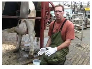
using of Benedict ,s test or urine strips
C. Protein: The main protein in the urine is
albumin which comes
under certain disease conditions such as
cystitis, glomerulonephritis, renal infarction,
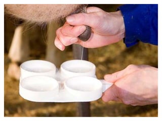
inflammation of genital organs and poisoning
(lead, arsenic and mercury). It can be detected
by using of sulfosalcylic acid test or urine
strips test.
D. Ketones bodies: Ketones bodies are
acetone, acetoacetic acid
and beta hydroxybutyric acid which are
formed as a result of breakdown of fatty acids.
It abnormally occurs in acetonemia,
pregnancy toxemia, fatty degeneration of the
liver and abomasal displacement. It can be
detected by using of Rother ,s test or urine
strips test
E. Bilirubin: It may be:
1) Pre-hepatic (hemolytic):It occurs in case of
bacillary
hemoglobinuria and leptospirosis, babesiosis,
anaplasma, infectious equine anemia, chronic
copper poisoning,hypophosphataemia and/or
heavy metal poisoning.
2) Hepatic It may be toxic, infective or obstructive
3) Post-hepatic: Obstruction by calculi or
compression by tumor.
F. Blood, hemoglobinuria & myoglobinuria:
1) Hematuria: The color is red and clowdy.lt is caused by
trauma
of the kidney, anthrax (pre-renal), acute glomerulonephritis,
tubular degeneration( by bacterial toxins and sulfanilamide
intoxication (renal) ; cystitis, urolithiasis, rough manipulation
of the catheter, tumor of renal tract and also hemorrhage of
genital tract (post-renal). If the blood comes during onset of
urination, the source of hemorrhage is coming from urethra. If
the whole urine is mixed with blood the possible source may
be kidneys but if only last portion of urine is red and
containing blood , it will be come from the bladder.
2. Hemoglobinuria: The color is brown to red,
caused by water
intoxication, babesiosis, bacillary
hemoglonburin, leptospirosis, chronic copper
poisoning and hypophosphatemia.
3) Myoglobinuria: Brown to black color of urine,
occurs in case of Azouturia disease. Blood,
hemoglobin & myoglobin can be detected by
using of Benzidine test or urine strips test.
2. Microscopical examination
Take 5-10 ml urine in a centrifuge tube and
Centrifuge it at 1000 rpm/10minutes. Discard
the supernatant and place a drop of sediment
on dry glass slide. Cover it with a cover slip
and examine under microscope
I. Organized sediment:
A. Epithelial cells: They increases in cystitis, and
other inflammatory
condition of urinary tract.
B. Leucocytes (pus cells) : The number of
leucocytes increases in
nephritis, pyelonephritis, urothritis and cystitis.
Presence of 10 leucocytes per high power field of
15 ml urine sediment are considered as an
inflammatory condition. The leucocytes are larger
than erythrocytes and have granular appearance.
C. Erythrocytes: It is spherical in shape, like a faint
colorless ring (Shadow cells). Presence of large
number of erythrocytes is an indication of
hemorrhage from urogenital system.
D. Casts: Presence of casts indicate mild form of
renal irritation
(hyaline casts), nephritis and degeneration of
kidney (epithilialcasts), hemorrhage in the renal
tubules (red cell cast) and suppurative infection
as in pyelonephritis and kidney abscess
(leucocytic cast)
E. Parasites: Capillaria plica (bladder worm of
dog and cat),
dioctophyma renale (kidney worm of dog).
Other worms/or ova may be present in the
urine sediment as a fecal contamination.
I. Un-organized sediment:
A. Crystals: Crystals occur as a result of acute
liver disease, carbon tetrachloride poisoning
and phosphorus poisoning. In alkaline
urine, the crystal may be triple‘ and
amorphous phosphates or calcium carbonate
and ammonium urates. In acidic urine the
crystals present may be amorphous urate,
uric acid or calcium oxalate
3. Cultural examination
Urine is collected in a sterile container,
inculated on culture media directly or after
centrifugation. Antibiotic sensitivity tests can
be done.
Skin scraping examination
The scrapings must be collected deeply from
the most affected part of the skin, the affected
part should be moistened with mineral oil.
A. Direct method: The skin scrapings are
placed on clean and dry
glass slide with one drop 10 sodium hydroxide
and cover it with cover slipe. Examine under
low power of microscope.
B. Sedimentation methods: The skin scrapings
are kept in 10%
potassium or sodium hydroxide, 2-4 hours
then transfer to centrifuge tube and
certrifugated at 3000 rpm/10 minutes. The
supernatant is discarded and one drop of the
sediment examine under microscope
C. Examination of skin scrapings for fungi:
Examination by Wood is lamp :The Wood 's
lamp has UV
light, which is directed on the skin or scrapings
collected in petridishes. If the fungus
microsporum is present, it gives yellow green
fluorescence, while no fluorescence in
negative infection
Microscopic examination: Collection of skin scrapings
should be from the center as well as from the periphery of the
lesion. Swab the lesion with 95% alcohol to remove any
saprophytic organism. The skin scrapings are collected in
sterilized petridishes containing 10% sodium hydroxide or
potassium hydroxide. Put one drop on clean slide and cover it
with cover slip and apply Vaseline around the rim of cover
slip. The slide is warmed gently for few second. Then examine
for the presence of chains of hyphae and spores.
Examination of milk
Collection of the samples: The udder of the
animal should be cleaned with water and
antiseptic solution like potasium permanganate
(1:1000).
The hands of examiner should be cleaned with
soap and antiseptics. Disinfect the teats with
alcohol 70%, collect the milk sample from each
teat in separate tube (5-10 ml) and discards the
first 3-4 streams of milk.
Tube should be stoppered and transported to the
laboratory in ice for examination.
I. Physical examination of milk:
A. Color: Normally the color of milk is white but in
acute mastitis it
may become reddish (presence of blood). Yellowish
coloration occur during colostral period, feeding of
carrots and tetracycline therapy.
B. Reaction: The pH of normal milk is 6.4 to 6.6 but in
mastitis it
becomes alkaline up to 7.4 due to presence of sodium
bicarbonate in the milk. The reaction can be determined
by using pH strips or pH meter.
C. Odor: Normally the odor of milk is pleasant but
in mastitis due
to Actinomyces pyogenes it becomes obnoxious. In
ketosis the odor of milk becomes sweet and
fruity.
C. Consistency: The colostrums is viscous. In
acute and sub acute mastitis the milk contains
fine and large flakes.
The watery consistency occurs due to poor
feeding and chronic mastitis.
II. Chemical examination of milk:
A. White slide test: 4-5 drops of milk are placed on a
clean dry slide. Add a drop of 4% sodium hydroxide
and mix with glass rod. In mastitis it becomes
thickened and flakes appear.
B. California mastitis test: This test is based on
increased the number of leucocytes and increased
alkalinity in mastitic milk. take 0.5 ml milk from each
quarter in plastic peddle cups and add equal quantity
of California reagent, mix well by circular movement of
peddle on a horizontal plane
1. Liquid milk with no streaks or precipitation:
negative for mastitis
2. Streaky milk: the weak positive
3. Slimy: ++.
4. Gelatinous: +++
C. Leucocytic count: Mark the area on central
portion of slide
(1squar cm). Put 0.1 ml, spread the milk
sample by bacteriological loop in this area. Dry
the smear and dissolve the fat by rinsing it in
xylene for 2-5 minute. Fix the smear with
alcohol 4-5 minutes and stain with methylene
blue for 1 minute. Remove the excess stain by
immersing the slide in alcohol
Count the leukocyte under oil immersion lens.
The cell of 10 field are counted and averaged
and multiplied by 500.000 to get the total
number of leucocytes in the milk.
III. Bacteriological examination of milk:
A. Direct microscopical examination.
B. Cultural examination: The milk is collected
in sterilized vials and stores in refrigerator and
send to the laboratory, for isolation of
the organism and antibiotic sensitivity test.

More Related Content

What's hot

What's hot (20)

Postmortem
PostmortemPostmortem
Postmortem
 
Lumpy skin disease
Lumpy skin diseaseLumpy skin disease
Lumpy skin disease
 
PPR (Peste des Petits Ruminants)
PPR (Peste des Petits Ruminants)PPR (Peste des Petits Ruminants)
PPR (Peste des Petits Ruminants)
 
Veterolegal cases and forensics
Veterolegal cases and forensicsVeterolegal cases and forensics
Veterolegal cases and forensics
 
Theileriosis
TheileriosisTheileriosis
Theileriosis
 
Canine Distemper
Canine DistemperCanine Distemper
Canine Distemper
 
Blue tongue in India
Blue tongue in IndiaBlue tongue in India
Blue tongue in India
 
Animal parameters lecture
Animal parameters lectureAnimal parameters lecture
Animal parameters lecture
 
Strangles
StranglesStrangles
Strangles
 
Bloat & tympany
Bloat & tympanyBloat & tympany
Bloat & tympany
 
Trypanosomiosis
TrypanosomiosisTrypanosomiosis
Trypanosomiosis
 
Pox diseases
Pox diseasesPox diseases
Pox diseases
 
GLANDERS
GLANDERSGLANDERS
GLANDERS
 
Anaplasma
AnaplasmaAnaplasma
Anaplasma
 
Bovine mastitis
Bovine mastitisBovine mastitis
Bovine mastitis
 
Swine fever
Swine feverSwine fever
Swine fever
 
Newcastle Disease
Newcastle DiseaseNewcastle Disease
Newcastle Disease
 
Bovine Viral Diarrhea
Bovine Viral DiarrheaBovine Viral Diarrhea
Bovine Viral Diarrhea
 
Babesia
BabesiaBabesia
Babesia
 
Presentation on jd final
Presentation on jd finalPresentation on jd final
Presentation on jd final
 

Viewers also liked

Post parturient-hemoglobinuria
Post parturient-hemoglobinuriaPost parturient-hemoglobinuria
Post parturient-hemoglobinuria
Jalal Syooti
 
Bacteriological measurement and its physical characteristics
Bacteriological measurement and its physical characteristicsBacteriological measurement and its physical characteristics
Bacteriological measurement and its physical characteristics
NIT MEGHALAYA
 

Viewers also liked (20)

MASTITIS DIAGNOSIS by ADIL RASOOL PARAY
MASTITIS DIAGNOSIS by ADIL RASOOL PARAYMASTITIS DIAGNOSIS by ADIL RASOOL PARAY
MASTITIS DIAGNOSIS by ADIL RASOOL PARAY
 
Milk defination, properties and nutrition
Milk defination, properties and nutritionMilk defination, properties and nutrition
Milk defination, properties and nutrition
 
Milk chemistry and composition - Basics for Dairy processing
Milk chemistry and composition - Basics for Dairy processingMilk chemistry and composition - Basics for Dairy processing
Milk chemistry and composition - Basics for Dairy processing
 
Mastitis
MastitisMastitis
Mastitis
 
Post parturient-hemoglobinuria
Post parturient-hemoglobinuriaPost parturient-hemoglobinuria
Post parturient-hemoglobinuria
 
DogNostics Career College.Module # 202 Canine and Feline Health & Handling
DogNostics Career College.Module # 202 Canine and Feline Health & HandlingDogNostics Career College.Module # 202 Canine and Feline Health & Handling
DogNostics Career College.Module # 202 Canine and Feline Health & Handling
 
Dr. Dan Givens - Bovine Viral Diarrhoea (BVD) Diagnostics - Goals, Strategies...
Dr. Dan Givens - Bovine Viral Diarrhoea (BVD) Diagnostics - Goals, Strategies...Dr. Dan Givens - Bovine Viral Diarrhoea (BVD) Diagnostics - Goals, Strategies...
Dr. Dan Givens - Bovine Viral Diarrhoea (BVD) Diagnostics - Goals, Strategies...
 
A proposal on_impact_of_mastitis_on_milk_productio(1)
A proposal on_impact_of_mastitis_on_milk_productio(1)A proposal on_impact_of_mastitis_on_milk_productio(1)
A proposal on_impact_of_mastitis_on_milk_productio(1)
 
Physico chemical and microbiological analysis of fermented cow milk (nono) co...
Physico chemical and microbiological analysis of fermented cow milk (nono) co...Physico chemical and microbiological analysis of fermented cow milk (nono) co...
Physico chemical and microbiological analysis of fermented cow milk (nono) co...
 
Diagnostic in cattle and buffalo ( SACHIN SUBEDI)
Diagnostic in cattle and buffalo ( SACHIN SUBEDI)Diagnostic in cattle and buffalo ( SACHIN SUBEDI)
Diagnostic in cattle and buffalo ( SACHIN SUBEDI)
 
Development of a Cheddar Cheese Using Biological Preservatives as Substitute ...
Development of a Cheddar Cheese Using Biological Preservatives as Substitute ...Development of a Cheddar Cheese Using Biological Preservatives as Substitute ...
Development of a Cheddar Cheese Using Biological Preservatives as Substitute ...
 
Bacteriological measurement and its physical characteristics
Bacteriological measurement and its physical characteristicsBacteriological measurement and its physical characteristics
Bacteriological measurement and its physical characteristics
 
Livestock Protective Fence (LPF) for enhanced milk production and mastitis co...
Livestock Protective Fence (LPF) for enhanced milk production and mastitis co...Livestock Protective Fence (LPF) for enhanced milk production and mastitis co...
Livestock Protective Fence (LPF) for enhanced milk production and mastitis co...
 
Bovine Viral Diarrhoea
Bovine Viral Diarrhoea Bovine Viral Diarrhoea
Bovine Viral Diarrhoea
 
Micro milk
Micro milkMicro milk
Micro milk
 
Applications of in vitro gas production technique. Avijit Dey. 4th June.2014
Applications of in vitro gas production technique. Avijit Dey. 4th June.2014Applications of in vitro gas production technique. Avijit Dey. 4th June.2014
Applications of in vitro gas production technique. Avijit Dey. 4th June.2014
 
Backcasting the Future of Animal Health Research
Backcasting the Future of Animal Health ResearchBackcasting the Future of Animal Health Research
Backcasting the Future of Animal Health Research
 
How to Control Mastitis and Lower Herd Milk Somatic Cell Count- Larry Fox
How to Control Mastitis and Lower Herd Milk Somatic Cell Count- Larry FoxHow to Control Mastitis and Lower Herd Milk Somatic Cell Count- Larry Fox
How to Control Mastitis and Lower Herd Milk Somatic Cell Count- Larry Fox
 
Dairy Cattle Introduction
Dairy Cattle Introduction Dairy Cattle Introduction
Dairy Cattle Introduction
 
Mastitis and its effect on milk processing
Mastitis and its effect on milk processingMastitis and its effect on milk processing
Mastitis and its effect on milk processing
 

Similar to Animal Sample Collection

Urine dr.hamada elsadaway
Urine dr.hamada elsadawayUrine dr.hamada elsadaway
Urine dr.hamada elsadaway
FarragBahbah
 
Microscopic examination of urine
Microscopic examination of urineMicroscopic examination of urine
Microscopic examination of urine
globalsoin
 
STOOL_EXAMINATION.pptx
STOOL_EXAMINATION.pptxSTOOL_EXAMINATION.pptx
STOOL_EXAMINATION.pptx
KnqAutlawzvFb
 

Similar to Animal Sample Collection (20)

Vpl 605 clinical pathology
Vpl 605  clinical pathology Vpl 605  clinical pathology
Vpl 605 clinical pathology
 
Physical examination of urine (4)
Physical examination of urine (4)Physical examination of urine (4)
Physical examination of urine (4)
 
sanjana.pptx
sanjana.pptxsanjana.pptx
sanjana.pptx
 
Urine dr.hamada elsadaway
Urine dr.hamada elsadawayUrine dr.hamada elsadaway
Urine dr.hamada elsadaway
 
Urine Interpretation / Test / Analysis
Urine Interpretation / Test / AnalysisUrine Interpretation / Test / Analysis
Urine Interpretation / Test / Analysis
 
Stool (1)
Stool (1)Stool (1)
Stool (1)
 
URINE SPECIMEN.pptx
URINE SPECIMEN.pptxURINE SPECIMEN.pptx
URINE SPECIMEN.pptx
 
Kamal
KamalKamal
Kamal
 
ROUTINE STOOLE EXAMINATIN
ROUTINE STOOLE EXAMINATIN ROUTINE STOOLE EXAMINATIN
ROUTINE STOOLE EXAMINATIN
 
Urine R/E and RFTs
Urine R/E and RFTsUrine R/E and RFTs
Urine R/E and RFTs
 
Microscopic examination of urine
Microscopic examination of urineMicroscopic examination of urine
Microscopic examination of urine
 
Urine.pptx
Urine.pptxUrine.pptx
Urine.pptx
 
Urinary Tract Infections and Urine Analysis .pptx
Urinary Tract Infections and Urine Analysis .pptxUrinary Tract Infections and Urine Analysis .pptx
Urinary Tract Infections and Urine Analysis .pptx
 
STOOL_EXAMINATION.pptx
STOOL_EXAMINATION.pptxSTOOL_EXAMINATION.pptx
STOOL_EXAMINATION.pptx
 
Microscopic examination of urine
Microscopic examination of urineMicroscopic examination of urine
Microscopic examination of urine
 
Urine Examination (Practical).pdf
Urine Examination (Practical).pdfUrine Examination (Practical).pdf
Urine Examination (Practical).pdf
 
Laboratory and diagnostic examination(urine analysis)
Laboratory and diagnostic examination(urine analysis)Laboratory and diagnostic examination(urine analysis)
Laboratory and diagnostic examination(urine analysis)
 
Urine examination
Urine examinationUrine examination
Urine examination
 
Feline urological syndrome 2018
Feline urological syndrome 2018Feline urological syndrome 2018
Feline urological syndrome 2018
 
Renal function tests
Renal function testsRenal function tests
Renal function tests
 

More from Shafi'i Abdullahi

More from Shafi'i Abdullahi (16)

Toxoplasmosis
ToxoplasmosisToxoplasmosis
Toxoplasmosis
 
African swine fever
African swine feverAfrican swine fever
African swine fever
 
Anaplasmosis
AnaplasmosisAnaplasmosis
Anaplasmosis
 
Rabies
RabiesRabies
Rabies
 
Trypanosomiosis
TrypanosomiosisTrypanosomiosis
Trypanosomiosis
 
Theileriosis
TheileriosisTheileriosis
Theileriosis
 
Ringworm
RingwormRingworm
Ringworm
 
FOOT AND MOUTH DISEASE ( FMD)
FOOT AND MOUTH DISEASE ( FMD)FOOT AND MOUTH DISEASE ( FMD)
FOOT AND MOUTH DISEASE ( FMD)
 
Fasciolosis
FasciolosisFasciolosis
Fasciolosis
 
BOVINE SPONGIFORM ENCEPHALOPHATHY (BSE), MAD COW
BOVINE SPONGIFORM ENCEPHALOPHATHY (BSE), MAD COWBOVINE SPONGIFORM ENCEPHALOPHATHY (BSE), MAD COW
BOVINE SPONGIFORM ENCEPHALOPHATHY (BSE), MAD COW
 
Camel pox
Camel poxCamel pox
Camel pox
 
Babesiosis
BabesiosisBabesiosis
Babesiosis
 
Anaplasmosis
Anaplasmosis Anaplasmosis
Anaplasmosis
 
Animal parameters
Animal parametersAnimal parameters
Animal parameters
 
Animal clinical parameters
Animal clinical parametersAnimal clinical parameters
Animal clinical parameters
 
Genome editing
Genome editingGenome editing
Genome editing
 

Animal Sample Collection

  • 1. 27th , April , 2016 SHAFI’I A. Mohamed
  • 2. Urine examination Urine samples can be collected in cows and sheep either by stimulation of the urethra through vulva or by catheterization. In delay exam, store the urine in refrigerator at 4 °C.
  • 3.
  • 4. 1. Chemical examination: A. Reaction (pH): The reaction of urine is determined by using pH strips or pH meter. Normal urine is alkaline in cattle and horse (7.4-8.4) and acidic (6-7) in dogs and cats. Acidic urine is abnormally observed in cases of starvation, fever, treatment with sodium acid phosphate, while the alkaline urine is abnormally observed in cases of cystitis, urine retention and treatment by carbonate, acetate and nitrate of sodium or potassium
  • 5. B. Glucose: Normally there is no any glucose content in the urine. Glucosuria occurs due to hyperglycemia and in diabetes mellitus, acute or chronic pancreatitis, hyper adrenaline and certain drugs (penicillin, tetracycline and chloramphenicol). Glucosuria can be detected in the urine by using of Benedict ,s test or urine strips
  • 6. C. Protein: The main protein in the urine is albumin which comes under certain disease conditions such as cystitis, glomerulonephritis, renal infarction, inflammation of genital organs and poisoning (lead, arsenic and mercury). It can be detected by using of sulfosalcylic acid test or urine strips test.
  • 7. D. Ketones bodies: Ketones bodies are acetone, acetoacetic acid and beta hydroxybutyric acid which are formed as a result of breakdown of fatty acids. It abnormally occurs in acetonemia, pregnancy toxemia, fatty degeneration of the liver and abomasal displacement. It can be detected by using of Rother ,s test or urine strips test
  • 8. E. Bilirubin: It may be: 1) Pre-hepatic (hemolytic):It occurs in case of bacillary hemoglobinuria and leptospirosis, babesiosis, anaplasma, infectious equine anemia, chronic copper poisoning,hypophosphataemia and/or heavy metal poisoning. 2) Hepatic It may be toxic, infective or obstructive 3) Post-hepatic: Obstruction by calculi or compression by tumor.
  • 9. F. Blood, hemoglobinuria & myoglobinuria: 1) Hematuria: The color is red and clowdy.lt is caused by trauma of the kidney, anthrax (pre-renal), acute glomerulonephritis, tubular degeneration( by bacterial toxins and sulfanilamide intoxication (renal) ; cystitis, urolithiasis, rough manipulation of the catheter, tumor of renal tract and also hemorrhage of genital tract (post-renal). If the blood comes during onset of urination, the source of hemorrhage is coming from urethra. If the whole urine is mixed with blood the possible source may be kidneys but if only last portion of urine is red and containing blood , it will be come from the bladder.
  • 10. 2. Hemoglobinuria: The color is brown to red, caused by water intoxication, babesiosis, bacillary hemoglonburin, leptospirosis, chronic copper poisoning and hypophosphatemia. 3) Myoglobinuria: Brown to black color of urine, occurs in case of Azouturia disease. Blood, hemoglobin & myoglobin can be detected by using of Benzidine test or urine strips test.
  • 11.
  • 12.
  • 13. 2. Microscopical examination Take 5-10 ml urine in a centrifuge tube and Centrifuge it at 1000 rpm/10minutes. Discard the supernatant and place a drop of sediment on dry glass slide. Cover it with a cover slip and examine under microscope
  • 14. I. Organized sediment: A. Epithelial cells: They increases in cystitis, and other inflammatory condition of urinary tract. B. Leucocytes (pus cells) : The number of leucocytes increases in nephritis, pyelonephritis, urothritis and cystitis. Presence of 10 leucocytes per high power field of 15 ml urine sediment are considered as an inflammatory condition. The leucocytes are larger than erythrocytes and have granular appearance.
  • 15. C. Erythrocytes: It is spherical in shape, like a faint colorless ring (Shadow cells). Presence of large number of erythrocytes is an indication of hemorrhage from urogenital system. D. Casts: Presence of casts indicate mild form of renal irritation (hyaline casts), nephritis and degeneration of kidney (epithilialcasts), hemorrhage in the renal tubules (red cell cast) and suppurative infection as in pyelonephritis and kidney abscess (leucocytic cast)
  • 16. E. Parasites: Capillaria plica (bladder worm of dog and cat), dioctophyma renale (kidney worm of dog). Other worms/or ova may be present in the urine sediment as a fecal contamination.
  • 17. I. Un-organized sediment: A. Crystals: Crystals occur as a result of acute liver disease, carbon tetrachloride poisoning and phosphorus poisoning. In alkaline urine, the crystal may be triple‘ and amorphous phosphates or calcium carbonate and ammonium urates. In acidic urine the crystals present may be amorphous urate, uric acid or calcium oxalate
  • 18. 3. Cultural examination Urine is collected in a sterile container, inculated on culture media directly or after centrifugation. Antibiotic sensitivity tests can be done.
  • 19.
  • 20. Skin scraping examination The scrapings must be collected deeply from the most affected part of the skin, the affected part should be moistened with mineral oil. A. Direct method: The skin scrapings are placed on clean and dry glass slide with one drop 10 sodium hydroxide and cover it with cover slipe. Examine under low power of microscope.
  • 21.
  • 22.
  • 23. B. Sedimentation methods: The skin scrapings are kept in 10% potassium or sodium hydroxide, 2-4 hours then transfer to centrifuge tube and certrifugated at 3000 rpm/10 minutes. The supernatant is discarded and one drop of the sediment examine under microscope
  • 24. C. Examination of skin scrapings for fungi: Examination by Wood is lamp :The Wood 's lamp has UV light, which is directed on the skin or scrapings collected in petridishes. If the fungus microsporum is present, it gives yellow green fluorescence, while no fluorescence in negative infection
  • 25.
  • 26. Microscopic examination: Collection of skin scrapings should be from the center as well as from the periphery of the lesion. Swab the lesion with 95% alcohol to remove any saprophytic organism. The skin scrapings are collected in sterilized petridishes containing 10% sodium hydroxide or potassium hydroxide. Put one drop on clean slide and cover it with cover slip and apply Vaseline around the rim of cover slip. The slide is warmed gently for few second. Then examine for the presence of chains of hyphae and spores.
  • 27. Examination of milk Collection of the samples: The udder of the animal should be cleaned with water and antiseptic solution like potasium permanganate (1:1000). The hands of examiner should be cleaned with soap and antiseptics. Disinfect the teats with alcohol 70%, collect the milk sample from each teat in separate tube (5-10 ml) and discards the first 3-4 streams of milk. Tube should be stoppered and transported to the laboratory in ice for examination.
  • 28.
  • 29.
  • 30. I. Physical examination of milk: A. Color: Normally the color of milk is white but in acute mastitis it may become reddish (presence of blood). Yellowish coloration occur during colostral period, feeding of carrots and tetracycline therapy. B. Reaction: The pH of normal milk is 6.4 to 6.6 but in mastitis it becomes alkaline up to 7.4 due to presence of sodium bicarbonate in the milk. The reaction can be determined by using pH strips or pH meter.
  • 31. C. Odor: Normally the odor of milk is pleasant but in mastitis due to Actinomyces pyogenes it becomes obnoxious. In ketosis the odor of milk becomes sweet and fruity. C. Consistency: The colostrums is viscous. In acute and sub acute mastitis the milk contains fine and large flakes. The watery consistency occurs due to poor feeding and chronic mastitis.
  • 32. II. Chemical examination of milk: A. White slide test: 4-5 drops of milk are placed on a clean dry slide. Add a drop of 4% sodium hydroxide and mix with glass rod. In mastitis it becomes thickened and flakes appear. B. California mastitis test: This test is based on increased the number of leucocytes and increased alkalinity in mastitic milk. take 0.5 ml milk from each quarter in plastic peddle cups and add equal quantity of California reagent, mix well by circular movement of peddle on a horizontal plane
  • 33. 1. Liquid milk with no streaks or precipitation: negative for mastitis 2. Streaky milk: the weak positive 3. Slimy: ++. 4. Gelatinous: +++
  • 34.
  • 35. C. Leucocytic count: Mark the area on central portion of slide (1squar cm). Put 0.1 ml, spread the milk sample by bacteriological loop in this area. Dry the smear and dissolve the fat by rinsing it in xylene for 2-5 minute. Fix the smear with alcohol 4-5 minutes and stain with methylene blue for 1 minute. Remove the excess stain by immersing the slide in alcohol
  • 36. Count the leukocyte under oil immersion lens. The cell of 10 field are counted and averaged and multiplied by 500.000 to get the total number of leucocytes in the milk.
  • 37.
  • 38.
  • 39. III. Bacteriological examination of milk: A. Direct microscopical examination. B. Cultural examination: The milk is collected in sterilized vials and stores in refrigerator and send to the laboratory, for isolation of the organism and antibiotic sensitivity test.