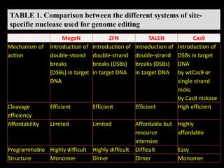
Welcome
Md Abu Kawochar (201730506003)
Mohammad Abdul Latif (2017302060007)
Shafi’i Abdullahi Mohamed (2016302160001)
Saleem Muhammad Hamzah (2017301160017)
Sunny Ahmar (2017301160015)
• Group # 07
GENOME EDITING
● Strategies and techniques developed for the targeted, specific
modification of the genetic information or genome of living
organisms.
- disruption, insertion, replacement at locus in the genome
- control gene expression
- create SNP
- create reporter fusions while maintaining endogenous gene
regulation
Importance of genome editing
Used in
• Scientific research
• Crops and livestock
• Industrial biotechnology
• Biomedicine
• Reproduction
The major advantages
– which uses more recent knowledge and technology,
– it enables a specific area of the DNA to be modified
– increasing the precision of the correction or insertion
– preventing any cell toxicity and offering perfect reproducibility.
Fig. 1. Timeline of and genome engineering research fields
•Mechanism in cells to repair
double strand DNA lesions.
• The most common form :
homologous recombination.
• Only used by the cell when there
is a homologue piece of DNA
present in the nucleus
• mostly in G2 and late S phase
• Important for suppressing
cancer
• maintains genomic stability by
repairing broken DNA strands
• Assumed to be error free
because of the use of a template
Repairs double-strand breaks in
DNA in the absence of
homologous templates
• G0/G1 and early S-phases
• "non-homologous" : the break
ends directly ligated
• "non-homologous end joining“ :
Moore and Haber(1996).
• utilizes short homologous DNA
sequences called microhomologies
to guide repair
• Imprecise repair when the
overhangs are not compatible
• Lead to translocations and
telomere fusion :hallmarks of
tumor cells.
• Also known as alternative non homologous end-joining (Alt-
NHEJ):More error-prone pathway
• A homology of 5 - 25 complementary base pairs
(microhomology) on both strands –align the strands
• Ligating the mismatched hanging strands of DNA, removing
overhanging nucleotides, and filling in the missing base pairs.
• overhanging bases (flaps) and mismatched bases on the strands
are removed and any missing nucleotides are inserted.
• Chromosome abnormalities and other complex rearrangements
● Mega nucleases first identified in 80s, they target large DNA
sequences of about 12 to 40 base pairs long which lead them to be
highly specific in the utmost of genomes (Gallagher et al., 2014).
●Meganucleases :
Endonucleases family
• best known meganucleases
proteins in the LAGLIDADG
family,
• Natural meganucleases :slight
variations in recognition sites
• Two methods for creating custom meganucleases:
– Mutagenesis involves generating collections of variants using a
meganuclease
– Combinatorial assembly is a method whereby protein subunits from
different enzymes can be associated or fused.
DNA binding domain and catalytic domain in mega nuclease are
linked so its construction is either expensive or labour-intensive as
compared with other genome editing tools. Therefore, mega
nucleases have major drawbacks which lead them to have a
considerably low priority as option amongst genome engineering
tools to work with.
Zinc Fingers
• Coordinate Zn with
Cysteine or Histidine
• Bind to
DNA/RNA/Proteins
• Hard to target for
specific binding sites
• Can target every ~500
BP
• Difficulty targeting AT
rich sequences
http://en.wikipedia.org/wiki/Zinc_finger
Zinc finger motifs occur in several transcription factors. The C-
terminal part of each finger is responsible for the specific
recognition of the DNA sequence.
 Several approaches are used to design specific zinc finger
nucleases for the chosen sequences.
Zinc finger nucleases consists of zinc finger proteins which in the
interest of targeting a particular DNA array to generate double-
stranded breaks by fusing it with non-specific Fok1 endonuclease.
(A) Designed zinc-finger protein in complex with target DNA (grey).
Each zinc-finger consists of approximately 30 amino acids in
arrangement (inset). Surface residues (-1, 2, 3 and 6) that contact DNA
are shown as sticks. Each zinc-finger domain contacts 3 or 4 bp in the
major groove of DNA. The side chains of the conserved Cys and His
residues are depicted as sticks in complex with a Zn2+ ion (purple).
Figure 1. Structure of zinc-finger
(B) Cartoon of a zinc-finger nuclease (ZFN) dimer bound to
DNA. ZFN target sites consist of two zinc-finger binding sites
separated by a 5–7-bp spacer sequence recognized by the FokI
cleavage domain. Zinc-finger proteins can be designed to
recognize unique ‘left’ and ‘right’ half-sites.
Construction of zinc finger nucleases is difficult as compared to
TALENs and CRISPR/CAS systems.
However, there are several disadvantages for using ZFN-based
technology, including the complexity and high cost of protein
domains construction for each particular genome locus and the
probability of inaccurate cleavage of target DNA (off-target
effects) due to single nucleotide substitutions or inappropriate
interaction between domains (Puchta & Hohn, 2010).
• TALENs were named as a method of year by nature methods in
2011 (Baker & Becker 2012).
• Class of specific DNA binding protein
• Produced by plant bacteria in Xantomonas
– Modulates host gene expression
• RVD: Repeat-variable di-residue
– Residues at position 12 & 13 that give target specificity
• Double Strand Break Repair:
– Non-homologous End Joining (NHEJ)
– Homologous Recombination (HR)
TALENs compose of transcription activator-like effectors
(TALEs) fused with the non-specific Fok1 endonuclease naturally
found in Flavobacterium okenkoides.
These TALEs proteins are naturally exuded by bacteria,
Xanthomonas spp, which gets bind to the targeted DNA sequence
with the help of DNA-binding domain.
Each duplication identifies a single nucleotide in target DNA.
TALE protein comprises of N terminal domain, central repetitive
regions and the C terminal domain.
Middle repetitive regions consists of 34 amino acids which are
identical to each other except for two amino acids at situations12
and 13 called as Repeat Variable di-residues (RVD) that
determines specificity of TALEs repeat.
Structure of TAL Effector & TALEN
RVD’s
TALEN Design Guidelines
1. TALEN monomer
binding sites must be
preceded by 5’-T
2. No T at position 1
3. No A at position 2
4. Must end with a T
5. Base composition
within two S.D. of
observed average
(C) TALE protein in
complex with target DNA
(grey). Individual TALE
repeats contain 33–35
amino acids that recognize
a single base pair via two
hypervariable residues
(repeat-variable diresidues;
RVDs) (shown as sticks) .
(D) Cartoon of a TALE nuclease (TALEN) dimer bound to DNA.
TALEN target sites consist of two TALE binding sites separated by
a spacer sequence of varying length (12–20 bp). TALEs can be
designed to recognize unique left and right half-sites. RVD
compositions are indicated.
Goals & Motivation
• Develop tools for
Genome Engineering
• DNA Targeting
• Goal: Develop system
for assembly of custom
TALEN & TAL
Effectors
http://www.newswise.com/images/uploads/2012/01/4/TAL_effector_DNA_color_1.JPG
Continuous thrust for the precise, advanced and
easier tools for genome editing, resulted in the
development of CRISPRs.
- genetic elements that bacteria use as a kind of acquired
immunity to protect against viruses
CRISPRs
First described (1987)
48% bacteria; 84% archaea
Short Regularly Spaced Repeats (SRSR) (2000)
Renamed CRISPR (2002)
A prokaryotic adaptive immune system (2005-12)
Identify Cas9 (2012)
Genome Engineering by CRISPR/Cas system (2013)
CRISPR Interference (2013)
Other applications of CRISPR /Cas system (2013 to…………)
CRISPRs
Action of CRISPR in bacteria
Different CRISPR-Cas system in Bacterial Adaptive
Immunity
Class 1- type I (CRISPR-Cas3) and type III
(CRISPRCas10)
uses several Cas proteins and the crRNA
Class 2- type II (CRISPR-Cas9) and type V
(CRISPRCpf1)
employ a large single-component Cas-9
protein in conjunction with crRNA and
tracerRNA.
Fig. 2. Biology of the type II-A CRISPR-Cas system. The type II-A system from S.
pyogenes is shown as an example.
The cas gene operon with tracrRNA and the CRISPR array.
The natural pathway of antiviral defense involves association of Cas9 with the
antirepeat-repeat RNA (tracrRNA: crRNA) duplexes, RNA co-processing by ribonuclease
III, further trimming, R-loop formation, and target DNA cleavage.
Fig. 2. Biology of the type II-A CRISPR-Cas system. The type II-A system from S.
pyogenes is shown as an example.
Details of the natural DNA cleavage with the duplex tracrRNA:crRNA.
Fig. 3. Evolution and structure of Cas9. The structure of S. pyogenes Cas9 in the unliganded and
RNA-DNA–bound forms
CRISPR-Cas9 Mechanism
Limitations
The delivery of g-RNA and the Cas9 protein has been constant
challenge.
In Cas9 system, a small percentage off-target effects was appeared.
However CRISPR-Cpf1 has recently been shown to diminish these
effects.
New Version of Cas9:
Cpf1 (CRISPR from Prevotella and Francisella 1)
CRISPR-Cpf1 is a class 2 CRISPR system
Cpf1 is a CRISPR-associated two-component RNA programmable
DNA nuclease
Does not require tracerRNA and the gene is 1kb smaller
Targeted DNA is cleaved as a 5 nt staggered cut distal to a 5’ T-
rich PAM
Cpf1 exhibit robust nuclease activity in human cells
Figure 1. General procedure of plant genome editing by CRISPR–Cas9.
Plant genome editing can typically be divided into four continuous
steps, and the estimated time needed for each step is indicated.
PCR/RE, polymerase chain reaction/restriction enzyme digestion.
Figure 2. DNA-free genome editing with CRISPR–Cas9 RNAs
TABLE 1. Comparison between the different systems of site-
specific nuclease used for genome editing
MegaN ZFN TALEN Cas9
Recognition site Between 14 and
40 bp
Typically 9–18
bp per ZFN
monomer,
18–36 bp per
ZFN pair
Typically 14–20
bp per
TALEN
monomer,
28–40 bp per
TALEN pair
22 bp (20-bp
guide sequence
C 2-bp
protospacer
adjacent motif
(PAM)
for Cas9; up to
44 bp for double
nicking
Specificity Small number
of positional
mismatches
tolerated
Small number of
positional
mismatches
tolerated
Small number of
positional
mismatches
tolerated
Positional and
multiple
consecutive
mismatches
tolerated
TABLE 1. Comparison between the different systems of site-
specific nuclease used for genome editing
MegaN ZFN TALEN Cas9
Targeting Targeting novel
sequences often
results in low
efficiency
Difficult to
target
non-G-rich
sequences
5ʹ targeted base
must
be a T for each
TALEN
monomer
Targeted
sequence must
pre- cede
a PAM
Cleavage
efficiency
Low efficiency Low efficiency Efficient Highly efficient
Off-target
effects
Possible off-
target
activities
Possible off-
target
activities
Limited off-
target
activities, not
fully studied
in plants
No off-targeted
activities
reported in
plants, but high
off-target levels
reported in
other systems.
TABLE 1. Comparison between the different systems of site-
specific nuclease used for genome editing
MegaN ZFN TALEN Cas9
Mechanism of
action
Introduction of
double-strand
breaks
(DSBs) in target
DNA
Introduction of
double-strand
breaks (DSBs)
in target DNA
Introduction of
double-strand
breaks (DSBs)
in target DNA
Introduction of
DSBs in target
DNA
by wtCas9 or
single strand
nicks
by Cas9 nickase
Cleavage
efficiency
Efficient Efficient Efficient High efficient
Affordability Limited Limited Affordable but
resource
intensive
Highly
affordable
Programmable Highly difficult Highly difficult Difficult Easy
Structure Monomer Dimer Dimer Monomer
Table 2. Examples of ZFN, TALEN, and CRISPR/Cas-mediated
genome editing in human cells and model organisms
Type of Modification: Gene disruption
Table 2. Examples of ZFN, TALEN, and CRISPR/Cas-mediated
genome editing in human cells and model organisms
Type of Modification: Gene addition
Type of Modification: Gene correction
Limitations
Ethical debate was emphasized by use of the technique on
human embryos.
The international bioethics committee of UNESCO considers
that the CRISPR Cas9 system only be used as a preventive,
diagnostic and therapeutic procedure without modifying the
descendants genome.
Concluding remarks and future directions
ZFNs, TALENs, and RNA-guided DNA endonucleases are
transformative tools that have the potential to revolutionize biological
research and affect personalized medicine.
Questions also remain regarding the optimal methods for delivering
these nucleases into cells and organisms.
Together, these technologies promise to expand our ability to
explore and alter any genome and constitute a new and promising
paradigm to understand and treat disease.
Genome editing

Genome editing

  • 1.
  • 2.
    Md Abu Kawochar(201730506003) Mohammad Abdul Latif (2017302060007) Shafi’i Abdullahi Mohamed (2016302160001) Saleem Muhammad Hamzah (2017301160017) Sunny Ahmar (2017301160015) • Group # 07
  • 3.
  • 4.
    ● Strategies andtechniques developed for the targeted, specific modification of the genetic information or genome of living organisms. - disruption, insertion, replacement at locus in the genome - control gene expression - create SNP - create reporter fusions while maintaining endogenous gene regulation
  • 5.
    Importance of genomeediting Used in • Scientific research • Crops and livestock • Industrial biotechnology • Biomedicine • Reproduction The major advantages – which uses more recent knowledge and technology, – it enables a specific area of the DNA to be modified – increasing the precision of the correction or insertion – preventing any cell toxicity and offering perfect reproducibility.
  • 6.
    Fig. 1. Timelineof and genome engineering research fields
  • 10.
    •Mechanism in cellsto repair double strand DNA lesions. • The most common form : homologous recombination. • Only used by the cell when there is a homologue piece of DNA present in the nucleus • mostly in G2 and late S phase • Important for suppressing cancer • maintains genomic stability by repairing broken DNA strands • Assumed to be error free because of the use of a template
  • 11.
    Repairs double-strand breaksin DNA in the absence of homologous templates • G0/G1 and early S-phases • "non-homologous" : the break ends directly ligated • "non-homologous end joining“ : Moore and Haber(1996). • utilizes short homologous DNA sequences called microhomologies to guide repair • Imprecise repair when the overhangs are not compatible • Lead to translocations and telomere fusion :hallmarks of tumor cells.
  • 12.
    • Also knownas alternative non homologous end-joining (Alt- NHEJ):More error-prone pathway • A homology of 5 - 25 complementary base pairs (microhomology) on both strands –align the strands • Ligating the mismatched hanging strands of DNA, removing overhanging nucleotides, and filling in the missing base pairs. • overhanging bases (flaps) and mismatched bases on the strands are removed and any missing nucleotides are inserted. • Chromosome abnormalities and other complex rearrangements
  • 18.
    ● Mega nucleasesfirst identified in 80s, they target large DNA sequences of about 12 to 40 base pairs long which lead them to be highly specific in the utmost of genomes (Gallagher et al., 2014). ●Meganucleases : Endonucleases family • best known meganucleases proteins in the LAGLIDADG family, • Natural meganucleases :slight variations in recognition sites
  • 19.
    • Two methodsfor creating custom meganucleases: – Mutagenesis involves generating collections of variants using a meganuclease – Combinatorial assembly is a method whereby protein subunits from different enzymes can be associated or fused. DNA binding domain and catalytic domain in mega nuclease are linked so its construction is either expensive or labour-intensive as compared with other genome editing tools. Therefore, mega nucleases have major drawbacks which lead them to have a considerably low priority as option amongst genome engineering tools to work with.
  • 20.
    Zinc Fingers • CoordinateZn with Cysteine or Histidine • Bind to DNA/RNA/Proteins • Hard to target for specific binding sites • Can target every ~500 BP • Difficulty targeting AT rich sequences http://en.wikipedia.org/wiki/Zinc_finger
  • 21.
    Zinc finger motifsoccur in several transcription factors. The C- terminal part of each finger is responsible for the specific recognition of the DNA sequence.  Several approaches are used to design specific zinc finger nucleases for the chosen sequences.
  • 22.
    Zinc finger nucleasesconsists of zinc finger proteins which in the interest of targeting a particular DNA array to generate double- stranded breaks by fusing it with non-specific Fok1 endonuclease.
  • 23.
    (A) Designed zinc-fingerprotein in complex with target DNA (grey). Each zinc-finger consists of approximately 30 amino acids in arrangement (inset). Surface residues (-1, 2, 3 and 6) that contact DNA are shown as sticks. Each zinc-finger domain contacts 3 or 4 bp in the major groove of DNA. The side chains of the conserved Cys and His residues are depicted as sticks in complex with a Zn2+ ion (purple). Figure 1. Structure of zinc-finger
  • 24.
    Figure 1. Structureof zinc-finger (B) Cartoon of a zinc-finger nuclease (ZFN) dimer bound to DNA. ZFN target sites consist of two zinc-finger binding sites separated by a 5–7-bp spacer sequence recognized by the FokI cleavage domain. Zinc-finger proteins can be designed to recognize unique ‘left’ and ‘right’ half-sites.
  • 26.
    Construction of zincfinger nucleases is difficult as compared to TALENs and CRISPR/CAS systems. However, there are several disadvantages for using ZFN-based technology, including the complexity and high cost of protein domains construction for each particular genome locus and the probability of inaccurate cleavage of target DNA (off-target effects) due to single nucleotide substitutions or inappropriate interaction between domains (Puchta & Hohn, 2010).
  • 27.
    • TALENs werenamed as a method of year by nature methods in 2011 (Baker & Becker 2012). • Class of specific DNA binding protein • Produced by plant bacteria in Xantomonas – Modulates host gene expression • RVD: Repeat-variable di-residue – Residues at position 12 & 13 that give target specificity • Double Strand Break Repair: – Non-homologous End Joining (NHEJ) – Homologous Recombination (HR)
  • 28.
    TALENs compose oftranscription activator-like effectors (TALEs) fused with the non-specific Fok1 endonuclease naturally found in Flavobacterium okenkoides. These TALEs proteins are naturally exuded by bacteria, Xanthomonas spp, which gets bind to the targeted DNA sequence with the help of DNA-binding domain.
  • 29.
    Each duplication identifiesa single nucleotide in target DNA. TALE protein comprises of N terminal domain, central repetitive regions and the C terminal domain. Middle repetitive regions consists of 34 amino acids which are identical to each other except for two amino acids at situations12 and 13 called as Repeat Variable di-residues (RVD) that determines specificity of TALEs repeat.
  • 30.
    Structure of TALEffector & TALEN RVD’s
  • 31.
    TALEN Design Guidelines 1.TALEN monomer binding sites must be preceded by 5’-T 2. No T at position 1 3. No A at position 2 4. Must end with a T 5. Base composition within two S.D. of observed average
  • 32.
    (C) TALE proteinin complex with target DNA (grey). Individual TALE repeats contain 33–35 amino acids that recognize a single base pair via two hypervariable residues (repeat-variable diresidues; RVDs) (shown as sticks) . (D) Cartoon of a TALE nuclease (TALEN) dimer bound to DNA. TALEN target sites consist of two TALE binding sites separated by a spacer sequence of varying length (12–20 bp). TALEs can be designed to recognize unique left and right half-sites. RVD compositions are indicated.
  • 33.
    Goals & Motivation •Develop tools for Genome Engineering • DNA Targeting • Goal: Develop system for assembly of custom TALEN & TAL Effectors http://www.newswise.com/images/uploads/2012/01/4/TAL_effector_DNA_color_1.JPG
  • 35.
    Continuous thrust forthe precise, advanced and easier tools for genome editing, resulted in the development of CRISPRs.
  • 36.
    - genetic elementsthat bacteria use as a kind of acquired immunity to protect against viruses CRISPRs
  • 38.
    First described (1987) 48%bacteria; 84% archaea Short Regularly Spaced Repeats (SRSR) (2000) Renamed CRISPR (2002) A prokaryotic adaptive immune system (2005-12) Identify Cas9 (2012) Genome Engineering by CRISPR/Cas system (2013) CRISPR Interference (2013) Other applications of CRISPR /Cas system (2013 to…………) CRISPRs
  • 39.
    Action of CRISPRin bacteria
  • 40.
    Different CRISPR-Cas systemin Bacterial Adaptive Immunity Class 1- type I (CRISPR-Cas3) and type III (CRISPRCas10) uses several Cas proteins and the crRNA Class 2- type II (CRISPR-Cas9) and type V (CRISPRCpf1) employ a large single-component Cas-9 protein in conjunction with crRNA and tracerRNA.
  • 44.
    Fig. 2. Biologyof the type II-A CRISPR-Cas system. The type II-A system from S. pyogenes is shown as an example. The cas gene operon with tracrRNA and the CRISPR array. The natural pathway of antiviral defense involves association of Cas9 with the antirepeat-repeat RNA (tracrRNA: crRNA) duplexes, RNA co-processing by ribonuclease III, further trimming, R-loop formation, and target DNA cleavage.
  • 45.
    Fig. 2. Biologyof the type II-A CRISPR-Cas system. The type II-A system from S. pyogenes is shown as an example. Details of the natural DNA cleavage with the duplex tracrRNA:crRNA. Fig. 3. Evolution and structure of Cas9. The structure of S. pyogenes Cas9 in the unliganded and RNA-DNA–bound forms
  • 46.
  • 47.
  • 48.
    Limitations The delivery ofg-RNA and the Cas9 protein has been constant challenge. In Cas9 system, a small percentage off-target effects was appeared. However CRISPR-Cpf1 has recently been shown to diminish these effects.
  • 49.
    New Version ofCas9: Cpf1 (CRISPR from Prevotella and Francisella 1) CRISPR-Cpf1 is a class 2 CRISPR system Cpf1 is a CRISPR-associated two-component RNA programmable DNA nuclease Does not require tracerRNA and the gene is 1kb smaller Targeted DNA is cleaved as a 5 nt staggered cut distal to a 5’ T- rich PAM Cpf1 exhibit robust nuclease activity in human cells
  • 53.
    Figure 1. Generalprocedure of plant genome editing by CRISPR–Cas9. Plant genome editing can typically be divided into four continuous steps, and the estimated time needed for each step is indicated. PCR/RE, polymerase chain reaction/restriction enzyme digestion.
  • 54.
    Figure 2. DNA-freegenome editing with CRISPR–Cas9 RNAs
  • 55.
    TABLE 1. Comparisonbetween the different systems of site- specific nuclease used for genome editing MegaN ZFN TALEN Cas9 Recognition site Between 14 and 40 bp Typically 9–18 bp per ZFN monomer, 18–36 bp per ZFN pair Typically 14–20 bp per TALEN monomer, 28–40 bp per TALEN pair 22 bp (20-bp guide sequence C 2-bp protospacer adjacent motif (PAM) for Cas9; up to 44 bp for double nicking Specificity Small number of positional mismatches tolerated Small number of positional mismatches tolerated Small number of positional mismatches tolerated Positional and multiple consecutive mismatches tolerated
  • 56.
    TABLE 1. Comparisonbetween the different systems of site- specific nuclease used for genome editing MegaN ZFN TALEN Cas9 Targeting Targeting novel sequences often results in low efficiency Difficult to target non-G-rich sequences 5ʹ targeted base must be a T for each TALEN monomer Targeted sequence must pre- cede a PAM Cleavage efficiency Low efficiency Low efficiency Efficient Highly efficient Off-target effects Possible off- target activities Possible off- target activities Limited off- target activities, not fully studied in plants No off-targeted activities reported in plants, but high off-target levels reported in other systems.
  • 57.
    TABLE 1. Comparisonbetween the different systems of site- specific nuclease used for genome editing MegaN ZFN TALEN Cas9 Mechanism of action Introduction of double-strand breaks (DSBs) in target DNA Introduction of double-strand breaks (DSBs) in target DNA Introduction of double-strand breaks (DSBs) in target DNA Introduction of DSBs in target DNA by wtCas9 or single strand nicks by Cas9 nickase Cleavage efficiency Efficient Efficient Efficient High efficient Affordability Limited Limited Affordable but resource intensive Highly affordable Programmable Highly difficult Highly difficult Difficult Easy Structure Monomer Dimer Dimer Monomer
  • 58.
    Table 2. Examplesof ZFN, TALEN, and CRISPR/Cas-mediated genome editing in human cells and model organisms Type of Modification: Gene disruption
  • 59.
    Table 2. Examplesof ZFN, TALEN, and CRISPR/Cas-mediated genome editing in human cells and model organisms Type of Modification: Gene addition Type of Modification: Gene correction
  • 60.
    Limitations Ethical debate wasemphasized by use of the technique on human embryos. The international bioethics committee of UNESCO considers that the CRISPR Cas9 system only be used as a preventive, diagnostic and therapeutic procedure without modifying the descendants genome.
  • 61.
    Concluding remarks andfuture directions ZFNs, TALENs, and RNA-guided DNA endonucleases are transformative tools that have the potential to revolutionize biological research and affect personalized medicine. Questions also remain regarding the optimal methods for delivering these nucleases into cells and organisms. Together, these technologies promise to expand our ability to explore and alter any genome and constitute a new and promising paradigm to understand and treat disease.