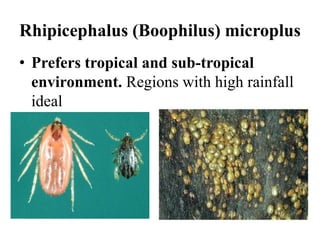
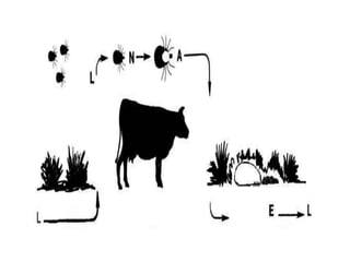
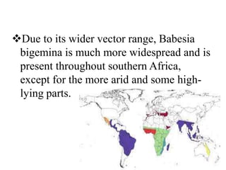
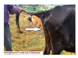
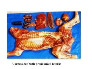
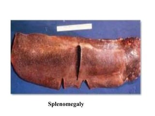
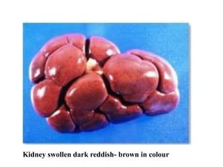
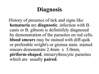
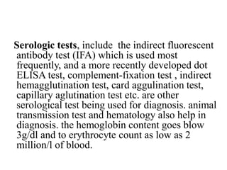
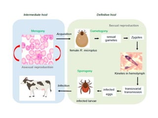
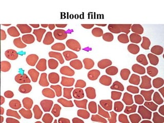
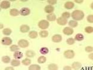
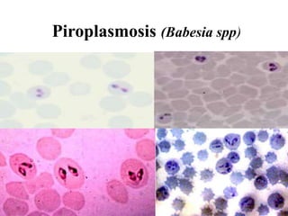
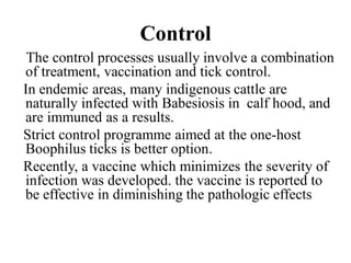
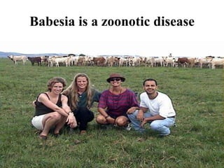
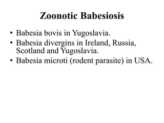
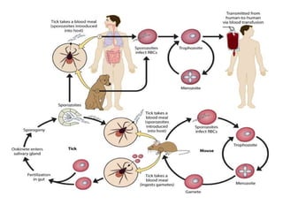
Faculty of Veterinary Medicine &
Animal Husbandry
Somali National University
Mogadishu, Gaheyr Campus
Nov. 01. 2019
INFECTIOUS DISEASES II (Protozoal Diseases)
Bovine Babesiosis
Bovine Babesiosis Common Names
Tick Fever
Cattle Fever
Texas Fever
Redwater
 Piroplasmosis
INTRODUCTION
Bovine babesiosis or redwater is a tick-
borne disease caused by the intra-
erythrocytic protozoan parasite Babesia.
Two species are economically important
in tropical and subtropical regions of
the world, including southern Africa:
Babesia bovis, which causes Asiatic
redwater, and Babesia bigemina, which
causes African redwater.
Babesia divergens causes an
economically important disease in the
British Isles and northern Europe.
The acute disease is characterized by
haemolysis and circulatory disorders (in
the case of B. bovis); death may follow in
some instances.
European breeds introduced to
tropical/subtropical regions are particularly
susceptible to Asiatic and African redwater.
A clinically inapparent form of the disease
is common in young animals, and recovered
animals become latent carriers for variable
periods. Recovery is followed by a lasting
immunity. Cross-immunity between the two
organisms is limited.
Babesia bovis was probably introduced into
southern Africa with the Asian blue tick
(Rhipicephalus (Boophilus) microplus)
during the latter part of the 19th century.
Babesia bigemina is principally transmitted
by the common, indigenous African blue
tick (Rhipicephalus (Boophilus)
decoloratus), as well as by R. (B.)
microplus.
Other tick vectors may also be involved.
• The diseases caused by B. bovis, B.
bigemina and B. divergens are clinically
very similar but it is important to
differentiate between them for a number
of reasons.
While B. bovis is the more virulent of the
two parasites, B. bigemina is probably
more important in southern African
because of its wider distribution.
Etiology
Babesia are protozoa in the family
Babesiidae, order Piroplasmida.
Bovine babesiosis is usually caused by
Babesia bovis, B. bigemina and B.
divergens. B. major, B. ovata, B.
occultans, B. jakimovi and some unnamed
organisms also infect cattle, but seem to
be much less virulent.
Principle Tick Vectors
Rhipicephalus (Boophilus)
microplus, known as the Southern or
tropical cattle tick
Rhipicephalus (Boophilus)
annulatus, known as the cattle tick
Rhipicephalus (Boophilus) microplus
• Prefers tropical and sub-tropical
environment. Regions with high rainfall
ideal
One Host Tick Biology
Larvae, nymphs and adult ticks feed, molt,
and mate on one host
The on-host sequence requires 20-25 days
Engorged female ticks drop to ground to lay
eggs and produce 6-legged larvae
The off-host sequence may last 6-9 months
without a subsequent host
Multiple generations of ticks are produced
each year.
Epidemiology
This disease is present in whole world., but is
prevalence is more in hot and humid areas.
ticks that acquire Babesia infections from
infected animals and then pass it to other
health animals as a subsequent blood meal
transmit the disease. the infection in ticks can
be passed onto the next generation through
the eggs. this disease can occur almost
everywhere around the world.
it is a major threat to cattle development
in many tropical and sub-tropical
countries, especially when exotic breeds
are used . disease mostly occurs
sporadically and can be treated easily so
mortality is not so common.
DISTRIBUTION
Both Babesia species occur in Central
and South America, parts of Europe and
Asia, Australia and Africa.
Babesia bigemina has been eradicated
from the United States of America. In
southern Africa Babesia bovis is
restricted to certain areas, usually the
higher rainfall areas in the eastern parts.
Due to its wider vector range, Babesia
bigemina is much more widespread and is
present throughout southern Africa,
except for the more arid and some high-
lying parts.
Hosts
Other hosts
• Man
• Camel
SUCEPTIBLE HOST/
RESERVIORS
European, Sanga and Zebu breeds are all
susceptible, and all develop latent
infections after recovery. European
breeds can retain B. bovis infections for
life and remain infective for ticks for up
to two years, while most cattle with a
significant Zebu content lose the
infection within two years.
Babesia bigemina infections rarely
persist for more than a year, regardless of
the host, and infected cattle remain
infective for ticks for only four to seven
weeks.
• It is possible to have Babesia spp. and
their vectors present in a cattle population
without measurable economic losses or
clinical disease.
Transmission
• Sporozoites pass to cattle from infected ticks
through blood feeding & enter red blood cells
• Reproduce asexually rupturing red blood
cells upon release of merozoites
• Re-enter gut of subsequent ticks through
feeding process
• Reproduce sexually within tick
• Infect developing eggs of next tick generation
Pathogenesis
Animals are effected after an infected tick
bites and feeds on a susceptible host for a
minimum of three days.
when the Babesia organism is introduced into
the host, it attaches to erythrocyte membranes
and endocytosed.
Hemolytic anemia and hypotensive shock are
typical clinical syndromes of infection.
Hemolytic anemia results from direct
erythrocyte damage by the parasite, and both
intravascular and extravascular immune-
mediated destruction of red blood cells.
infection can produce thrombocytopenia, the
mechanism of which consists of immune-
mediated destruction and sequestration in the
spleen.
Clinical findings
Fever is the first clinical sign one to three
weeks after an infective tick bite.
following the bite , the parasites invade red
blood cells where they multiply and invade
more red blood cells the destruction of
increasing number of red blood cells causes
anemia, shock and also release large
quantities of haemoglobin into the circulation
the sick animals are usually depressed, loss
appetite, and their ayes and gums are pale
from anemia and jaundiced due to bile
pigments in their circulation. red coloured
urine can usually be observed in these
animals. pregnant animals often abort. in
severe B. bovis infection cases, nervous signs
such as incoordination. paralysis and coma
are presented which often lead to death.
Babesia infections can range from in
appearance to acute severe diseases.
In severe cases, animals often die within one
or two days of the appearance of clinical
signs. however in less severe cases, animals
can have a fever for about a weak and it may
remain sick for about three weeks. after that ,
they can have slow recovery, but will remain
infected carriers for the rest of their lives.
Necropsy findings
Physical examination reveals splenomegaly,
lymphadenomagely, hematuria, and icterus.
hypotensive shock results from the release and
production of vasoactive amines and cytokines
which produce vasodilatation.
There will be enlarged kidney and red-brown
colored urine filled bladder. on the different layers
of heart muscle there will echymotic hemorrhage.
there will be intravascular clotting
Haemoglobinuria: Opened urinary bladder
with dark, reddish-brown urine
Carcass calf with pronounced Icterus
Splenomegaly
Kidney swollen dark reddish- brown in colour
Diagnosis
History of presence of tick and signs like
hematuria are diagnostic. infection with B.
canis or B. gibsoni is definitifely diagnosed
by demonstration of the parasites on red cells.
blood smears may be stained with diff-quik
or preferable wright's or giemsa stain. stained
smears demonstrate 2.4mm x 5.0mm,
piriform-shaped, intraerythrocytic parasites
which are usually paired.
Serologic tests, include the indirect fluorescent
antibody test (IFA) which is used most
frequently, and a more recently developed dot
ELISA test, complement-fixation test , indirect
hemagglutination test, card aggulination test,
capillary aglutination test etc. are other
serological test being used for diagnosis. animal
transmission test and hematology also help in
diagnosis. the hemoglobin content goes blow
3g/dl and to erythrocyte count as low as 2
million/l of blood.
Blood film
Piroplasmosis (Babesia spp)
Differential Diagnosis
Babesiosis in bovines could be confused
with anaplasmosis, but the latter
generally leads to rumen stasis and
constipation. Other causes of haematuria
or haemoglobinuria may lead to a
suspicion of babesiosis. Cerebral
babesiosis could be confused with
heartwater.
Control
The control processes usually involve a combination
of treatment, vaccination and tick control.
In endemic areas, many indigenous cattle are
naturally infected with Babesiosis in calf hood, and
are immuned as a results.
Strict control programme aimed at the one-host
Boophilus ticks is better option.
Recently, a vaccine which minimizes the severity of
infection was developed. the vaccine is reported to
be effective in diminishing the pathologic effects
Babesia is a zoonotic disease
Zoonotic Babesiosis
• Babesia bovis in Yugoslavia.
• Babesia divergins in Ireland, Russia,
Scotland and Yugoslavia.
• Babesia microti (rodent parasite) in USA.
Babesia
Babesia

Babesia

  • 1.
    Faculty of VeterinaryMedicine & Animal Husbandry Somali National University Mogadishu, Gaheyr Campus Nov. 01. 2019 INFECTIOUS DISEASES II (Protozoal Diseases) Bovine Babesiosis
  • 2.
    Bovine Babesiosis CommonNames Tick Fever Cattle Fever Texas Fever Redwater  Piroplasmosis
  • 3.
    INTRODUCTION Bovine babesiosis orredwater is a tick- borne disease caused by the intra- erythrocytic protozoan parasite Babesia.
  • 4.
    Two species areeconomically important in tropical and subtropical regions of the world, including southern Africa: Babesia bovis, which causes Asiatic redwater, and Babesia bigemina, which causes African redwater.
  • 5.
    Babesia divergens causesan economically important disease in the British Isles and northern Europe. The acute disease is characterized by haemolysis and circulatory disorders (in the case of B. bovis); death may follow in some instances.
  • 6.
    European breeds introducedto tropical/subtropical regions are particularly susceptible to Asiatic and African redwater. A clinically inapparent form of the disease is common in young animals, and recovered animals become latent carriers for variable periods. Recovery is followed by a lasting immunity. Cross-immunity between the two organisms is limited.
  • 7.
    Babesia bovis wasprobably introduced into southern Africa with the Asian blue tick (Rhipicephalus (Boophilus) microplus) during the latter part of the 19th century. Babesia bigemina is principally transmitted by the common, indigenous African blue tick (Rhipicephalus (Boophilus) decoloratus), as well as by R. (B.) microplus. Other tick vectors may also be involved.
  • 8.
    • The diseasescaused by B. bovis, B. bigemina and B. divergens are clinically very similar but it is important to differentiate between them for a number of reasons. While B. bovis is the more virulent of the two parasites, B. bigemina is probably more important in southern African because of its wider distribution.
  • 9.
    Etiology Babesia are protozoain the family Babesiidae, order Piroplasmida. Bovine babesiosis is usually caused by Babesia bovis, B. bigemina and B. divergens. B. major, B. ovata, B. occultans, B. jakimovi and some unnamed organisms also infect cattle, but seem to be much less virulent.
  • 10.
    Principle Tick Vectors Rhipicephalus(Boophilus) microplus, known as the Southern or tropical cattle tick Rhipicephalus (Boophilus) annulatus, known as the cattle tick
  • 11.
    Rhipicephalus (Boophilus) microplus •Prefers tropical and sub-tropical environment. Regions with high rainfall ideal
  • 12.
    One Host TickBiology Larvae, nymphs and adult ticks feed, molt, and mate on one host The on-host sequence requires 20-25 days Engorged female ticks drop to ground to lay eggs and produce 6-legged larvae The off-host sequence may last 6-9 months without a subsequent host Multiple generations of ticks are produced each year.
  • 14.
    Epidemiology This disease ispresent in whole world., but is prevalence is more in hot and humid areas. ticks that acquire Babesia infections from infected animals and then pass it to other health animals as a subsequent blood meal transmit the disease. the infection in ticks can be passed onto the next generation through the eggs. this disease can occur almost everywhere around the world.
  • 15.
    it is amajor threat to cattle development in many tropical and sub-tropical countries, especially when exotic breeds are used . disease mostly occurs sporadically and can be treated easily so mortality is not so common.
  • 16.
    DISTRIBUTION Both Babesia speciesoccur in Central and South America, parts of Europe and Asia, Australia and Africa. Babesia bigemina has been eradicated from the United States of America. In southern Africa Babesia bovis is restricted to certain areas, usually the higher rainfall areas in the eastern parts.
  • 17.
    Due to itswider vector range, Babesia bigemina is much more widespread and is present throughout southern Africa, except for the more arid and some high- lying parts.
  • 18.
  • 19.
  • 20.
    SUCEPTIBLE HOST/ RESERVIORS European, Sangaand Zebu breeds are all susceptible, and all develop latent infections after recovery. European breeds can retain B. bovis infections for life and remain infective for ticks for up to two years, while most cattle with a significant Zebu content lose the infection within two years.
  • 21.
    Babesia bigemina infectionsrarely persist for more than a year, regardless of the host, and infected cattle remain infective for ticks for only four to seven weeks. • It is possible to have Babesia spp. and their vectors present in a cattle population without measurable economic losses or clinical disease.
  • 22.
    Transmission • Sporozoites passto cattle from infected ticks through blood feeding & enter red blood cells • Reproduce asexually rupturing red blood cells upon release of merozoites • Re-enter gut of subsequent ticks through feeding process • Reproduce sexually within tick • Infect developing eggs of next tick generation
  • 23.
    Pathogenesis Animals are effectedafter an infected tick bites and feeds on a susceptible host for a minimum of three days. when the Babesia organism is introduced into the host, it attaches to erythrocyte membranes and endocytosed. Hemolytic anemia and hypotensive shock are typical clinical syndromes of infection.
  • 24.
    Hemolytic anemia resultsfrom direct erythrocyte damage by the parasite, and both intravascular and extravascular immune- mediated destruction of red blood cells. infection can produce thrombocytopenia, the mechanism of which consists of immune- mediated destruction and sequestration in the spleen.
  • 25.
    Clinical findings Fever isthe first clinical sign one to three weeks after an infective tick bite. following the bite , the parasites invade red blood cells where they multiply and invade more red blood cells the destruction of increasing number of red blood cells causes anemia, shock and also release large quantities of haemoglobin into the circulation
  • 26.
    the sick animalsare usually depressed, loss appetite, and their ayes and gums are pale from anemia and jaundiced due to bile pigments in their circulation. red coloured urine can usually be observed in these animals. pregnant animals often abort. in severe B. bovis infection cases, nervous signs such as incoordination. paralysis and coma are presented which often lead to death.
  • 27.
    Babesia infections canrange from in appearance to acute severe diseases. In severe cases, animals often die within one or two days of the appearance of clinical signs. however in less severe cases, animals can have a fever for about a weak and it may remain sick for about three weeks. after that , they can have slow recovery, but will remain infected carriers for the rest of their lives.
  • 29.
    Necropsy findings Physical examinationreveals splenomegaly, lymphadenomagely, hematuria, and icterus. hypotensive shock results from the release and production of vasoactive amines and cytokines which produce vasodilatation. There will be enlarged kidney and red-brown colored urine filled bladder. on the different layers of heart muscle there will echymotic hemorrhage. there will be intravascular clotting
  • 30.
    Haemoglobinuria: Opened urinarybladder with dark, reddish-brown urine
  • 31.
    Carcass calf withpronounced Icterus
  • 32.
  • 33.
    Kidney swollen darkreddish- brown in colour
  • 34.
    Diagnosis History of presenceof tick and signs like hematuria are diagnostic. infection with B. canis or B. gibsoni is definitifely diagnosed by demonstration of the parasites on red cells. blood smears may be stained with diff-quik or preferable wright's or giemsa stain. stained smears demonstrate 2.4mm x 5.0mm, piriform-shaped, intraerythrocytic parasites which are usually paired.
  • 35.
    Serologic tests, includethe indirect fluorescent antibody test (IFA) which is used most frequently, and a more recently developed dot ELISA test, complement-fixation test , indirect hemagglutination test, card aggulination test, capillary aglutination test etc. are other serological test being used for diagnosis. animal transmission test and hematology also help in diagnosis. the hemoglobin content goes blow 3g/dl and to erythrocyte count as low as 2 million/l of blood.
  • 37.
  • 39.
  • 40.
    Differential Diagnosis Babesiosis inbovines could be confused with anaplasmosis, but the latter generally leads to rumen stasis and constipation. Other causes of haematuria or haemoglobinuria may lead to a suspicion of babesiosis. Cerebral babesiosis could be confused with heartwater.
  • 41.
    Control The control processesusually involve a combination of treatment, vaccination and tick control. In endemic areas, many indigenous cattle are naturally infected with Babesiosis in calf hood, and are immuned as a results. Strict control programme aimed at the one-host Boophilus ticks is better option. Recently, a vaccine which minimizes the severity of infection was developed. the vaccine is reported to be effective in diminishing the pathologic effects
  • 42.
    Babesia is azoonotic disease
  • 43.
    Zoonotic Babesiosis • Babesiabovis in Yugoslavia. • Babesia divergins in Ireland, Russia, Scotland and Yugoslavia. • Babesia microti (rodent parasite) in USA.