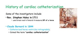
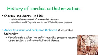
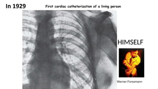
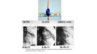
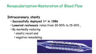
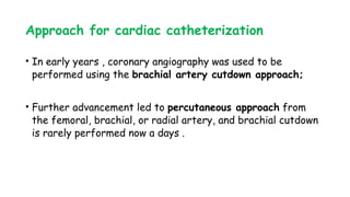
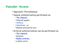
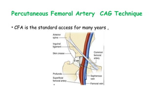
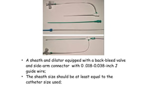
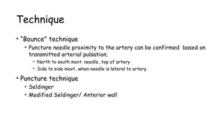
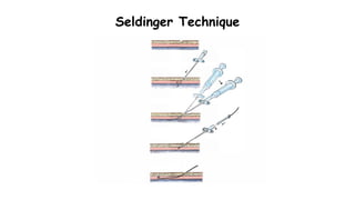
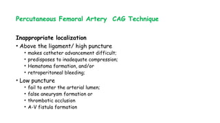
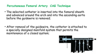
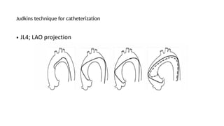
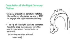
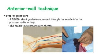
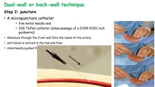
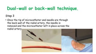
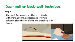
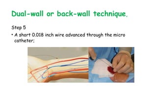
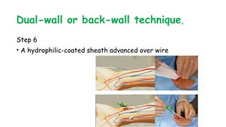
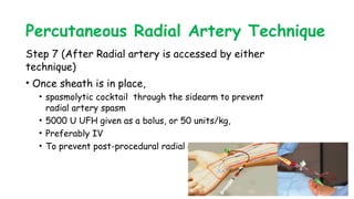
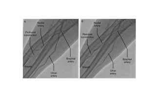
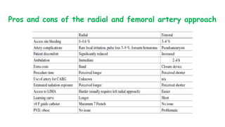
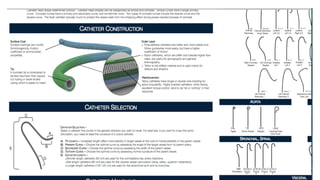
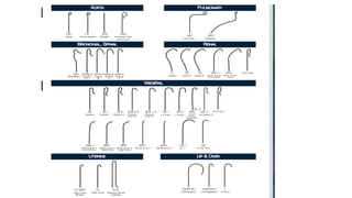
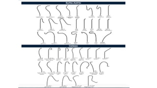
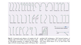
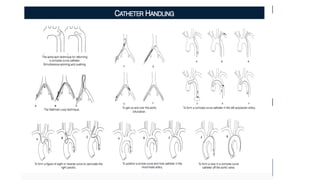
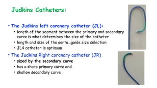
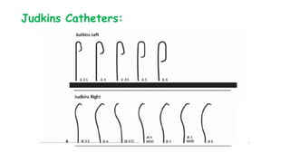
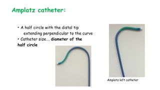
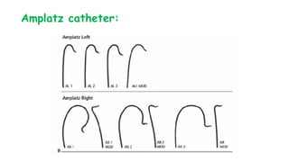
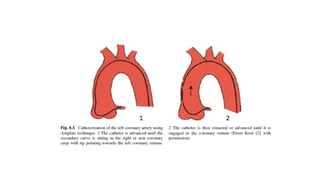
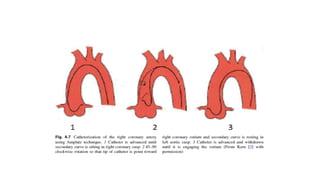
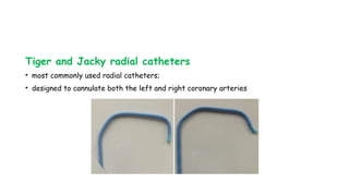
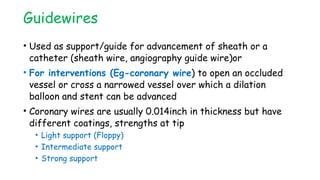
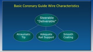
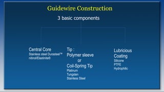
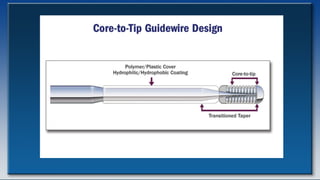
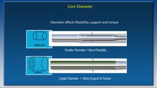
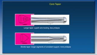
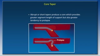
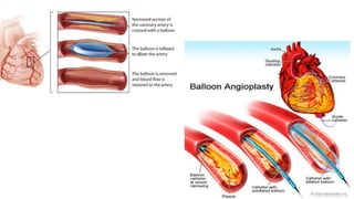
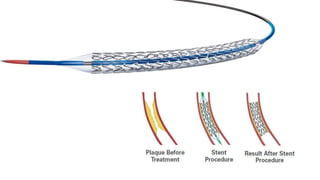
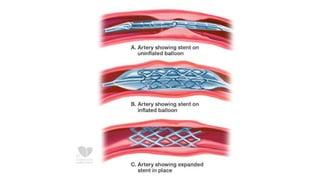
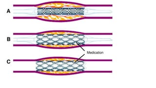
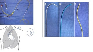
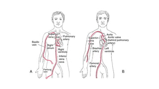
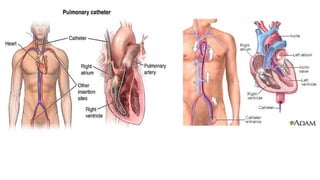
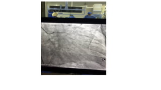
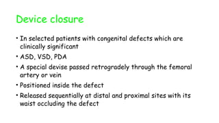
Common Devices and procedures in
Interventional/Invasive Cardiology
By: Seifu Bacha; MD, Internist & Cardiologist
SPHMMC Cardiology Division
November 2023
Outline
โ€ข Brief History of invasive cardiology
โ€ข Vascular access techniques
โ€ข Guide catheters and Guide wires
โ€ข Balloon catheters and Stents
โ€ข Common Invasive Cardiac procedures
History of cardiac catheterization
Some of the investigators include
โ€ข Rev. Stephan Hales in 1711
โ€ข used brass road in femoral A measured BP of a horse
โ€ข Claude Bernard in 1844
โ€ข Catheterized horse ventricles retrogradely;
โ€ข Coined the term โ€œcardiac catheterizationโ€
History of cardiac catheterization
โ€ข Chaveau and Marey in 1861
โ€ข published measurement of intracardiac pressure;
โ€ข apical beat and LV systole; aortic and LV simultaneous pressure
โ€ข Andre Cournand and Dickinson Richards at Columbia
University
โ€ข Hemodynamic exploration and intracardiac pressure measurement in
normal subjects and congenital heart disease
HIMSELF
Werner Forssmann
First cardiac catheterization of a living person
In 1929
History of cardiac catheterization
โ€ข Retrograde left heart catheterization
โ€ข first reported by Zimmerman and Limon-Lason in 1950
โ€ข The percutaneous (rather than cut-down) technique
โ€ข developed by Seldinger (radiologist) in 1953
History of cardiac catheterization
โ€ข Selective coronary arteriography
โ€ข 1st accidentally by Mason Sones (pediatric cardiologist) in
1959
โ€ข 1967- Melvin Judkins- specially shaped catheters to perform
selective angiography
โ€ข Modified CAG for Percutaneous approach
โ€ข in 1959 and 1967, By Ricketts, Abrams and Judkins
โ€ข Swan and Ganz
โ€ข In 1970
โ€ข Developed a flow guided balloon tipped catheter
Revascularizationโ€“Restoration of Blood Flow
โ€ข 1964 โ€“First concept of percutaneous revascularization
(Charles Dotter and Judkins)
(used spring-coil guide wire with large rigid dilator)
โ€ข 1974 -Percutaneous transluminal peripheral
angioplasty
Andreas Gruentzig- series of experiments in animals,
cadavers)
โ€ข 1977 -Percutaneous transluminal coronary angioplasty
(Andreas Gruentzig) On a conscious human
The patient, 38-year-old Adolph Bachman, underwent successful angioplasty to a proximal left
coronary artery lesion in Sep.16, 1977
Revascularizationโ€“Restoration of Blood Flow
Intracoronary stents
โ€ข Successfully deployed 1st in 1986
โ€ข Lowered restenosis rates from 30โ€“50% to 15โ€“30% ,
โ€ข By markedly reducing
โ€ข elastic recoil and
โ€ข negative remodeling
Approach for cardiac catheterization
โ€ข In early years , coronary angiography was used to be
performed using the brachial artery cutdown approach;
โ€ข Further advancement led to percutaneous approach from
the femoral, brachial, or radial artery, and brachial cutdown
is rarely performed now a days .
Preparations and premedication
โ€ข NPO after midnight except for medication
โ€ข Shave inguinal area
โ€ข Light shower
โ€ข Consent
โ€ข Conscious Sedation
โ€ข Midazolam 0.5mg IV PRN
โ€ข Fentanyl 25-50mg IV
Vascular Access
โ€ข Approach= Percutaneous
โ€ข Venous catheterization performed via
โ€ข The femoral,
โ€ข Internal jugular,
โ€ข Axillary,
โ€ข Subclavian , or
โ€ข Median antecubital vein;
โ€ข Arterial catheterization can be performed via
โ€ข The femoral,
โ€ข Brachial,
โ€ข Radial arteries,
โ€ข Lumbar aorta.
Percutaneous Femoral Artery CAG Technique
โ€ข CFA is the standard access for many years ,
Percutaneous Femoral Artery CAG Technique
โ€ข Patient preparation for femoral A/V catheterization
โ€ข Localize pulsation FA/ Shave 10cm diameter/
โ€ข Prepare both groins
โ€ข Clean puncture area with chlorhexidine-alcohol based antiseptic
โ€ข Drape from clavicles to below the feet leaving groin exposed
โ€ข Conscious sedation
โ€ข Locate femoral pulsation
โ€ข Local Anesthetics 10-20 ml 1% or 2% Lidocaine/ Xylocaine
Percutaneous Femoral Artery CAG Technique
โ€ข Locate Inguinal ligament
โ€ข And then femoral artery
โ€ข 1-2 cm below inguinal ligament
โ€ข Donโ€™t use skin crease
โ€ข Aim for mid common femoral
artery
facilitates vessel entry &
effective compression;
Percutaneous Femoral Artery CAG Technique
โ€ข Fluoroscopy
โ€ข AP view
โ€ข Localize Ibfh (inferior border of femoral
head) by forceps
โ€ข Or intra-procedure ultrasound
โ€ข FA: Pulsating, circular, non-compressible
Materials for puncture
โ€ข 18-gauge single-wall puncture
needle
โ€ข 18-21 gauge thin-walled
Seldinger needle with
Obturator;
โ€ข Micropunctureยฎ Introducer Set
:
โ€ข 21 gauge needle ,
โ€ข 5F introducer and dilator and
โ€ข 0 .018-inch nitinol wire
โ€ข A sheath and dilator equipped with a back-bleed valve
and side-arm connector with 0 .018-0.038-inch J
guide wire;
โ€ข The sheath size should be at least equal to the
catheter size used;
Technique
โ€ข โ€œBounce" technique
โ€ข Puncture needle proximity to the artery can be confirmed based on
transmitted arterial pulsation;
โ€ข North to south movt. needleโ€ฆtop of artery
โ€ข Side to side movtโ€ฆwhen needle is lateral to artery
โ€ข Puncture technique
โ€ข Seldinger
โ€ข Modified Seldinger/ Anterior wall
Seldinger Technique
Modified Seldinger Technique
Percutaneous Femoral Artery CAG Technique
โ€ข Never use force when advancing is difficult;
โ€ข Fluoroscopy โ€ฆ.may show tip in branchโ€ฆslightly withdraw and
reorient;
โ€ข Tip of needle may be back wallโ€ฆdepress the unit
โ€ข Control proximal end of the guide-wire and that it is held in a
fixed position as the sheath and dilator are introduced;
โ€ข Sheath and dilator are rotated as a unit to ease insertion;
Percutaneous Femoral Artery CAG Technique
Inappropriate localization
โ€ข Above the ligament/ high puncture
โ€ข makes catheter advancement difficult;
โ€ข predisposes to inadequate compression;
โ€ข Hematoma formation, and/or
โ€ข retroperitoneal bleeding;
โ€ข Low puncture
โ€ข fail to enter the arterial lumen;
โ€ข false aneurysm formation or
โ€ข thrombotic occlusion
โ€ข A-V fistula formation
Percutaneous Femoral Artery CAG Technique
โ€ข The selected catheter is inserted into the femoral sheath
and advanced around the arch and into the ascending aorta
before the guidewire is removed;
โ€ข After removal of the guidewire, the catheter is attached to
a specially designed manifold system that permits the
maintenance of a closed system;
Percutaneous Femoral Artery CAG Technique
โ€ข Catheter is immediately double-flushed-----blood is withdrawn
and discarded, after which heparinized saline flush is injected
through the catheter lumen;
โ€ข Difficulty in blood withdrawal suggests apposition of the
catheter tip to the aortic wallโ€ฆcorrected by slight withdrawal
or rotation of the catheter until free blood aspiration is
possible.
โ€ข If notโ€ฆ.remove catheter and flush it in towel to see any
thrombus collected in the catheter;
Percutaneous Femoral Artery CAG Technique
โ€ข The lumen of the introducing sheath should also be flushed
immediately before and after each catheter insertion and
every 5 minutes thereafter;
โ€ข Alternatively, the side arm of the sheath may be connected
to a 30 ml/hour continuous flow regulator.
Percutaneous Femoral Artery CAG Technique
โ€ข Always start by โ€œZeroingโ€ the pressure to Atmospheric
pressure
โ€ข Once the catheter has been flushed with saline solution, tip
pressure should be displayed on the physiologic monitor at all
times (except during actual contrast injections);
โ€ข Recording this baseline pressure before contrast
administration serves as an important baseline reference
point;
โ€ข Any subsequent alteration in that waveform during coronary
angiography may signify
? Damping
โ€ข A fall in overall catheter tip pressure (because of an ostial coronary stenosis or
an unfavorable catheter position within the coronary artery)
? Ventricularization
โ€ข Diastolic pressure drops while systolic remains high
Cannulation of the Left Coronary Ostium
โ€ข Once the above measures are completed and all is in order,
the coronary angiographic catheter is advanced into the
aortic root in preparation for selective engagement of the
desired coronary ostium;
โ€ข Engagement of the left coronary ostium is usually quite easy
with the Judkins technique;
"the catheters know where to go if not thwarted by the operator"
Judkins technique for catheterization
โ€ข JL4; LAO projection
Cannulation of right coronary Ostium
โ€ข Requires slightly more catheter
manipulation;
โ€ข JR4 is brought around the aortic
arch with the tip facing inward
until it comes to lie against right
side of the aortic root with its
tip aimed toward the left
coronary ostium;
Cannulation of the Right Coronary
Ostium
โ€ข In LAO projection, carefully rotates
the catheter clockwise by nearly 180ยฐ
to engage the right coronary artery;
โ€ข The tip of the right Judkins catheter
tends to drop more deeply into the
aortic root when the catheter is
rotated;
(as tertiary arm aligns with roof
aortic arch);
Percutaneous Radial Artery Technique
โ€ข Increased patient safety from
โ€ข virtual elimination of access site bleeding and vascular
complications
โ€ข associated with early sheath removal,
โ€ข improved patient comfort,
โ€ข faster recovery
Percutaneous Radial Artery Technique
โ€ข Reasons for not adopting TRA
โ€ข Higher access failure rate
โ€ข increased radiation exposure
โ€ข incompatibility of sheaths larger than 6F
โ€ข propensity for vasospasm,
โ€ข thrombotic radial artery occlusion,
โ€ข dissections
โ€ข compartment syndrome,
โ€ข limited catheter stability,
โ€ข poor coronary engagement
Percutaneous Radial Artery Technique
โ€ข Average diameter of radial artery (RA)
โ€ข 2.8 mm in females and 3.1 mm in males (compatible with 6F sheaths);
โ€ข Advantages of TRA over femoral access
โ€ข ease of compression and hemostasis;
โ€ข vast collateralization of the radial artery;
โ€ข no risk of neurologic sequelae (cff Ulnar)
โ€ข Ulnar artery
โ€ข deep lying, mobile, adjacent to the ulnar nerve NOT IDEAL for
first-line vascular access
Percutaneous Radial Artery Technique
โ€ข Positioning
โ€ข left or the right radial artery
โ€ข Comfortable position to patient and operator
โ€ข If RIGHT
โ€ข Abduct right arm by 30 degree
โ€ข Wrist hyperextended
โ€ข Rolled towel/ dedicated splint
โ€ข Fingers taped to the arm board
โ€ข Oximeter
Percutaneous Radial Artery Technique
Radial Puncture
โ€ข Access techniques
โ€ข single-wall versus double-wall or back-wall technique
โ€ข kits include
โ€ข a micropuncture needle or a 20 gauge angiocath needle
โ€ข a short 0.018-0.021 inch wire,
โ€ข arterial sheath with/ without hydrophilic coating
โ€ข less spasm and patient discomfort
โ€ข 5F-6F
โ€ข shorter ( 10 to 13 cm) or longer (23 cm) length
โ€ข Adequate Sedation
Anterior-wall technique
โ€ข Step 1: LA
โ€ข Approximately 2 cm proximal to the radial styloid process,
โ€ข not at the wrist;
โ€ข anesthetized with
โ€ข 2-3 cc of 1% lidocaine with a small (3cc) syringe 25 gauge needle;
Anterior-wall technique
โ€ข Step 2: arterial puncture in front wall technique
โ€ข a short 2.5 cm, stainless steel, 21 G needle/ micro
puncture catheter needle
front wall technique
Anterior-wall technique
โ€ข Step 3: Access techniques-front wall technique
โ€ข Needle advanced into radial artery
โ€ข Blood return indicates intraluminal needle position
โ€ข The blood return is rarely pulsatile or brisk.
Anterior-wall technique
โ€ข Step 4: guide wire
โ€ข A 0.018in short guidewire advanced through the needle into the
proximal radial artery,
โ€ข The needle is exchanged with sheath
Dual-wall or back-wall technique,
Step 2: puncture
โ€ข A micropuncture catheter
โ€ข fine metal needle and
โ€ข 22G Teflon catheter (allow passage of a 0.018-0.021 inch
guidewire);
โ€ข Advanced through the front wall Into the lumen of the artery
โ€ข until blood is noticed in the hub and then
โ€ข intentionally pushed through the back wall of the Artery
Dual-wall or back-wall technique,
Step 3
โ€ข Once the tip of microcatheter and needle are through
the back wall of the radial artery, the needle is
removed and the microcatheter left in place across the
radial artery
Dual-wall or back-wall technique,
Step 4
โ€ข the small Teflon microcatheter is slowly
withdrawn until the appearance of brisk
pulsatile flow that confirms the distal tip is in
lumen
Dual-wall or back-wall technique,
Step 5
โ€ข A short 0.018 inch wire advanced through the micro
catheter;
Dual-wall or back-wall technique,
Step 6
โ€ข A hydrophilic-coated sheath advanced over wire
Percutaneous Radial Artery Technique
Step 7 (After Radial artery is accessed by either
technique)
โ€ข Once sheath is in place,
โ€ข spasmolytic cocktail through the sidearm to prevent
radial artery spasm
โ€ข 5000 U UFH given as a bolus, or 50 units/kg,
โ€ข Preferably IV
โ€ข To prevent post-procedural radial artery occlusion;
Pros and cons of the radial and femoral artery approach
Vascular Catheters
โ€ข Selection of catheter is an elementary decision, however, it
can be the difference between success and failure
โ€ข Diagnostic catheters (for diagnostic purposes) and
โ€ข Guiding catheters (for intervention)
โžขcome in multiple preformed shapes and sizes
Guide catheters
โ€ข Constructed from polyethylene or polyurethane;
(resist softening in the body);
โ€ข Distal wire mesh that allows torque creation site
โ€ข Have a stiffer shaft, larger internal diameter,
โ€ข Has a soft distal tip to minimize the risk of arterial dissection;
โ€ข MOST procedure are performed through a 6 F guide (5-8 F)
catheters;
โ€ข As a rule, the outer diameter (OD) of the guiding catheter is
the same dimension as the inner diameter (ID) of the sheath;
Basic Functions of a Guiding Catheter
โ€ข Provide a supportive conduit for advancement
of guidewires and devices
โ€ข Serve as a vehicle for contrast injection
โ€ข Measurement of blood pressure
Important Characteristics of a Guiding Catheter
โ€ข Atraumatic tip
โ€ข Proper preformed shape ( co-axial with vessel)
โ€ข Torque control
โ€ข Kink resistance
โ€ข Radiopacity
Guide catheters
โ€ข Measured in French (Fr), referring to the outer diameter
(Note: refers to inner diameter for sheaths)
โ€ข 1Fr= 3mm [Eg- 6Fr catheter has an outer diameter of
2mm(6 divided by 3)
โ€ข Length usually 100cm
โ€ข Choice of size of the curve depends on the aortic root
diameter- usually ranges from 3.5 to 5.0cm)
โ€ข Left/Right indicates the side of the coronary artery
โ€ข Eg: 6Fr JL4.0 indicates a Judkinโ€™s Left catheter with
outer diameter of 6Fr and curve length of 4.0cm
Judkins Catheters:
โ€ข The Judkins left coronary catheter (JL):
โ€ข length of the segment between the primary and secondary
curve is what determines the size of the catheter
โ€ข length and size of the aortaโ€ฆguide size selection
โ€ข JL4 catheter is optimum
โ€ข The Judkins Right coronary catheter (JR)
โ€ข sized by the secondary curve
โ€ข has a sharp primary curve and
โ€ข shallow secondary curve
Judkins Catheters:
Amplatz catheter:
โ€ข A half circle with the distal tip
extending perpendicular to the curve
โ€ข Catheter sizeโ€ฆ. diameter of the
half circle
Amplatz left catheter
Amplatz catheter:
Tiger and Jacky radial catheters
โ€ข most commonly used radial catheters;
โ€ข designed to cannulate both the left and right coronary arteries
Guidewires
โ€ข Used as support/guide for advancement of sheath or a
catheter (sheath wire, angiography guide wire)or
โ€ข For interventions (Eg-coronary wire) to open an occluded
vessel or cross a narrowed vessel over which a dilation
balloon and stent can be advanced
โ€ข Coronary wires are usually 0.014inch in thickness but have
different coatings, strengths at tip
โ€ข Light support (Floppy)
โ€ข Intermediate support
โ€ข Strong support
Basic Coronary Guide Wire Characteristics
Adequate
Rail Support
Atraumatic
Tip
Smooth
Coating
Steerable
โ€œDeliverableโ€
Guidewire Construction
Central Core
Stainless steel Durasteelโ„ข
nitinol/Elastiniteยฎ
Tip :
Polymer sleeve
or
Coil-Spring Tip
Platinum
Tungsten
Stainless Steel
Lubricious
Coating
Silicone
PTFE
Hydrophilic
3 basic components
Core Diameter
Diameter affects flexibility, support and torque
Smaller Diameter = More Flexibility
Larger Diameter = More Support & Torque
75
Core Taper
Longer taper- superb wire tracking, less prolapse
Shorter taper- longer segments of consistent support, more prolapse
76
Core Taper
โ€ข Abrupt or short tapers produce a core which provides
greater segment length of support but also greater
tendency to prolapse
Prolapse
77
Core Taper
โ€ข Broad, gradual or long tapers produce a core which
offers greater tracking and wire which prolapses less
Successful
Tracking
78
Core Material
โ€ข Stainless steel
โ€“ Original core material technology
โ€“ Good support, push force and torque
โ€“ Less flexible than newer core materials
Core Material
โ€ข Nitinol/Elastiniteยฎ
โ€“ Super-elastic alloy designed for kink resistance
โ€“ Excellent flexibility and steering
โ€“ Durable nature may facilitate treatment of
multiple lesions and/or tortuous vessels
โ€“ No memory
Work-Horse Guide Wire Characteristics
โ€ข Intermediate Core Diameter
โ€ข Gentle Core Taper
โ€ข Resilient Core with good torque control
โ€ข Soft Tip
โ€ข Coils or Covers
โ€ข Smooth Coating
Change Coronary Guide Wire Characteristics
Adequate
Rail Support
Atraumatic
Spring Tip
Smooth
Coating
Steerable
โ€œDeliverableโ€
Stiff
Tip
Dissections &
Perforations
Increased
Rail Support
Straightening
Artifacts
Hydrophilic
Coating
Perforation
Balloon Catheters (Balloons)
โ€ข Used for dilation of a narrowed vessel
โ€ข Both for coronary and peripheral arteries
โ€ข Over the wire vs Monorail/rapid exchange (widely used)
โ€ข Different types, mainly 3
โ€ข Differ on the distribution and amount of pressure applied on
the diseased and surrounding normal vessel when inflated
Balloon Catheters (Balloons)
โ€ข Complaint = made of polyurethane or silicone. Inflated by
volume instead of pressure from 100-800%- rarely used
โ€ข Semicomplaint = mid pressure balloons- more compliance and
more flexibility than NC
โ€ข Non Complaint (NC)= high pressure balloons- made of
polyster or nylon. Used when balloon needs to expand to a
specific diameter and exert high pressure
Balloon Catheters (Balloons)
โ€ข Size variable from small to large
โ€ข Labelled with the format of โ€œXโ€ mm by โ€œYโ€ mm where the
first number (X) refers to the diameter when inflated and
the second (Y) the length of the balloon
โ€ข Eg; โ€œ3.5 * 12mm NCโ€ labelled balloon implies- a non complaint
balloon of diameter 3.5mm and length 12mm (1.2cm)
โ€ข Inflated using an inflator deflator device with a pressure
gauge indicating the amount of pressure applied (in unit of
atmosphere)
Balloon Catheters (Balloons)
โ€ข The balloon pack is labelled with the amount of pressure
required to achieve a given diameter
โ€ข Nominal Pressure (NP)- the pressure at which the balloon
reaches its labeled diameter
โ€ข Rated Burst Pressure (RBP)- pressure at which 99.9%of
balloons can survive
Stents
โ€ข A very thin but strong wire mesh tubes wrapped around a
balloon and placed at the site of the dilated lesion after
which the balloon is inflated and deflated leaving the stent in
place
โ€ข Prevents recoil and re stenosis of the artery
โ€ข Nomenclature of size (diameter and length), delivery
mechanism similar to balloons
Stents
โ€ข Gradually evolved since first discovery to this date
โ€ข Mainly two types
โ€ข BMS (bare metal stent)- used in the past with a relatively
higher rate of restenosis
โ€ข DES (drug eluting stent)- coated with different types of
drugs which are released gradually and prevent inflammation,
hence reduces risk of restenosis better than BMS
Common Invasive Procedures
โ€ข Less commonly used, not widely available, expensive, high
complication rates
โ–When non invasive tests are inconclusive or not available or
intervention is planned at same time
โ€ข May include:
oLeft Heart catheterization
oRight Heart catheterization
oElectrophysiological studies
o
Left Heart Catheterization
โ€ข Coronary Angiography
โ€ข LV angiogram
โ€ข Measurement of gradients, valve regurgitation
quantifications-less commonly used after advent of modern
echo machines
Right Heart catheterization
โ€ข Monitoring of central venous pressure, guide fluid therapy
โ€ข For measurement of pressures (PA, PCWP)
โ€ข Measurement of Cardiac output
โ€ข Diagnosis of shunts
โ€ข Tests of drug effectiveness (pulmonary hypertension)
CORONARY ANGIOGRAPHY
โ€ข Gold standard for diagnosis of CAD
โ€ข Through femoral or radial arteries
โ€ข Passage of special catheters retrograde into aortic root,
cannulation of the left and right coronanry arteries
โ€ข Injection of a radioopaque agent to replace blood
โ€ข Xray taken to see the arteries in different views
PCI
โ€ข After doing CAG if a treatable lesion is found
โ€ข A special thin (0.014) inch coronary wire is advanced and the
diseased part crossed
โ€ข A balloon is passed over the wire and inflated at the lesion site
to crash the atherosclerotic plaque and expand the artery
โ€ข Can be left here but recoil and restenosis common
โ€ข A stent is placed at the site
โ€ข Bare Metal Stent- BMS (in the past), Drug Eluting Stent- DES
(current practice)
PTMC
โ€ข Percutaneous valvotomy- commonly done for mitral stenosis
โ€ข Commonly through femoral vein
โ€ข Septal puncture done with a special needle to cross from the
right atrium to left atrium
โ€ข Septum will be dilated after putting a special wire into LA
โ€ข Balloon in deflated position is passed over the wire and pushed to
cross the mitral valve an enter LV
โ€ข It will be placed across the narrowed valve and inflated which
results in tear of the mitral commissure and widening of the valve
PPI
โ€ข In patients with symptomatic bradycardia
โ€ข Single chamber (ventricles only) or dual chambers (atrium and
ventricle)
โ€ข Leads are placed inside the cardiac chambers- through cephalic
vein, axillary or subclavian vein
โ€ข Fixed at appropriate sites
โ€ข Connected to the pulse generator which is placed in a subcutaneous
pocket made just below the clavicle
โ€ข Battery usually lasts for 10 years and may need to be replaced
after that
Device closure
โ€ข In selected patients with congenital defects which are
clinically significant
โ€ข ASD, VSD, PDA
โ€ข A special devise passed retrogradely through the femoral
artery or vein
โ€ข Positioned inside the defect
โ€ข Released sequentially at distal and proximal sites with its
waist occluding the defect
Thank you

Invasive_Cardio-Devices_procedures[1].pdf

  • 1.
    Common Devices andprocedures in Interventional/Invasive Cardiology By: Seifu Bacha; MD, Internist & Cardiologist SPHMMC Cardiology Division November 2023
  • 2.
    Outline โ€ข Brief Historyof invasive cardiology โ€ข Vascular access techniques โ€ข Guide catheters and Guide wires โ€ข Balloon catheters and Stents โ€ข Common Invasive Cardiac procedures
  • 3.
    History of cardiaccatheterization Some of the investigators include โ€ข Rev. Stephan Hales in 1711 โ€ข used brass road in femoral A measured BP of a horse โ€ข Claude Bernard in 1844 โ€ข Catheterized horse ventricles retrogradely; โ€ข Coined the term โ€œcardiac catheterizationโ€
  • 4.
    History of cardiaccatheterization โ€ข Chaveau and Marey in 1861 โ€ข published measurement of intracardiac pressure; โ€ข apical beat and LV systole; aortic and LV simultaneous pressure โ€ข Andre Cournand and Dickinson Richards at Columbia University โ€ข Hemodynamic exploration and intracardiac pressure measurement in normal subjects and congenital heart disease
  • 5.
    HIMSELF Werner Forssmann First cardiaccatheterization of a living person In 1929
  • 6.
    History of cardiaccatheterization โ€ข Retrograde left heart catheterization โ€ข first reported by Zimmerman and Limon-Lason in 1950 โ€ข The percutaneous (rather than cut-down) technique โ€ข developed by Seldinger (radiologist) in 1953
  • 7.
    History of cardiaccatheterization โ€ข Selective coronary arteriography โ€ข 1st accidentally by Mason Sones (pediatric cardiologist) in 1959 โ€ข 1967- Melvin Judkins- specially shaped catheters to perform selective angiography โ€ข Modified CAG for Percutaneous approach โ€ข in 1959 and 1967, By Ricketts, Abrams and Judkins โ€ข Swan and Ganz โ€ข In 1970 โ€ข Developed a flow guided balloon tipped catheter
  • 8.
    Revascularizationโ€“Restoration of BloodFlow โ€ข 1964 โ€“First concept of percutaneous revascularization (Charles Dotter and Judkins) (used spring-coil guide wire with large rigid dilator) โ€ข 1974 -Percutaneous transluminal peripheral angioplasty Andreas Gruentzig- series of experiments in animals, cadavers) โ€ข 1977 -Percutaneous transluminal coronary angioplasty (Andreas Gruentzig) On a conscious human
  • 9.
    The patient, 38-year-oldAdolph Bachman, underwent successful angioplasty to a proximal left coronary artery lesion in Sep.16, 1977
  • 10.
    Revascularizationโ€“Restoration of BloodFlow Intracoronary stents โ€ข Successfully deployed 1st in 1986 โ€ข Lowered restenosis rates from 30โ€“50% to 15โ€“30% , โ€ข By markedly reducing โ€ข elastic recoil and โ€ข negative remodeling
  • 11.
    Approach for cardiaccatheterization โ€ข In early years , coronary angiography was used to be performed using the brachial artery cutdown approach; โ€ข Further advancement led to percutaneous approach from the femoral, brachial, or radial artery, and brachial cutdown is rarely performed now a days .
  • 12.
    Preparations and premedication โ€ขNPO after midnight except for medication โ€ข Shave inguinal area โ€ข Light shower โ€ข Consent โ€ข Conscious Sedation โ€ข Midazolam 0.5mg IV PRN โ€ข Fentanyl 25-50mg IV
  • 13.
    Vascular Access โ€ข Approach=Percutaneous โ€ข Venous catheterization performed via โ€ข The femoral, โ€ข Internal jugular, โ€ข Axillary, โ€ข Subclavian , or โ€ข Median antecubital vein; โ€ข Arterial catheterization can be performed via โ€ข The femoral, โ€ข Brachial, โ€ข Radial arteries, โ€ข Lumbar aorta.
  • 14.
    Percutaneous Femoral ArteryCAG Technique โ€ข CFA is the standard access for many years ,
  • 15.
    Percutaneous Femoral ArteryCAG Technique โ€ข Patient preparation for femoral A/V catheterization โ€ข Localize pulsation FA/ Shave 10cm diameter/ โ€ข Prepare both groins โ€ข Clean puncture area with chlorhexidine-alcohol based antiseptic โ€ข Drape from clavicles to below the feet leaving groin exposed โ€ข Conscious sedation โ€ข Locate femoral pulsation โ€ข Local Anesthetics 10-20 ml 1% or 2% Lidocaine/ Xylocaine
  • 16.
    Percutaneous Femoral ArteryCAG Technique โ€ข Locate Inguinal ligament โ€ข And then femoral artery โ€ข 1-2 cm below inguinal ligament โ€ข Donโ€™t use skin crease โ€ข Aim for mid common femoral artery facilitates vessel entry & effective compression;
  • 17.
    Percutaneous Femoral ArteryCAG Technique โ€ข Fluoroscopy โ€ข AP view โ€ข Localize Ibfh (inferior border of femoral head) by forceps โ€ข Or intra-procedure ultrasound โ€ข FA: Pulsating, circular, non-compressible
  • 18.
    Materials for puncture โ€ข18-gauge single-wall puncture needle โ€ข 18-21 gauge thin-walled Seldinger needle with Obturator; โ€ข Micropunctureยฎ Introducer Set : โ€ข 21 gauge needle , โ€ข 5F introducer and dilator and โ€ข 0 .018-inch nitinol wire
  • 19.
    โ€ข A sheathand dilator equipped with a back-bleed valve and side-arm connector with 0 .018-0.038-inch J guide wire; โ€ข The sheath size should be at least equal to the catheter size used;
  • 20.
    Technique โ€ข โ€œBounce" technique โ€ขPuncture needle proximity to the artery can be confirmed based on transmitted arterial pulsation; โ€ข North to south movt. needleโ€ฆtop of artery โ€ข Side to side movtโ€ฆwhen needle is lateral to artery โ€ข Puncture technique โ€ข Seldinger โ€ข Modified Seldinger/ Anterior wall
  • 21.
  • 22.
  • 23.
    Percutaneous Femoral ArteryCAG Technique โ€ข Never use force when advancing is difficult; โ€ข Fluoroscopy โ€ฆ.may show tip in branchโ€ฆslightly withdraw and reorient; โ€ข Tip of needle may be back wallโ€ฆdepress the unit โ€ข Control proximal end of the guide-wire and that it is held in a fixed position as the sheath and dilator are introduced; โ€ข Sheath and dilator are rotated as a unit to ease insertion;
  • 24.
    Percutaneous Femoral ArteryCAG Technique Inappropriate localization โ€ข Above the ligament/ high puncture โ€ข makes catheter advancement difficult; โ€ข predisposes to inadequate compression; โ€ข Hematoma formation, and/or โ€ข retroperitoneal bleeding; โ€ข Low puncture โ€ข fail to enter the arterial lumen; โ€ข false aneurysm formation or โ€ข thrombotic occlusion โ€ข A-V fistula formation
  • 25.
    Percutaneous Femoral ArteryCAG Technique โ€ข The selected catheter is inserted into the femoral sheath and advanced around the arch and into the ascending aorta before the guidewire is removed; โ€ข After removal of the guidewire, the catheter is attached to a specially designed manifold system that permits the maintenance of a closed system;
  • 26.
    Percutaneous Femoral ArteryCAG Technique โ€ข Catheter is immediately double-flushed-----blood is withdrawn and discarded, after which heparinized saline flush is injected through the catheter lumen; โ€ข Difficulty in blood withdrawal suggests apposition of the catheter tip to the aortic wallโ€ฆcorrected by slight withdrawal or rotation of the catheter until free blood aspiration is possible. โ€ข If notโ€ฆ.remove catheter and flush it in towel to see any thrombus collected in the catheter;
  • 27.
    Percutaneous Femoral ArteryCAG Technique โ€ข The lumen of the introducing sheath should also be flushed immediately before and after each catheter insertion and every 5 minutes thereafter; โ€ข Alternatively, the side arm of the sheath may be connected to a 30 ml/hour continuous flow regulator.
  • 28.
    Percutaneous Femoral ArteryCAG Technique โ€ข Always start by โ€œZeroingโ€ the pressure to Atmospheric pressure โ€ข Once the catheter has been flushed with saline solution, tip pressure should be displayed on the physiologic monitor at all times (except during actual contrast injections); โ€ข Recording this baseline pressure before contrast administration serves as an important baseline reference point;
  • 29.
    โ€ข Any subsequentalteration in that waveform during coronary angiography may signify ? Damping โ€ข A fall in overall catheter tip pressure (because of an ostial coronary stenosis or an unfavorable catheter position within the coronary artery) ? Ventricularization โ€ข Diastolic pressure drops while systolic remains high
  • 30.
    Cannulation of theLeft Coronary Ostium โ€ข Once the above measures are completed and all is in order, the coronary angiographic catheter is advanced into the aortic root in preparation for selective engagement of the desired coronary ostium; โ€ข Engagement of the left coronary ostium is usually quite easy with the Judkins technique; "the catheters know where to go if not thwarted by the operator"
  • 31.
    Judkins technique forcatheterization โ€ข JL4; LAO projection
  • 32.
    Cannulation of rightcoronary Ostium โ€ข Requires slightly more catheter manipulation; โ€ข JR4 is brought around the aortic arch with the tip facing inward until it comes to lie against right side of the aortic root with its tip aimed toward the left coronary ostium;
  • 33.
    Cannulation of theRight Coronary Ostium โ€ข In LAO projection, carefully rotates the catheter clockwise by nearly 180ยฐ to engage the right coronary artery; โ€ข The tip of the right Judkins catheter tends to drop more deeply into the aortic root when the catheter is rotated; (as tertiary arm aligns with roof aortic arch);
  • 34.
    Percutaneous Radial ArteryTechnique โ€ข Increased patient safety from โ€ข virtual elimination of access site bleeding and vascular complications โ€ข associated with early sheath removal, โ€ข improved patient comfort, โ€ข faster recovery
  • 35.
    Percutaneous Radial ArteryTechnique โ€ข Reasons for not adopting TRA โ€ข Higher access failure rate โ€ข increased radiation exposure โ€ข incompatibility of sheaths larger than 6F โ€ข propensity for vasospasm, โ€ข thrombotic radial artery occlusion, โ€ข dissections โ€ข compartment syndrome, โ€ข limited catheter stability, โ€ข poor coronary engagement
  • 36.
    Percutaneous Radial ArteryTechnique โ€ข Average diameter of radial artery (RA) โ€ข 2.8 mm in females and 3.1 mm in males (compatible with 6F sheaths); โ€ข Advantages of TRA over femoral access โ€ข ease of compression and hemostasis; โ€ข vast collateralization of the radial artery; โ€ข no risk of neurologic sequelae (cff Ulnar) โ€ข Ulnar artery โ€ข deep lying, mobile, adjacent to the ulnar nerve NOT IDEAL for first-line vascular access
  • 37.
    Percutaneous Radial ArteryTechnique โ€ข Positioning โ€ข left or the right radial artery โ€ข Comfortable position to patient and operator โ€ข If RIGHT โ€ข Abduct right arm by 30 degree โ€ข Wrist hyperextended โ€ข Rolled towel/ dedicated splint โ€ข Fingers taped to the arm board โ€ข Oximeter
  • 38.
    Percutaneous Radial ArteryTechnique Radial Puncture โ€ข Access techniques โ€ข single-wall versus double-wall or back-wall technique โ€ข kits include โ€ข a micropuncture needle or a 20 gauge angiocath needle โ€ข a short 0.018-0.021 inch wire, โ€ข arterial sheath with/ without hydrophilic coating โ€ข less spasm and patient discomfort โ€ข 5F-6F โ€ข shorter ( 10 to 13 cm) or longer (23 cm) length โ€ข Adequate Sedation
  • 39.
    Anterior-wall technique โ€ข Step1: LA โ€ข Approximately 2 cm proximal to the radial styloid process, โ€ข not at the wrist; โ€ข anesthetized with โ€ข 2-3 cc of 1% lidocaine with a small (3cc) syringe 25 gauge needle;
  • 40.
    Anterior-wall technique โ€ข Step2: arterial puncture in front wall technique โ€ข a short 2.5 cm, stainless steel, 21 G needle/ micro puncture catheter needle front wall technique
  • 41.
    Anterior-wall technique โ€ข Step3: Access techniques-front wall technique โ€ข Needle advanced into radial artery โ€ข Blood return indicates intraluminal needle position โ€ข The blood return is rarely pulsatile or brisk.
  • 42.
    Anterior-wall technique โ€ข Step4: guide wire โ€ข A 0.018in short guidewire advanced through the needle into the proximal radial artery, โ€ข The needle is exchanged with sheath
  • 43.
    Dual-wall or back-walltechnique, Step 2: puncture โ€ข A micropuncture catheter โ€ข fine metal needle and โ€ข 22G Teflon catheter (allow passage of a 0.018-0.021 inch guidewire); โ€ข Advanced through the front wall Into the lumen of the artery โ€ข until blood is noticed in the hub and then โ€ข intentionally pushed through the back wall of the Artery
  • 44.
    Dual-wall or back-walltechnique, Step 3 โ€ข Once the tip of microcatheter and needle are through the back wall of the radial artery, the needle is removed and the microcatheter left in place across the radial artery
  • 45.
    Dual-wall or back-walltechnique, Step 4 โ€ข the small Teflon microcatheter is slowly withdrawn until the appearance of brisk pulsatile flow that confirms the distal tip is in lumen
  • 46.
    Dual-wall or back-walltechnique, Step 5 โ€ข A short 0.018 inch wire advanced through the micro catheter;
  • 47.
    Dual-wall or back-walltechnique, Step 6 โ€ข A hydrophilic-coated sheath advanced over wire
  • 48.
    Percutaneous Radial ArteryTechnique Step 7 (After Radial artery is accessed by either technique) โ€ข Once sheath is in place, โ€ข spasmolytic cocktail through the sidearm to prevent radial artery spasm โ€ข 5000 U UFH given as a bolus, or 50 units/kg, โ€ข Preferably IV โ€ข To prevent post-procedural radial artery occlusion;
  • 50.
    Pros and consof the radial and femoral artery approach
  • 51.
    Vascular Catheters โ€ข Selectionof catheter is an elementary decision, however, it can be the difference between success and failure โ€ข Diagnostic catheters (for diagnostic purposes) and โ€ข Guiding catheters (for intervention) โžขcome in multiple preformed shapes and sizes
  • 52.
    Guide catheters โ€ข Constructedfrom polyethylene or polyurethane; (resist softening in the body); โ€ข Distal wire mesh that allows torque creation site โ€ข Have a stiffer shaft, larger internal diameter, โ€ข Has a soft distal tip to minimize the risk of arterial dissection; โ€ข MOST procedure are performed through a 6 F guide (5-8 F) catheters; โ€ข As a rule, the outer diameter (OD) of the guiding catheter is the same dimension as the inner diameter (ID) of the sheath;
  • 53.
    Basic Functions ofa Guiding Catheter โ€ข Provide a supportive conduit for advancement of guidewires and devices โ€ข Serve as a vehicle for contrast injection โ€ข Measurement of blood pressure
  • 54.
    Important Characteristics ofa Guiding Catheter โ€ข Atraumatic tip โ€ข Proper preformed shape ( co-axial with vessel) โ€ข Torque control โ€ข Kink resistance โ€ข Radiopacity
  • 55.
    Guide catheters โ€ข Measuredin French (Fr), referring to the outer diameter (Note: refers to inner diameter for sheaths) โ€ข 1Fr= 3mm [Eg- 6Fr catheter has an outer diameter of 2mm(6 divided by 3) โ€ข Length usually 100cm โ€ข Choice of size of the curve depends on the aortic root diameter- usually ranges from 3.5 to 5.0cm) โ€ข Left/Right indicates the side of the coronary artery โ€ข Eg: 6Fr JL4.0 indicates a Judkinโ€™s Left catheter with outer diameter of 6Fr and curve length of 4.0cm
  • 62.
    Judkins Catheters: โ€ข TheJudkins left coronary catheter (JL): โ€ข length of the segment between the primary and secondary curve is what determines the size of the catheter โ€ข length and size of the aortaโ€ฆguide size selection โ€ข JL4 catheter is optimum โ€ข The Judkins Right coronary catheter (JR) โ€ข sized by the secondary curve โ€ข has a sharp primary curve and โ€ข shallow secondary curve
  • 63.
  • 64.
    Amplatz catheter: โ€ข Ahalf circle with the distal tip extending perpendicular to the curve โ€ข Catheter sizeโ€ฆ. diameter of the half circle Amplatz left catheter
  • 65.
  • 68.
    Tiger and Jackyradial catheters โ€ข most commonly used radial catheters; โ€ข designed to cannulate both the left and right coronary arteries
  • 69.
    Guidewires โ€ข Used assupport/guide for advancement of sheath or a catheter (sheath wire, angiography guide wire)or โ€ข For interventions (Eg-coronary wire) to open an occluded vessel or cross a narrowed vessel over which a dilation balloon and stent can be advanced โ€ข Coronary wires are usually 0.014inch in thickness but have different coatings, strengths at tip โ€ข Light support (Floppy) โ€ข Intermediate support โ€ข Strong support
  • 70.
    Basic Coronary GuideWire Characteristics Adequate Rail Support Atraumatic Tip Smooth Coating Steerable โ€œDeliverableโ€
  • 71.
    Guidewire Construction Central Core Stainlesssteel Durasteelโ„ข nitinol/Elastiniteยฎ Tip : Polymer sleeve or Coil-Spring Tip Platinum Tungsten Stainless Steel Lubricious Coating Silicone PTFE Hydrophilic 3 basic components
  • 74.
    Core Diameter Diameter affectsflexibility, support and torque Smaller Diameter = More Flexibility Larger Diameter = More Support & Torque
  • 75.
    75 Core Taper Longer taper-superb wire tracking, less prolapse Shorter taper- longer segments of consistent support, more prolapse
  • 76.
    76 Core Taper โ€ข Abruptor short tapers produce a core which provides greater segment length of support but also greater tendency to prolapse Prolapse
  • 77.
    77 Core Taper โ€ข Broad,gradual or long tapers produce a core which offers greater tracking and wire which prolapses less Successful Tracking
  • 78.
    78 Core Material โ€ข Stainlesssteel โ€“ Original core material technology โ€“ Good support, push force and torque โ€“ Less flexible than newer core materials
  • 79.
    Core Material โ€ข Nitinol/Elastiniteยฎ โ€“Super-elastic alloy designed for kink resistance โ€“ Excellent flexibility and steering โ€“ Durable nature may facilitate treatment of multiple lesions and/or tortuous vessels โ€“ No memory
  • 80.
    Work-Horse Guide WireCharacteristics โ€ข Intermediate Core Diameter โ€ข Gentle Core Taper โ€ข Resilient Core with good torque control โ€ข Soft Tip โ€ข Coils or Covers โ€ข Smooth Coating
  • 81.
    Change Coronary GuideWire Characteristics Adequate Rail Support Atraumatic Spring Tip Smooth Coating Steerable โ€œDeliverableโ€ Stiff Tip Dissections & Perforations Increased Rail Support Straightening Artifacts Hydrophilic Coating Perforation
  • 82.
    Balloon Catheters (Balloons) โ€ขUsed for dilation of a narrowed vessel โ€ข Both for coronary and peripheral arteries โ€ข Over the wire vs Monorail/rapid exchange (widely used) โ€ข Different types, mainly 3 โ€ข Differ on the distribution and amount of pressure applied on the diseased and surrounding normal vessel when inflated
  • 83.
    Balloon Catheters (Balloons) โ€ขComplaint = made of polyurethane or silicone. Inflated by volume instead of pressure from 100-800%- rarely used โ€ข Semicomplaint = mid pressure balloons- more compliance and more flexibility than NC โ€ข Non Complaint (NC)= high pressure balloons- made of polyster or nylon. Used when balloon needs to expand to a specific diameter and exert high pressure
  • 84.
    Balloon Catheters (Balloons) โ€ขSize variable from small to large โ€ข Labelled with the format of โ€œXโ€ mm by โ€œYโ€ mm where the first number (X) refers to the diameter when inflated and the second (Y) the length of the balloon โ€ข Eg; โ€œ3.5 * 12mm NCโ€ labelled balloon implies- a non complaint balloon of diameter 3.5mm and length 12mm (1.2cm) โ€ข Inflated using an inflator deflator device with a pressure gauge indicating the amount of pressure applied (in unit of atmosphere)
  • 85.
    Balloon Catheters (Balloons) โ€ขThe balloon pack is labelled with the amount of pressure required to achieve a given diameter โ€ข Nominal Pressure (NP)- the pressure at which the balloon reaches its labeled diameter โ€ข Rated Burst Pressure (RBP)- pressure at which 99.9%of balloons can survive
  • 88.
    Stents โ€ข A verythin but strong wire mesh tubes wrapped around a balloon and placed at the site of the dilated lesion after which the balloon is inflated and deflated leaving the stent in place โ€ข Prevents recoil and re stenosis of the artery โ€ข Nomenclature of size (diameter and length), delivery mechanism similar to balloons
  • 89.
    Stents โ€ข Gradually evolvedsince first discovery to this date โ€ข Mainly two types โ€ข BMS (bare metal stent)- used in the past with a relatively higher rate of restenosis โ€ข DES (drug eluting stent)- coated with different types of drugs which are released gradually and prevent inflammation, hence reduces risk of restenosis better than BMS
  • 93.
    Common Invasive Procedures โ€ขLess commonly used, not widely available, expensive, high complication rates โ–When non invasive tests are inconclusive or not available or intervention is planned at same time โ€ข May include: oLeft Heart catheterization oRight Heart catheterization oElectrophysiological studies o
  • 94.
    Left Heart Catheterization โ€ขCoronary Angiography โ€ข LV angiogram โ€ข Measurement of gradients, valve regurgitation quantifications-less commonly used after advent of modern echo machines
  • 95.
    Right Heart catheterization โ€ขMonitoring of central venous pressure, guide fluid therapy โ€ข For measurement of pressures (PA, PCWP) โ€ข Measurement of Cardiac output โ€ข Diagnosis of shunts โ€ข Tests of drug effectiveness (pulmonary hypertension)
  • 99.
    CORONARY ANGIOGRAPHY โ€ข Goldstandard for diagnosis of CAD โ€ข Through femoral or radial arteries โ€ข Passage of special catheters retrograde into aortic root, cannulation of the left and right coronanry arteries โ€ข Injection of a radioopaque agent to replace blood โ€ข Xray taken to see the arteries in different views
  • 101.
    PCI โ€ข After doingCAG if a treatable lesion is found โ€ข A special thin (0.014) inch coronary wire is advanced and the diseased part crossed โ€ข A balloon is passed over the wire and inflated at the lesion site to crash the atherosclerotic plaque and expand the artery โ€ข Can be left here but recoil and restenosis common โ€ข A stent is placed at the site โ€ข Bare Metal Stent- BMS (in the past), Drug Eluting Stent- DES (current practice)
  • 102.
    PTMC โ€ข Percutaneous valvotomy-commonly done for mitral stenosis โ€ข Commonly through femoral vein โ€ข Septal puncture done with a special needle to cross from the right atrium to left atrium โ€ข Septum will be dilated after putting a special wire into LA โ€ข Balloon in deflated position is passed over the wire and pushed to cross the mitral valve an enter LV โ€ข It will be placed across the narrowed valve and inflated which results in tear of the mitral commissure and widening of the valve
  • 103.
    PPI โ€ข In patientswith symptomatic bradycardia โ€ข Single chamber (ventricles only) or dual chambers (atrium and ventricle) โ€ข Leads are placed inside the cardiac chambers- through cephalic vein, axillary or subclavian vein โ€ข Fixed at appropriate sites โ€ข Connected to the pulse generator which is placed in a subcutaneous pocket made just below the clavicle โ€ข Battery usually lasts for 10 years and may need to be replaced after that
  • 104.
    Device closure โ€ข Inselected patients with congenital defects which are clinically significant โ€ข ASD, VSD, PDA โ€ข A special devise passed retrogradely through the femoral artery or vein โ€ข Positioned inside the defect โ€ข Released sequentially at distal and proximal sites with its waist occluding the defect
  • 105.