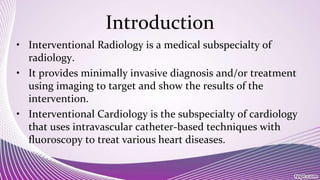
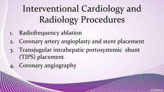
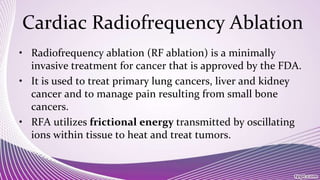
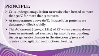
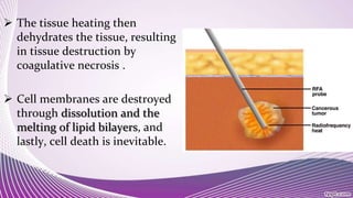
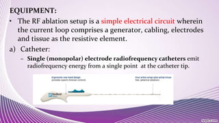
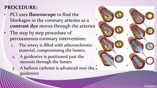
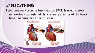
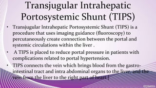
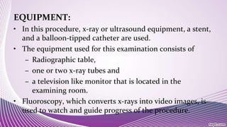
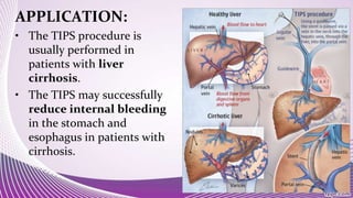
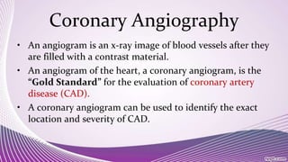
Interventional cardiology and radiology use minimally invasive techniques guided by imaging to diagnose and treat diseases. Some common procedures include radiofrequency ablation to treat cancers, coronary angioplasty and stent placement to open blocked arteries, transjugular intrahepatic portosystemic shunt placement to reduce portal hypertension, and coronary angiography to image heart arteries using contrast dye. These procedures use catheters and stents along with imaging like fluoroscopy to precisely deliver treatments to diseased areas while minimizing patient trauma.
Angioplasty uses imaging guidance to insert a balloon-tipped catheter into a narrow or blocked blood vessel where the balloon is inflated to open the vessel and improve blood flow. It may be done with vascular stenting – the placement of a small wire mesh tube within the blood vessel to help keep it open. The procedure is much less invasive than other surgical interventions and usually does not require general anesthesia.
This slide will provide illustrative information regarding coronary angioplasty . It also focus on practical area knowledge of cardiac catheterization which one should focus while caring patient with coronary angioplasty.
The CVP catheter is an important tool used to assess right ventricular function and systemic fluid status. Normal CVP is 2-6 mm Hg. CVP is elevated by : overhydration which increases venous return.
Advanced Cardiovascular Life Support (ACLS) is the pre-eminent resuscitation course for the recognition and intervention of cardiopulmonary arrest or other cardiovascular emergencies.
SELDINGER TECHNIQUE & INTERVENTIONAL RADIOLOGYRiyas M K
its a basic introduction about Seldinger technique and Intervetional radiology.In interventional radiology, procedures generally start with the Seldinger technique to access the vasculature, using a needle through which a guidewire is inserted, followed by navigation of catheters within the vessels.
invasive non invasive procedures.pdf for bsc nursing studentsshanmukhadevi
Chest X-ray:
The chest X-ray is a noninvasive tool used to visualize internal structures, such as the heart, lungs, soft tissues, and bones.
Most chest X-rays are taken while the patient is inhaling so that the lungs are fully expanded.
Several types of chest X-rays can be used to assess heart size, contour, and position; other types reveal cardiac and pericardial calcification as well as physiologic alterations in pulmonary circulation.
Title: Sense of Smell
Presenter: Dr. Faiza, Assistant Professor of Physiology
Qualifications:
MBBS (Best Graduate, AIMC Lahore)
FCPS Physiology
ICMT, CHPE, DHPE (STMU)
MPH (GC University, Faisalabad)
MBA (Virtual University of Pakistan)
Learning Objectives:
Describe the primary categories of smells and the concept of odor blindness.
Explain the structure and location of the olfactory membrane and mucosa, including the types and roles of cells involved in olfaction.
Describe the pathway and mechanisms of olfactory signal transmission from the olfactory receptors to the brain.
Illustrate the biochemical cascade triggered by odorant binding to olfactory receptors, including the role of G-proteins and second messengers in generating an action potential.
Identify different types of olfactory disorders such as anosmia, hyposmia, hyperosmia, and dysosmia, including their potential causes.
Key Topics:
Olfactory Genes:
3% of the human genome accounts for olfactory genes.
400 genes for odorant receptors.
Olfactory Membrane:
Located in the superior part of the nasal cavity.
Medially: Folds downward along the superior septum.
Laterally: Folds over the superior turbinate and upper surface of the middle turbinate.
Total surface area: 5-10 square centimeters.
Olfactory Mucosa:
Olfactory Cells: Bipolar nerve cells derived from the CNS (100 million), with 4-25 olfactory cilia per cell.
Sustentacular Cells: Produce mucus and maintain ionic and molecular environment.
Basal Cells: Replace worn-out olfactory cells with an average lifespan of 1-2 months.
Bowman’s Gland: Secretes mucus.
Stimulation of Olfactory Cells:
Odorant dissolves in mucus and attaches to receptors on olfactory cilia.
Involves a cascade effect through G-proteins and second messengers, leading to depolarization and action potential generation in the olfactory nerve.
Quality of a Good Odorant:
Small (3-20 Carbon atoms), volatile, water-soluble, and lipid-soluble.
Facilitated by odorant-binding proteins in mucus.
Membrane Potential and Action Potential:
Resting membrane potential: -55mV.
Action potential frequency in the olfactory nerve increases with odorant strength.
Adaptation Towards the Sense of Smell:
Rapid adaptation within the first second, with further slow adaptation.
Psychological adaptation greater than receptor adaptation, involving feedback inhibition from the central nervous system.
Primary Sensations of Smell:
Camphoraceous, Musky, Floral, Pepperminty, Ethereal, Pungent, Putrid.
Odor Detection Threshold:
Examples: Hydrogen sulfide (0.0005 ppm), Methyl-mercaptan (0.002 ppm).
Some toxic substances are odorless at lethal concentrations.
Characteristics of Smell:
Odor blindness for single substances due to lack of appropriate receptor protein.
Behavioral and emotional influences of smell.
Transmission of Olfactory Signals:
From olfactory cells to glomeruli in the olfactory bulb, involving lateral inhibition.
Primitive, less old, and new olfactory systems with different path
micro teaching on communication m.sc nursing.pdfAnurag Sharma
Microteaching is a unique model of practice teaching. It is a viable instrument for the. desired change in the teaching behavior or the behavior potential which, in specified types of real. classroom situations, tends to facilitate the achievement of specified types of objectives.
Title: Sense of Taste
Presenter: Dr. Faiza, Assistant Professor of Physiology
Qualifications:
MBBS (Best Graduate, AIMC Lahore)
FCPS Physiology
ICMT, CHPE, DHPE (STMU)
MPH (GC University, Faisalabad)
MBA (Virtual University of Pakistan)
Learning Objectives:
Describe the structure and function of taste buds.
Describe the relationship between the taste threshold and taste index of common substances.
Explain the chemical basis and signal transduction of taste perception for each type of primary taste sensation.
Recognize different abnormalities of taste perception and their causes.
Key Topics:
Significance of Taste Sensation:
Differentiation between pleasant and harmful food
Influence on behavior
Selection of food based on metabolic needs
Receptors of Taste:
Taste buds on the tongue
Influence of sense of smell, texture of food, and pain stimulation (e.g., by pepper)
Primary and Secondary Taste Sensations:
Primary taste sensations: Sweet, Sour, Salty, Bitter, Umami
Chemical basis and signal transduction mechanisms for each taste
Taste Threshold and Index:
Taste threshold values for Sweet (sucrose), Salty (NaCl), Sour (HCl), and Bitter (Quinine)
Taste index relationship: Inversely proportional to taste threshold
Taste Blindness:
Inability to taste certain substances, particularly thiourea compounds
Example: Phenylthiocarbamide
Structure and Function of Taste Buds:
Composition: Epithelial cells, Sustentacular/Supporting cells, Taste cells, Basal cells
Features: Taste pores, Taste hairs/microvilli, and Taste nerve fibers
Location of Taste Buds:
Found in papillae of the tongue (Fungiform, Circumvallate, Foliate)
Also present on the palate, tonsillar pillars, epiglottis, and proximal esophagus
Mechanism of Taste Stimulation:
Interaction of taste substances with receptors on microvilli
Signal transduction pathways for Umami, Sweet, Bitter, Sour, and Salty tastes
Taste Sensitivity and Adaptation:
Decrease in sensitivity with age
Rapid adaptation of taste sensation
Role of Saliva in Taste:
Dissolution of tastants to reach receptors
Washing away the stimulus
Taste Preferences and Aversions:
Mechanisms behind taste preference and aversion
Influence of receptors and neural pathways
Impact of Sensory Nerve Damage:
Degeneration of taste buds if the sensory nerve fiber is cut
Abnormalities of Taste Detection:
Conditions: Ageusia, Hypogeusia, Dysgeusia (parageusia)
Causes: Nerve damage, neurological disorders, infections, poor oral hygiene, adverse drug effects, deficiencies, aging, tobacco use, altered neurotransmitter levels
Neurotransmitters and Taste Threshold:
Effects of serotonin (5-HT) and norepinephrine (NE) on taste sensitivity
Supertasters:
25% of the population with heightened sensitivity to taste, especially bitterness
Increased number of fungiform papillae
Acute scrotum is a general term referring to an emergency condition affecting the contents or the wall of the scrotum.
There are a number of conditions that present acutely, predominantly with pain and/or swelling
A careful and detailed history and examination, and in some cases, investigations allow differentiation between these diagnoses. A prompt diagnosis is essential as the patient may require urgent surgical intervention
Testicular torsion refers to twisting of the spermatic cord, causing ischaemia of the testicle.
Testicular torsion results from inadequate fixation of the testis to the tunica vaginalis producing ischemia from reduced arterial inflow and venous outflow obstruction.
The prevalence of testicular torsion in adult patients hospitalized with acute scrotal pain is approximately 25 to 50 percent
These lecture slides, by Dr Sidra Arshad, offer a quick overview of physiological basis of a normal electrocardiogram.
Learning objectives:
1. Define an electrocardiogram (ECG) and electrocardiography
2. Describe how dipoles generated by the heart produce the waveforms of the ECG
3. Describe the components of a normal electrocardiogram of a typical bipolar leads (limb II)
4. Differentiate between intervals and segments
5. Enlist some common indications for obtaining an ECG
Study Resources:
1. Chapter 11, Guyton and Hall Textbook of Medical Physiology, 14th edition
2. Chapter 9, Human Physiology - From Cells to Systems, Lauralee Sherwood, 9th edition
3. Chapter 29, Ganong’s Review of Medical Physiology, 26th edition
4. Electrocardiogram, StatPearls - https://www.ncbi.nlm.nih.gov/books/NBK549803/
5. ECG in Medical Practice by ABM Abdullah, 4th edition
6. ECG Basics, http://www.nataliescasebook.com/tag/e-c-g-basics
Tom Selleck Health: A Comprehensive Look at the Iconic Actor’s Wellness Journeygreendigital
Tom Selleck, an enduring figure in Hollywood. has captivated audiences for decades with his rugged charm, iconic moustache. and memorable roles in television and film. From his breakout role as Thomas Magnum in Magnum P.I. to his current portrayal of Frank Reagan in Blue Bloods. Selleck's career has spanned over 50 years. But beyond his professional achievements. fans have often been curious about Tom Selleck Health. especially as he has aged in the public eye.
Follow us on: Pinterest
Introduction
Many have been interested in Tom Selleck health. not only because of his enduring presence on screen but also because of the challenges. and lifestyle choices he has faced and made over the years. This article delves into the various aspects of Tom Selleck health. exploring his fitness regimen, diet, mental health. and the challenges he has encountered as he ages. We'll look at how he maintains his well-being. the health issues he has faced, and his approach to ageing .
Early Life and Career
Childhood and Athletic Beginnings
Tom Selleck was born on January 29, 1945, in Detroit, Michigan, and grew up in Sherman Oaks, California. From an early age, he was involved in sports, particularly basketball. which played a significant role in his physical development. His athletic pursuits continued into college. where he attended the University of Southern California (USC) on a basketball scholarship. This early involvement in sports laid a strong foundation for his physical health and disciplined lifestyle.
Transition to Acting
Selleck's transition from an athlete to an actor came with its physical demands. His first significant role in "Magnum P.I." required him to perform various stunts and maintain a fit appearance. This role, which he played from 1980 to 1988. necessitated a rigorous fitness routine to meet the show's demands. setting the stage for his long-term commitment to health and wellness.
Fitness Regimen
Workout Routine
Tom Selleck health and fitness regimen has evolved. adapting to his changing roles and age. During his "Magnum, P.I." days. Selleck's workouts were intense and focused on building and maintaining muscle mass. His routine included weightlifting, cardiovascular exercises. and specific training for the stunts he performed on the show.
Selleck adjusted his fitness routine as he aged to suit his body's needs. Today, his workouts focus on maintaining flexibility, strength, and cardiovascular health. He incorporates low-impact exercises such as swimming, walking, and light weightlifting. This balanced approach helps him stay fit without putting undue strain on his joints and muscles.
Importance of Flexibility and Mobility
In recent years, Selleck has emphasized the importance of flexibility and mobility in his fitness regimen. Understanding the natural decline in muscle mass and joint flexibility with age. he includes stretching and yoga in his routine. These practices help prevent injuries, improve posture, and maintain mobilit
NVBDCP.pptx Nation vector borne disease control programSapna Thakur
NVBDCP was launched in 2003-2004 . Vector-Borne Disease: Disease that results from an infection transmitted to humans and other animals by blood-feeding arthropods, such as mosquitoes, ticks, and fleas. Examples of vector-borne diseases include Dengue fever, West Nile Virus, Lyme disease, and malaria.
Report Back from SGO 2024: What’s the Latest in Cervical Cancer?bkling
Are you curious about what’s new in cervical cancer research or unsure what the findings mean? Join Dr. Emily Ko, a gynecologic oncologist at Penn Medicine, to learn about the latest updates from the Society of Gynecologic Oncology (SGO) 2024 Annual Meeting on Women’s Cancer. Dr. Ko will discuss what the research presented at the conference means for you and answer your questions about the new developments.
Pulmonary Thromboembolism - etilogy, types, medical- Surgical and nursing man...VarunMahajani
Disruption of blood supply to lung alveoli due to blockage of one or more pulmonary blood vessels is called as Pulmonary thromboembolism. In this presentation we will discuss its causes, types and its management in depth.
New Directions in Targeted Therapeutic Approaches for Older Adults With Mantl...i3 Health
i3 Health is pleased to make the speaker slides from this activity available for use as a non-accredited self-study or teaching resource.
This slide deck presented by Dr. Kami Maddocks, Professor-Clinical in the Division of Hematology and
Associate Division Director for Ambulatory Operations
The Ohio State University Comprehensive Cancer Center, will provide insight into new directions in targeted therapeutic approaches for older adults with mantle cell lymphoma.
STATEMENT OF NEED
Mantle cell lymphoma (MCL) is a rare, aggressive B-cell non-Hodgkin lymphoma (NHL) accounting for 5% to 7% of all lymphomas. Its prognosis ranges from indolent disease that does not require treatment for years to very aggressive disease, which is associated with poor survival (Silkenstedt et al, 2021). Typically, MCL is diagnosed at advanced stage and in older patients who cannot tolerate intensive therapy (NCCN, 2022). Although recent advances have slightly increased remission rates, recurrence and relapse remain very common, leading to a median overall survival between 3 and 6 years (LLS, 2021). Though there are several effective options, progress is still needed towards establishing an accepted frontline approach for MCL (Castellino et al, 2022). Treatment selection and management of MCL are complicated by the heterogeneity of prognosis, advanced age and comorbidities of patients, and lack of an established standard approach for treatment, making it vital that clinicians be familiar with the latest research and advances in this area. In this activity chaired by Michael Wang, MD, Professor in the Department of Lymphoma & Myeloma at MD Anderson Cancer Center, expert faculty will discuss prognostic factors informing treatment, the promising results of recent trials in new therapeutic approaches, and the implications of treatment resistance in therapeutic selection for MCL.
Target Audience
Hematology/oncology fellows, attending faculty, and other health care professionals involved in the treatment of patients with mantle cell lymphoma (MCL).
Learning Objectives
1.) Identify clinical and biological prognostic factors that can guide treatment decision making for older adults with MCL
2.) Evaluate emerging data on targeted therapeutic approaches for treatment-naive and relapsed/refractory MCL and their applicability to older adults
3.) Assess mechanisms of resistance to targeted therapies for MCL and their implications for treatment selection
New Drug Discovery and Development .....NEHA GUPTA
The "New Drug Discovery and Development" process involves the identification, design, testing, and manufacturing of novel pharmaceutical compounds with the aim of introducing new and improved treatments for various medical conditions. This comprehensive endeavor encompasses various stages, including target identification, preclinical studies, clinical trials, regulatory approval, and post-market surveillance. It involves multidisciplinary collaboration among scientists, researchers, clinicians, regulatory experts, and pharmaceutical companies to bring innovative therapies to market and address unmet medical needs.
- Video recording of this lecture in English language: https://youtu.be/lK81BzxMqdo
- Video recording of this lecture in Arabic language: https://youtu.be/Ve4P0COk9OI
- Link to download the book free: https://nephrotube.blogspot.com/p/nephrotube-nephrology-books.html
- Link to NephroTube website: www.NephroTube.com
- Link to NephroTube social media accounts: https://nephrotube.blogspot.com/p/join-nephrotube-on-social-media.html
Maxilla, Mandible & Hyoid Bone & Clinical Correlations by Dr. RIG.pptx
Interventional Radiology And Cardiology
1.
2. Introduction
• Interventional Radiology is a medical subspecialty of
radiology.
• It provides minimally invasive diagnosis and/or treatment
using imaging to target and show the results of the
intervention.
• Interventional Cardiology is the subspecialty of cardiology
that uses intravascular catheter-based techniques with
fluoroscopy to treat various heart diseases.
4. Cardiac Radiofrequency Ablation
• Radiofrequency ablation (RF ablation) is a minimally
invasive treatment for cancer that is approved by the FDA.
• It is used to treat primary lung cancers, liver and kidney
cancer and to manage pain resulting from small bone
cancers.
• RFA utilizes frictional energy transmitted by oscillating
ions within tissue to heat and treat tumors.
5. PRINCIPLE:
Cells undergo coagulation necrosis when heated to more
than 50°C for more than 5 minutes.
At temperatures above 60°C, intracellular proteins are
denatured (killed) rapidly.
The AC current (350-500 kHz) of RF waves passing down
from an un-insulated electrode tip into the surrounding
tissues generates changes in the direction of ions and
creates ionic agitation and frictional heating.
6. The tissue heating then
dehydrates the tissue, resulting
in tissue destruction by
coagulative necrosis .
Cell membranes are destroyed
through dissolution and the
melting of lipid bilayers, and
lastly, cell death is inevitable.
7. EQUIPMENT:
• The RF ablation setup is a simple electrical circuit wherein
the current loop comprises a generator, cabling, electrodes
and tissue as the resistive element.
a) Catheter:
– Single (monopolar) electrode radiofrequency catheters emit
radiofrequency energy from a single point at the catheter tip.
8. • Multi electrode radiofrequency
catheters have several electrodes, each of
which can deliver radiofrequency energy.
• These catheters can ablate a larger area of
tissue than single point radiofrequency
energy catheters, which could decrease
procedure time.
• These catheters can also deliver superficial
bipolar radiofrequency energy, which my
reduce injury to deeper tissues in certain
areas of the heart.
9. • Irrigated tip catheters:
– Closed Irrigation: Saline is perfused via a
pump mechanism through the catheter tip,
turns around within the catheter tip, and
returns back to the pump.
– Open Irrigation: Open irrigation overcomes
the limitations of non-irrigated ablation by
lowering electrode and tissue surface
temperature.
– The efficacy of power delivery is increased
along with the safety of reduced char
formation.
10. Three types of RF electrodes are currently available
commercially:
– Two brands of Multi polar retractable needle electrodes:
1. RITA Medical Systems, Mountain View, CA;
2. LeVeen needle electrode, Boston Scientific, Boston, MA
– Internally cooled electrode:
1. Cool-Tip RF electrode; Radionics, Burlington, MA
11. Generator and ground pads:
• The RF ablation generator provides three essential
functions: power generation, control, and user interface.
• The ground pad closes the electrical current path and is
designed to disperse energy over a large surface area to
reduce the likelihood of thermal injury to the skin.
12. WORKING:
• The procedure can be done in a variety of ways.
• RFA can be performed percutaneously.
• In this technique, under ultrasound guidance the LeVeen®
Electrode is inserted through a small puncture in the
chest and positioned within the tumor and opened
allowing the multiple tines to spread out.
13. • Then, the generator is turned on and the radiofrequency
energy is passed from the tines of the electrode into the
tumor.
• The energy creates heat which destroys the cells in the area.
• The result is destruction (ablation) of the tumor.
14. APPLICATIONS:
• RF ablation is used in recurrent atrial flutter (AFL), atrial
fibrillation (AF), Supraventricular Tachycardia (SVT).
• It is used in the treatment of varicose veins.
• RFA is used to treat severe chronic pain the lower (lumbar) back.
• RFA is also used in radio frequency lesioning, for vein closure in
areas where intrusive surgery is contraindicated by trauma.
• RFA is used in liver resection to control bleeding (homeostasis)
and facilitate the transaction process.
• RFA is also used to ablate tumors in patients where surgery is
not possible due to health condition.
15.
16. Coronary Angioplasty And
Stent Placement
• Coronary angioplasty is a procedure used to open blocked
coronary arteries (caused by coronary artery disease).
• It restores blood flow to the heart muscle without open-heart-
surgery.
• Coronary angioplasty is also known as Percutaneous
transluminal coronary angioplasty (PICA).
• The combination of coronary angioplasty with stenting is
usually referred to as Percutaneous Coronary Intervention
(PCI).
17. EQUIPMENT:
• The most commonly used
PCI equipment consists of
four basic elements:
1. a guiding catheter,
2. a balloon catheter,
3. a coronary guidewire,
4. a stent.
18. Guiding Catheter:
• A special large lumen catheter is used to
deliver the coronary balloon catheter and
other interventional devices to the vessel
that contains the lesion to be dilated.
Function:
• A guiding catheter serves three major
functions during angioplasty:
– Balloon/Stent catheter delivery and guidance
– Backup support for balloon/Stent
advancement
– Pressure monitoring and contrast injections
(1)Stiffer body (2)variable
softer primary curve (3) wire
braiding (4)atraumatic tip (5)
large lumen(6) lubrious
coating
19. Balloon Dilatation Catheter System:
When a stent is placed inside of a coronary artery, it acts as a
support or scaffold, keeping the vessel open.
Stent procedures are usually used along with balloon
catheters.
There are three types of PCI balloon catheters:
Over-the-wire (OTW)
Monorail
Fixed-wire
20. Stents:
• A Stent is a tiny, expandable, metal mesh coil that is put
into the newly-opened area of the artery to help keep the
artery from narrowing or closing again.
• It helps re-narrowing of the blood vessel.
21. PROCEDURE:
• PCI uses fluoroscope to find the
blockages in the coronary arteries as a
contrast dye moves through the arteries.
• The step by step procedure of
percutaneous coronary intervention:
1. The artery is filled with atherosclerotic
material, compromising the lumen.
2. A guidewire is positioned past the
stenosis through the lumen
3. A balloon catheter is advanced over the
guidewire
22. 4. The balloon is inflated
5. The balloon catheter is exchanged
for a stent (on a balloon)
6. The stent is expanded
7. The expanded stent remains in
place after the deflated balloon is
withdrawn.
25. Transjugular Intrahepatic
Portosystemic Shunt (TIPS)
• Transjugular Intrahepatic Portosystemic Shunt (TIPS) is a
procedure that uses imaging guidance (fluoroscopy) to
percutaneously create connection between the portal and
systemic circulations within the liver .
• A TIPS is placed to reduce portal pressure in patients with
complications related to portal hypertension.
• TIPS connects the vein which brings blood from the gastro-
intestinal tract and intra abdominal organs to the liver, and the
vein from the liver to the right part of heart.[
26. EQUIPMENT:
• In this procedure, x-ray or ultrasound equipment, a stent,
and a balloon-tipped catheter are used.
• The equipment used for this examination consists of
– Radiographic table,
– one or two x-ray tubes and
– a television like monitor that is located in the
examining room.
• Fluoroscopy, which converts x-rays into video images, is
used to watch and guide progress of the procedure.
27. • A catheter is a long, thin, plastic tube that is approximately
1/8 inch in diameter.
• The Stent used in this procedure is a small wire mesh
tube, often covered with a fabric made of GORE-TEX®.
• Other equipment that may be used during the procedure
includes an intravenous line (IV), ultrasound machine
and devices that monitor heart beat and blood pressure
28. PROCEDURE:
• With the use of Fluoroscopy guidance
and an injection of contrast material
containing iodine, the location of the
hepatic vein is identified.
• From the hepatic vein, a connection
through the liver is made into the
portal vein using guiding catheter.
• Once the connection is made to the
portal vein, fluoroscopy guidance is
utilized to dilate the connection and
place a stent
29.
30. APPLICATION:
• The TIPS procedure is
usually performed in
patients with liver
cirrhosis.
• The TIPS may successfully
reduce internal bleeding
in the stomach and
esophagus in patients with
cirrhosis.
31. Coronary Angiography
• An angiogram is an x-ray image of blood vessels after they
are filled with a contrast material.
• An angiogram of the heart, a coronary angiogram, is the
“Gold Standard” for the evaluation of coronary artery
disease (CAD).
• A coronary angiogram can be used to identify the exact
location and severity of CAD.
32. EQUIPMENT:
The equipments needed for coronary angiogram are
Angiogram needle,
vascular sheath,
Guidewires,
Catheters,
Fluoroscopic machine,
Power injector
The catheter inserted into
angiogram needle.
33. PROCEDURE:
• Coronary angiography is
performed with the use of local
anesthesia and IV sedation, and is
generally not significantly
uncomfortable.
• In performing a coronary
angiogram, a small catheter is
inserted through the skin into an
artery in either the groin or the
arm.
34. • Under fluoroscopy guidance, the
catheter is then advanced to the
opening of the coronary arteries.
• A small amount of radiographic
contrast, is injected into each
coronary artery.
• The images that are produced are
called angiogram
• After the procedure, the catheter is
removed and the artery in the leg or
arm is sutured, sealed to prevent
bleeding
35. APPLICATION:
Coronary Angiography procedure is used to diagnose coronary
heart disease and coronary micro-vascular disease after
chest pain, sudden cardiac arrest, or abnormal results from
tests such as an electrocardiogram (EKG) of the heart or an
exercise stress test.
36.
37. Bibbliography
• Yasunori Minami and Masatoshi Kudo, “Radiofrequency Ablation of
Hepatocellular Carcinoma: A Literature Review,” International
Journal of Hepatology, vol. 2011, Article ID 104685, 9 pages, 2011.
doi:10.4061/2011/104685
• http://www.rfalung.com/rfa_brochure.pdf