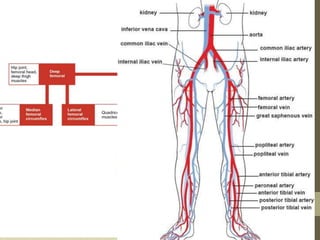
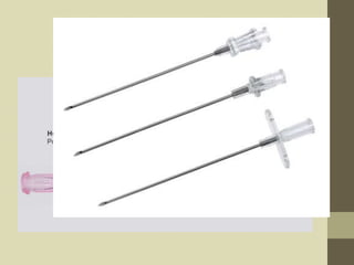
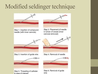
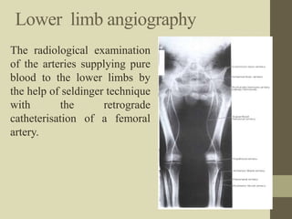
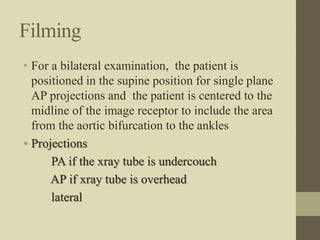
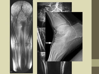
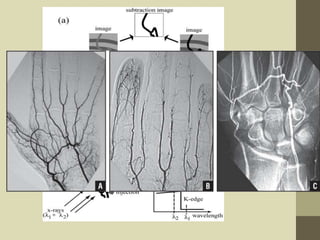
Peripheral
Angiography
SONI NAGARKOTI
B.Sc.MIT 2nd Year
NAMS,Bir Hospital
Overview
• Introduction
• General anatomy
• Indication
• Equipments and accesories
• Procedure
• Filming
• Aftercare of patient
• Advancements
Angiography
• Angiography is the one of the radiological imaging
procedure available for detailed investigation of heart
and blood vessels after injecting the contrast media.
• Angiography can be more specifically described as
follows:
•Arteriography: imaging of the arteries
•Venography: imaging of the veins/phlebogram
•Angiocardiography: imaging of the heart and
associated structures
•Lymphography: imaging of the lymphatic
vessels/nodes
Importances
• Shows blockage, deformities in the vessels
• Could be both therapeutic and diagnostic
• Angiography followed by angioplasty and
stenting could be life saving in case of arterial
blockage or narrowing of vessels
• Angioplasty is beneficial for reducing angina
• Could be beneficial to reduce ischemia due to
blockage in any peripheral or coronary arteries
Peripheral angiography
• Peripheral Angiography is a radiologic
examination of the peripheral vasculature (artery)
after the injection of contrast media.
• Could be upper limb angiography or lower limb
angiography
General anatomy
Vasculature of upper limb
Vasculature of lower limb
Peripheral arterial diseases(PAD)
Narrowing of peripheral arteries serving the legs,
arms, head
• Cause = atherosclerosis
(if severe blocked blood flow can cause ischemia,
necrosis or tissue death and may lead to
amputations)
• Symptoms= crampings, pain, tiredness in leg or
hip muscles , change in skin colour
• Risk factors= smoking, old age, diabetes, HTN,
high blood cholesterol, heart diseases
Indications
• Atherosclerotic disease
• Embolus and thrombus
• Arterial ischemia, stenosis or occlusion.
• Trauma to a limb with arterial involvement
• Congenital abnormalities
• Buerger’s disease or other forms of arteritis
• Prior to and following vascular surgery
• Neoplasm
• Angioma
• Popliteal artery entrapment syndrome
Contraindications
• Hypersensitivity to iodinated contrast media.
• Blood–clotting disorders or bleeding disorder
• Anti coagulant medication
• Impaired renal function
• Local infection of the puncture site.
• Unable to do vascular surgery.
• High grade fever
• Low Hb
Relative contraindications
• Blood dyscrasias
• Aneurysms or pseudoaneurysms
• Local soft tissue infections
• Severe hypertension
• Ehlers- Danlos syndrome
Equipments and accesories
Equipment includes:-
- High power x-ray generator.
- X-ray tube
- Floating/tilting type of x-ray table
- Fluoroscopic unit with II TV system
- Rapid serial film changer.
- Auto-injector
- Resuscitative apparatus
• Generator- required 3 phase 12-pulse unit with a
power rating of 85-100kw at 100k (to withstand
heat)
• Fast screen film combination.
• grid also necessary to reduce scatter radiation.
• Automatic injector is required for bolus injection
at predetermined amount and preset rate.
• physiologic monitoring equipment is required
for constant cardic and intravascular pressure
recording.
Accessories include
1. Local anesthesia
2. Puncture needle / introducer needle
3. Guide wire/ glide wire(glide wire is
hydrophilic or lubricated guide wire)
4. Angiography catheter
5. Angiographic sheath with dilator
6. Sterile angiographic tray
7. Surgical blade
8. Gloves
Introducer needle
• Consists of :
1. Outer thin walled blunt cannula
2. Inner needle
3. Stilette
• Size based on external diameter of needle. (18G)
• Internal diameters should be known to allow for
appropriate Guide-wires matching
• Proper needle gauge and guide wire diameter are
chosen because :
- large diameter could result in backflow of blood
through cannula of needle, blood loss
- small needle gauge cause difficult or impossible for
wire to pass through the cannula of the needle
Guide wires
• Used as a platform over which a catheter is to be
advanced.
• Is a stainless steel wire that acts as a guide until
the catheter reaches the area of interest.
• Outside of the guidewire is teflon.
(teflon reduces friction and clot formation)
• May saturated with heparin.
• Inner core is fixed or moveable.
• Available with straight
or j-shapped tips.
(J tipped wires is useful for negotiating with
vessels having irregular walls)
Contd…
• Guide-wires ranges
from 20-57 inches
(50-145cm)
• Diameter ranges from –
0.014 – 0.052 inches
(0.03 – 0.13 cm)
• Frequently used diameter –
0.035 inches(0.09cm)
and 0.038 inches (1.0cm).
(glide wires are slippery with excellent torque and
are useful for tortous vessels)
Angiographic sheath with dilator
• Sheath is short flexible plastic tube having hole
in one side and valve at another side.
• It is used to prevent the blood clot
• Dilator is rigid structures which gives support to
sheath during insertion.
Angiography catheters
• Serve as a principle pipeline through which contrast
media is transported.
• Made up of polyethylene, polyurethane, nylon and
teflon.
• Polyethylene catheter is mostly used.
• Catheters are vary in length, size and no. of side
holes.
• When using the femoral approach, short-length
catheters (60–80 cm) are adequate for angiography of
the structures below the diaphragm, whereas long
catheters (100–120 cm) are needed for carotid artery,
subclavian artery, or arm angiography.
• Five- to six-French (1-F catheter = 0.333 mm)
diameter catheters are most commonly used.(3F =
1mm)
• For DSA, 5-F catheters are sufficient.
• They may have an end hole, side holes, or both end
and side holes.
• Side holes are important as they affect the flow rate
and allow large volume of the contrast medium and
pressure exerted on the catheter.(safe)
• ‘ideal catheter ’ should be able to sustain high
pressure injection, to track well, be non-
thrombogenic, have good memory, and should
torque well.
Notes
• Introduction of catheter over a guide wire is
faciliated by dilation of path by dilator
• If the patient has large amount of subcutaneous
fat in the puncture area, catheter control will be
better by using introducer sheath
Types of catheter
Name of catheter function
1. Cobra visceral
2. Head hunter Carotid subclavian
3. Simmons Celiac trunk
4. Shephard hook Visceral , spinal
5.Renal double curve Renal
6. Bernstein left carotid
7. Straight Lower limb
8.Pigtail Non selective
9.Balloon angioplasty
Angiographic tray
• A sterile tray contains the basic equipment
necessary for a Seldinger catheterization of a
femoral artery. Basic sterile items include the
following:
1.Hemostats
2.Prep sponges and antiseptic solution
3.Scalpel blade
4.Syringe and needle for local anesthetic
5.Basins and medicine cup
6.Sterile drapes and towels
7.Band-Aids
8.Sterile image intensifier cover if C arm is used
Contrast media includes
non-ionic, low osmolar
- for lower extrimities:- 30-40ml of 300mgI/ml
- for upper extrimities:- 20-30ml of 300mgI/ml
For eg ultravist omnipaque
Drug includes
Local anesthesia ( inj. xylocane 2%)
- Heparin
- Atropin
- Adrenaline
- hydrocortisone
- inj. Cefazolin 1g
Angiographic team in Angio
room
• Radiologist ( or other specialist)
• Cardiovascular nurse
• Radiologic Technologists (CV)
• Sometimes Anesthesiologist depending on the
procedure
Role of CIT
• Proper positioning of the patient
• Exposure in adequate time
• Changing of cassette
• Moving the tube
• Lowering the radiation dose as much as possible.
• In case of c-arm the exposure is auto adjusted.
• Otherwise have to change the kv for each time
from reaching thicker part to thinner part (from
femur to ankle).
PATIENT PREPARATION
• Admission in hospital 24 hrs prior to examination,
• Prothrombine time and bleeding duration must be
checked
• Serological test (HIV, HCV, HbsAg) must performed.
• Puncture site should be shaved.
• Informed consent must be taken and explain about
the procedure.
• NPO at least 4-6 hrs prior to examination.
• Patients must bring all the previous records : x-ray,
ECG reports, CT reports, catheter, IV-cannula,
disposal syringes – 10ml and 20 ml, heparin
injection, normal saline, contrast media.
• Micturation should be done before examination.
Puncture site
1. Femoral artery (most frequently used)
2. Brachial artery
3. Axillary artery
4. Radial artery
Technique (Seldinger technique)
1. In this technique, both wall of vessel is
punctured
2. Stilette of needle is removed
3. Needle is withdrawn in such a way that the
bevel is within the lumen of vessel and blood
flows from hub
4. Guide wire is inserted through the needle
5. Needle is withdrawn keeping guide wire in situ
6. Catheter is threaded over guide wire
7. Guide wire is withdrawn
Modified seldinger technique
Lower limb angiography
The radiological examination
of the arteries supplying pure
blood to the lower limbs by
the help of seldinger technique
with the retrograde
catheterisation of a femoral
artery.
Patient and equipment
positioning
• Supine in the centre of the imaging couch.
• Head raised on a shallow pillow.
• Both knees are extended with leg straight and
positioned together.
• Feet pointing upward and rotate slightly inward (
to demonstration the gap between the tibia and
fibula )
• The feet are secured by a triangular wedge foam
pad and restraining straps.
• Image intensifier is parallel to the imaging coach.
• Tube is placed under the table.
Procedure
1. Generally, femoral artery is considered for
puncture
2. The femoral artery of leg opposite to the
symptomatic leg is chosen
3. Location and point to be punctured is cleaned
with betadine solution
4. Using aseptic technique, local anesthesia is
infiltrated at the puncture site
5. A skin incision is made on the puncture point to
reduce binding of soft tissue on catheter
• The introducer needle is then advanced through
soft tissue using Seldinger technique
• After puncture of artery, stillette of needle is
removed and needle hub is depressed so that it runs
parallel to the skin
• A guidewire is introduced through needle and
advanced gently along artery using intermittent
fluroscopy
• The guidewire is guided from femoral artery to the
bifurcation of two common iliac arteries
• The needle is then withdrawn over guidewire and
catheter is threaded on the free portion of
guidewire
• Generally, dilators matching the patency of
catheter is used to dilate the path prior using
catheter
• The tip of the catheter is positioned above the
aortic biforcation under fluoroscopic control.
• The contrast media is injected, the flow of
contrast is observed and timed as it traverses the
femoral arteries to the knees and lower down the
limbs.
• An image acquisition protocol is prescribed to
ensure complete visualization of the arterial
vessels
Filming
• For a bilateral examination, the patient is
positioned in the supine position for single plane
AP projections and the patient is centered to the
midline of the image receptor to include the area
from the aortic bifurcation to the ankles
• Projections
PA if the xray tube is undercouch
AP if xray tube is overhead
lateral
Upper limb angiography
• Radiological study of
arterial system
supplying the upper
limbs by the retrograde
introduction of contrast
media through the
femoral artery.
Patients and equipment position
• Patients lie supine on the table.
• Arms extended and placed along side the trunk.
• Palms of the hands placing upwards.
• In some cases arm raised above the head to
investigate the effects on arterial circulation
• Image intensifier/film changer is positioned
parallel to the imaging coach and above the
region under investigation.
Procedure
1. Generally, femoral artery is considered for
puncture
2. Both femoral arteries are palpated, if pulsations
are of similar strength, right femoral artery is
generally chosen as it is technically easier
3. Location and point to be punctured is cleaned
with betadine solution
4. Using aseptic technique, local anesthesia is
infiltrated at the puncture site
5. A skin incision is made on the puncture point to
reduce binding of soft tissue on catheter
• The introducer needle is then advanced through
soft tissue using Seldinger technique
• After puncture of artery, stylet of needle is
removed and needle hub is depressed so that it runs
parallel to the skin
• A guidewire is introduced through needle and
advanced gently along artery using intermittent
fluroscopy
• The guidewire is guided from femoral artery to the
bifurcation of two common iliac arteries
• The guidewire is then guided to the abdominal
aorta to thoracic aorta to the arch of aorta and
finally to the tip of subclavian artery
• Selective catheterization of the subclavian
arteries is done by using headhunter catheter
• Contrast is then injected and flow of contrast is
followed under fluoroscopy guidance
Filming
• Generally, true AP projections are taken with the
arms extended and hand supinated
• Hand arteriograms are obtained in supine or
prone arm positions
• Injections and imaging system depends on the
equipment used
• A representative program for a rapid imaging
system may be two films per second for 5
seconds followed by one per second for 5
seconds.
Aftercare
• the punctured site is pressed at least 15 mins.
• Dressing is applied over the punctured site.
• Puncture site should be taken care of for
haemorrhage
• Patient is advised to complete bed rest for 6hrs.
• Vital sign must be recorded upto 24 hrs
- every 15 mins for 4 hrs.
- every 4 hrs for 20 hrs.
• the patient is advised not to bend the limb for 24
hrs.
• Patients with larger catheter, anti platelet therapy, and
anticoagulation require longer observation
• Inj.cefazolin is given immediate after procedure
Complications
1.Due to anaesthesia
• Emesis
• Agitation
• Rash
2.Due to contrast media
• Mild(nausea, vomiting, dizziness, headache,
rashes)
• Moderate (utricaria, facial edema, hypotension,
hypertension, tachycardia, bradycardia)
• Severe ( pulmonary oedema, unconsiousss,
death)
3.Due to technique
1. Haemorrhage/haematoma
2. Arterial thrombus
3. Infection to puncture site
4. Pseudoaneurysms
5. Arteriovenous fistula
6. Air embolus
7. Artery dissection
8. Guidewire breakage
9. bacteraemia
Radiation protection
• Angio team should be provided adequate
protection i.e. with leaded glass observation
window.
• Use of radiation protection devices.
• Minimal fluoro use as possible
• Proper collimation
• Angio personnel should wear badges and ring
monitors.
• Avoidance of repeat exposure
• Cardinal rules i.e. time, distance and shielding
Advancements in angiography
1. Digital subtraction angiography
2. Rotational angiography
3. Computed tomography angiography
4. Magnetic resonance angiography
5. Ultrasound
Conventional
angiography
You can see the
bony skeleton
Digital
subtraction
The bony skeleton
is subtracted
CT angiography MR angiography
Digital subtraction angiography
• Fluoroscopy technique used in IR to clearly
visualize blood vessels in a bony and soft tissue
environment
• Compares xray image of a region of body before
and after radiopaque iodine based dye has been
injected
• Useful in diagnosis and treatment of arterial and
venous occulusion including artery stenosis,,
embolisms, ischaemia
Principle
• the non-contrast image (mask image) of the region is
taken before injecting contrast material and therefore
shows only anatomy, as well as any radiopaque
foreign bodies (surgical clips, stents, etc.) as would a
regular x-ray image.
• contrast images are taken in succession while
contrast material is being injected. These images
show the opacified vessels superimposed on the
anatomy and are stored on the computer.
• the mask image is then subtracted from the contrast
images pixel by pixel. The resulting subtraction
images shows the filled vessels only.
• recording can continue to provide a sequence of
subtracted images based on the initial mask. the
subtraction images can be viewed in real time.
• Even if the patient lies still, there is bound to be
some degree of misregistration of images due to
movement between the acquisition of the mask
image and the subsequent contrast images. The effect
is prominent at high-contrast interfaces, such as
bone-soft tissue, metal staples and coils, and bowel
air. Pixel shifting (either manual or automatic), i.e.
moving the mask retrospectively, can minimize
misregistration, but focal movement such as bowel
peristalsis, will not be corrected
Rotational angiography
• Rotational angiography is a medical imaging
technique based on x-ray, that allows to acquire
CT-like 3D volumes during interventional
catherization
• Also known as flat panel volume CT or cone
beam CT
• In order to acquire a 3D image with a fixed C-
Arm, the C-Arm is positioned at the body part in
question so that this body part is in the isocenter
between the x-ray tube and the detector.
• The C-Arm then rotates around that isocenter, the
rotation being between 200° and 360°
• Such a rotation takes between 5 and 20 seconds,
during which a few hundred 2D images are
acquired.
• A piece of software then performs a cone beam
reconstruction
• The resulting voxel data can then be viewed as
a multiplanar reconstruction, i.e. by scrolling
through the slices from three projection angles, or
as a 3D volume
Ceiling mounted c arm at catherizationlab for rotational
angiography
Refrences
1) A Guide to Radiological Procedures, Chapman
5th Edition
2) Merrill’s Atlas of Radiographic Positions
Volume 3-10th Edition.
3) www.radiopaedia.org
4) www.google.com
Questions
1. Which type of guide wire used in peripheral
angiogram? What is its length?
2. What is the size of catheter used in angio?
why?
3. There are more advanced modalities for angio.
Still conventional peripheral angio is in
practice. why?
4. Describe the pathway for upper limb
angiography whose puncture site is femoral
artery.
5. How does DSA works?

Peripheral angiography

  • 1.
  • 2.
    Overview • Introduction • Generalanatomy • Indication • Equipments and accesories • Procedure • Filming • Aftercare of patient • Advancements
  • 3.
    Angiography • Angiography isthe one of the radiological imaging procedure available for detailed investigation of heart and blood vessels after injecting the contrast media. • Angiography can be more specifically described as follows: •Arteriography: imaging of the arteries •Venography: imaging of the veins/phlebogram •Angiocardiography: imaging of the heart and associated structures •Lymphography: imaging of the lymphatic vessels/nodes
  • 4.
    Importances • Shows blockage,deformities in the vessels • Could be both therapeutic and diagnostic • Angiography followed by angioplasty and stenting could be life saving in case of arterial blockage or narrowing of vessels • Angioplasty is beneficial for reducing angina • Could be beneficial to reduce ischemia due to blockage in any peripheral or coronary arteries
  • 5.
    Peripheral angiography • PeripheralAngiography is a radiologic examination of the peripheral vasculature (artery) after the injection of contrast media. • Could be upper limb angiography or lower limb angiography
  • 6.
  • 7.
  • 8.
  • 10.
    Peripheral arterial diseases(PAD) Narrowingof peripheral arteries serving the legs, arms, head • Cause = atherosclerosis (if severe blocked blood flow can cause ischemia, necrosis or tissue death and may lead to amputations) • Symptoms= crampings, pain, tiredness in leg or hip muscles , change in skin colour • Risk factors= smoking, old age, diabetes, HTN, high blood cholesterol, heart diseases
  • 12.
    Indications • Atherosclerotic disease •Embolus and thrombus • Arterial ischemia, stenosis or occlusion. • Trauma to a limb with arterial involvement • Congenital abnormalities • Buerger’s disease or other forms of arteritis • Prior to and following vascular surgery • Neoplasm • Angioma • Popliteal artery entrapment syndrome
  • 13.
    Contraindications • Hypersensitivity toiodinated contrast media. • Blood–clotting disorders or bleeding disorder • Anti coagulant medication • Impaired renal function • Local infection of the puncture site. • Unable to do vascular surgery. • High grade fever • Low Hb
  • 14.
    Relative contraindications • Blooddyscrasias • Aneurysms or pseudoaneurysms • Local soft tissue infections • Severe hypertension • Ehlers- Danlos syndrome
  • 15.
    Equipments and accesories Equipmentincludes:- - High power x-ray generator. - X-ray tube - Floating/tilting type of x-ray table - Fluoroscopic unit with II TV system - Rapid serial film changer. - Auto-injector - Resuscitative apparatus
  • 16.
    • Generator- required3 phase 12-pulse unit with a power rating of 85-100kw at 100k (to withstand heat) • Fast screen film combination. • grid also necessary to reduce scatter radiation. • Automatic injector is required for bolus injection at predetermined amount and preset rate. • physiologic monitoring equipment is required for constant cardic and intravascular pressure recording.
  • 17.
    Accessories include 1. Localanesthesia 2. Puncture needle / introducer needle 3. Guide wire/ glide wire(glide wire is hydrophilic or lubricated guide wire) 4. Angiography catheter 5. Angiographic sheath with dilator 6. Sterile angiographic tray 7. Surgical blade 8. Gloves
  • 18.
    Introducer needle • Consistsof : 1. Outer thin walled blunt cannula 2. Inner needle 3. Stilette • Size based on external diameter of needle. (18G) • Internal diameters should be known to allow for appropriate Guide-wires matching • Proper needle gauge and guide wire diameter are chosen because : - large diameter could result in backflow of blood through cannula of needle, blood loss - small needle gauge cause difficult or impossible for wire to pass through the cannula of the needle
  • 20.
    Guide wires • Usedas a platform over which a catheter is to be advanced. • Is a stainless steel wire that acts as a guide until the catheter reaches the area of interest. • Outside of the guidewire is teflon. (teflon reduces friction and clot formation) • May saturated with heparin. • Inner core is fixed or moveable. • Available with straight or j-shapped tips. (J tipped wires is useful for negotiating with vessels having irregular walls)
  • 21.
    Contd… • Guide-wires ranges from20-57 inches (50-145cm) • Diameter ranges from – 0.014 – 0.052 inches (0.03 – 0.13 cm) • Frequently used diameter – 0.035 inches(0.09cm) and 0.038 inches (1.0cm). (glide wires are slippery with excellent torque and are useful for tortous vessels)
  • 23.
    Angiographic sheath withdilator • Sheath is short flexible plastic tube having hole in one side and valve at another side. • It is used to prevent the blood clot • Dilator is rigid structures which gives support to sheath during insertion.
  • 25.
    Angiography catheters • Serveas a principle pipeline through which contrast media is transported. • Made up of polyethylene, polyurethane, nylon and teflon. • Polyethylene catheter is mostly used. • Catheters are vary in length, size and no. of side holes. • When using the femoral approach, short-length catheters (60–80 cm) are adequate for angiography of the structures below the diaphragm, whereas long catheters (100–120 cm) are needed for carotid artery, subclavian artery, or arm angiography.
  • 26.
    • Five- tosix-French (1-F catheter = 0.333 mm) diameter catheters are most commonly used.(3F = 1mm) • For DSA, 5-F catheters are sufficient. • They may have an end hole, side holes, or both end and side holes. • Side holes are important as they affect the flow rate and allow large volume of the contrast medium and pressure exerted on the catheter.(safe) • ‘ideal catheter ’ should be able to sustain high pressure injection, to track well, be non- thrombogenic, have good memory, and should torque well.
  • 27.
    Notes • Introduction ofcatheter over a guide wire is faciliated by dilation of path by dilator • If the patient has large amount of subcutaneous fat in the puncture area, catheter control will be better by using introducer sheath
  • 28.
    Types of catheter Nameof catheter function 1. Cobra visceral 2. Head hunter Carotid subclavian 3. Simmons Celiac trunk 4. Shephard hook Visceral , spinal 5.Renal double curve Renal 6. Bernstein left carotid 7. Straight Lower limb 8.Pigtail Non selective 9.Balloon angioplasty
  • 30.
    Angiographic tray • Asterile tray contains the basic equipment necessary for a Seldinger catheterization of a femoral artery. Basic sterile items include the following: 1.Hemostats 2.Prep sponges and antiseptic solution 3.Scalpel blade 4.Syringe and needle for local anesthetic 5.Basins and medicine cup 6.Sterile drapes and towels 7.Band-Aids 8.Sterile image intensifier cover if C arm is used
  • 31.
    Contrast media includes non-ionic,low osmolar - for lower extrimities:- 30-40ml of 300mgI/ml - for upper extrimities:- 20-30ml of 300mgI/ml For eg ultravist omnipaque
  • 32.
    Drug includes Local anesthesia( inj. xylocane 2%) - Heparin - Atropin - Adrenaline - hydrocortisone - inj. Cefazolin 1g
  • 33.
    Angiographic team inAngio room • Radiologist ( or other specialist) • Cardiovascular nurse • Radiologic Technologists (CV) • Sometimes Anesthesiologist depending on the procedure
  • 34.
    Role of CIT •Proper positioning of the patient • Exposure in adequate time • Changing of cassette • Moving the tube • Lowering the radiation dose as much as possible. • In case of c-arm the exposure is auto adjusted. • Otherwise have to change the kv for each time from reaching thicker part to thinner part (from femur to ankle).
  • 35.
    PATIENT PREPARATION • Admissionin hospital 24 hrs prior to examination, • Prothrombine time and bleeding duration must be checked • Serological test (HIV, HCV, HbsAg) must performed. • Puncture site should be shaved. • Informed consent must be taken and explain about the procedure. • NPO at least 4-6 hrs prior to examination. • Patients must bring all the previous records : x-ray, ECG reports, CT reports, catheter, IV-cannula, disposal syringes – 10ml and 20 ml, heparin injection, normal saline, contrast media. • Micturation should be done before examination.
  • 36.
    Puncture site 1. Femoralartery (most frequently used) 2. Brachial artery 3. Axillary artery 4. Radial artery
  • 37.
    Technique (Seldinger technique) 1.In this technique, both wall of vessel is punctured 2. Stilette of needle is removed 3. Needle is withdrawn in such a way that the bevel is within the lumen of vessel and blood flows from hub 4. Guide wire is inserted through the needle 5. Needle is withdrawn keeping guide wire in situ 6. Catheter is threaded over guide wire 7. Guide wire is withdrawn
  • 39.
  • 40.
    Lower limb angiography Theradiological examination of the arteries supplying pure blood to the lower limbs by the help of seldinger technique with the retrograde catheterisation of a femoral artery.
  • 41.
    Patient and equipment positioning •Supine in the centre of the imaging couch. • Head raised on a shallow pillow. • Both knees are extended with leg straight and positioned together. • Feet pointing upward and rotate slightly inward ( to demonstration the gap between the tibia and fibula ) • The feet are secured by a triangular wedge foam pad and restraining straps. • Image intensifier is parallel to the imaging coach. • Tube is placed under the table.
  • 42.
    Procedure 1. Generally, femoralartery is considered for puncture 2. The femoral artery of leg opposite to the symptomatic leg is chosen 3. Location and point to be punctured is cleaned with betadine solution 4. Using aseptic technique, local anesthesia is infiltrated at the puncture site 5. A skin incision is made on the puncture point to reduce binding of soft tissue on catheter
  • 43.
    • The introducerneedle is then advanced through soft tissue using Seldinger technique • After puncture of artery, stillette of needle is removed and needle hub is depressed so that it runs parallel to the skin • A guidewire is introduced through needle and advanced gently along artery using intermittent fluroscopy • The guidewire is guided from femoral artery to the bifurcation of two common iliac arteries • The needle is then withdrawn over guidewire and catheter is threaded on the free portion of guidewire
  • 44.
    • Generally, dilatorsmatching the patency of catheter is used to dilate the path prior using catheter • The tip of the catheter is positioned above the aortic biforcation under fluoroscopic control. • The contrast media is injected, the flow of contrast is observed and timed as it traverses the femoral arteries to the knees and lower down the limbs. • An image acquisition protocol is prescribed to ensure complete visualization of the arterial vessels
  • 45.
    Filming • For abilateral examination, the patient is positioned in the supine position for single plane AP projections and the patient is centered to the midline of the image receptor to include the area from the aortic bifurcation to the ankles • Projections PA if the xray tube is undercouch AP if xray tube is overhead lateral
  • 47.
    Upper limb angiography •Radiological study of arterial system supplying the upper limbs by the retrograde introduction of contrast media through the femoral artery.
  • 48.
    Patients and equipmentposition • Patients lie supine on the table. • Arms extended and placed along side the trunk. • Palms of the hands placing upwards. • In some cases arm raised above the head to investigate the effects on arterial circulation • Image intensifier/film changer is positioned parallel to the imaging coach and above the region under investigation.
  • 49.
    Procedure 1. Generally, femoralartery is considered for puncture 2. Both femoral arteries are palpated, if pulsations are of similar strength, right femoral artery is generally chosen as it is technically easier 3. Location and point to be punctured is cleaned with betadine solution 4. Using aseptic technique, local anesthesia is infiltrated at the puncture site 5. A skin incision is made on the puncture point to reduce binding of soft tissue on catheter
  • 50.
    • The introducerneedle is then advanced through soft tissue using Seldinger technique • After puncture of artery, stylet of needle is removed and needle hub is depressed so that it runs parallel to the skin • A guidewire is introduced through needle and advanced gently along artery using intermittent fluroscopy • The guidewire is guided from femoral artery to the bifurcation of two common iliac arteries • The guidewire is then guided to the abdominal aorta to thoracic aorta to the arch of aorta and finally to the tip of subclavian artery
  • 51.
    • Selective catheterizationof the subclavian arteries is done by using headhunter catheter • Contrast is then injected and flow of contrast is followed under fluoroscopy guidance
  • 52.
    Filming • Generally, trueAP projections are taken with the arms extended and hand supinated • Hand arteriograms are obtained in supine or prone arm positions • Injections and imaging system depends on the equipment used • A representative program for a rapid imaging system may be two films per second for 5 seconds followed by one per second for 5 seconds.
  • 54.
    Aftercare • the puncturedsite is pressed at least 15 mins. • Dressing is applied over the punctured site. • Puncture site should be taken care of for haemorrhage • Patient is advised to complete bed rest for 6hrs. • Vital sign must be recorded upto 24 hrs - every 15 mins for 4 hrs. - every 4 hrs for 20 hrs. • the patient is advised not to bend the limb for 24 hrs. • Patients with larger catheter, anti platelet therapy, and anticoagulation require longer observation • Inj.cefazolin is given immediate after procedure
  • 55.
    Complications 1.Due to anaesthesia •Emesis • Agitation • Rash 2.Due to contrast media • Mild(nausea, vomiting, dizziness, headache, rashes) • Moderate (utricaria, facial edema, hypotension, hypertension, tachycardia, bradycardia) • Severe ( pulmonary oedema, unconsiousss, death)
  • 56.
    3.Due to technique 1.Haemorrhage/haematoma 2. Arterial thrombus 3. Infection to puncture site 4. Pseudoaneurysms 5. Arteriovenous fistula 6. Air embolus 7. Artery dissection 8. Guidewire breakage 9. bacteraemia
  • 57.
    Radiation protection • Angioteam should be provided adequate protection i.e. with leaded glass observation window. • Use of radiation protection devices. • Minimal fluoro use as possible • Proper collimation • Angio personnel should wear badges and ring monitors. • Avoidance of repeat exposure • Cardinal rules i.e. time, distance and shielding
  • 58.
    Advancements in angiography 1.Digital subtraction angiography 2. Rotational angiography 3. Computed tomography angiography 4. Magnetic resonance angiography 5. Ultrasound
  • 59.
    Conventional angiography You can seethe bony skeleton Digital subtraction The bony skeleton is subtracted CT angiography MR angiography
  • 60.
    Digital subtraction angiography •Fluoroscopy technique used in IR to clearly visualize blood vessels in a bony and soft tissue environment • Compares xray image of a region of body before and after radiopaque iodine based dye has been injected • Useful in diagnosis and treatment of arterial and venous occulusion including artery stenosis,, embolisms, ischaemia
  • 61.
    Principle • the non-contrastimage (mask image) of the region is taken before injecting contrast material and therefore shows only anatomy, as well as any radiopaque foreign bodies (surgical clips, stents, etc.) as would a regular x-ray image. • contrast images are taken in succession while contrast material is being injected. These images show the opacified vessels superimposed on the anatomy and are stored on the computer. • the mask image is then subtracted from the contrast images pixel by pixel. The resulting subtraction images shows the filled vessels only.
  • 62.
    • recording cancontinue to provide a sequence of subtracted images based on the initial mask. the subtraction images can be viewed in real time. • Even if the patient lies still, there is bound to be some degree of misregistration of images due to movement between the acquisition of the mask image and the subsequent contrast images. The effect is prominent at high-contrast interfaces, such as bone-soft tissue, metal staples and coils, and bowel air. Pixel shifting (either manual or automatic), i.e. moving the mask retrospectively, can minimize misregistration, but focal movement such as bowel peristalsis, will not be corrected
  • 64.
    Rotational angiography • Rotationalangiography is a medical imaging technique based on x-ray, that allows to acquire CT-like 3D volumes during interventional catherization • Also known as flat panel volume CT or cone beam CT • In order to acquire a 3D image with a fixed C- Arm, the C-Arm is positioned at the body part in question so that this body part is in the isocenter between the x-ray tube and the detector. • The C-Arm then rotates around that isocenter, the rotation being between 200° and 360°
  • 65.
    • Such arotation takes between 5 and 20 seconds, during which a few hundred 2D images are acquired. • A piece of software then performs a cone beam reconstruction • The resulting voxel data can then be viewed as a multiplanar reconstruction, i.e. by scrolling through the slices from three projection angles, or as a 3D volume
  • 66.
    Ceiling mounted carm at catherizationlab for rotational angiography
  • 68.
    Refrences 1) A Guideto Radiological Procedures, Chapman 5th Edition 2) Merrill’s Atlas of Radiographic Positions Volume 3-10th Edition. 3) www.radiopaedia.org 4) www.google.com
  • 69.
    Questions 1. Which typeof guide wire used in peripheral angiogram? What is its length? 2. What is the size of catheter used in angio? why? 3. There are more advanced modalities for angio. Still conventional peripheral angio is in practice. why? 4. Describe the pathway for upper limb angiography whose puncture site is femoral artery. 5. How does DSA works?