Downloaded 150 times

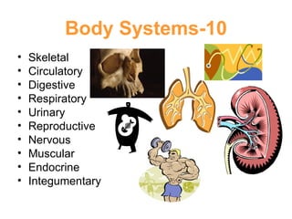
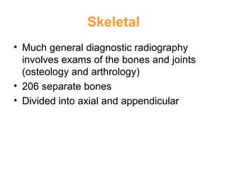
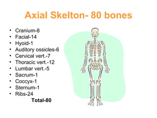
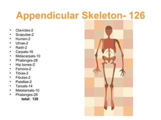
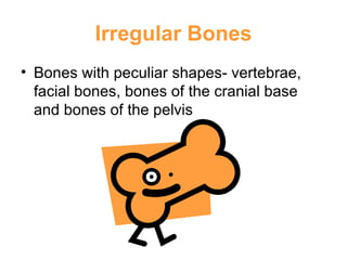
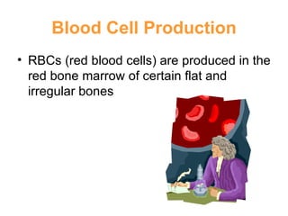

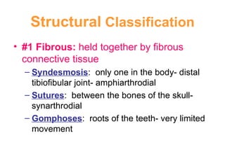

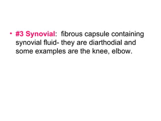
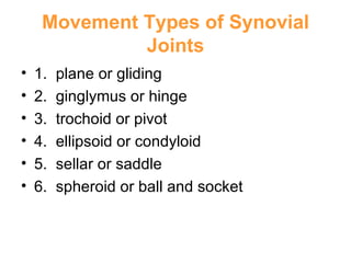
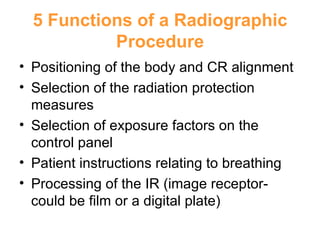
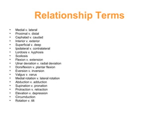
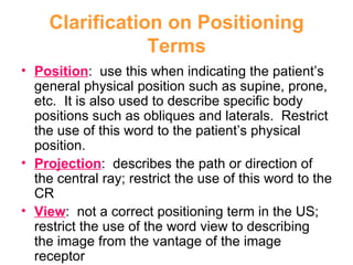
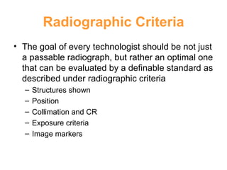
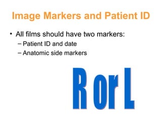


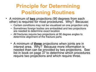
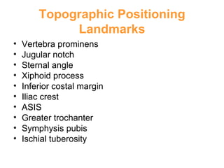

This chapter discusses general anatomy, terminology, and positioning procedures. It provides an overview of the skeletal system and bone classification. It also covers joints, radiographic projections, positioning terms, and criteria for optimal radiographic images. Key topics include the 206 bones of the human body, the 10 body systems, and important landmarks used for positioning.


