INTERVENTIONAL RADIOGRAPHY
Alerna Irene Cal, RRT, MAED
I. INTERVENTIONAL RADIOGRAPHY
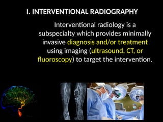
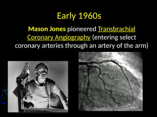
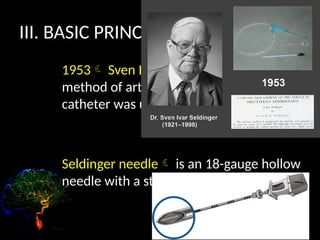
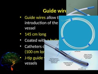
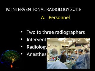
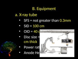
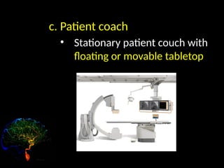
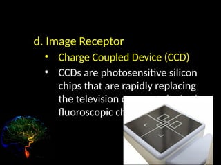
Interventional radiology is a
subspecialty which provides minimally
invasive diagnosis and/or treatment
using imaging (ultrasound, CT, or
fluoroscopy) to target the intervention.
II. HISTORY
1930
Interventional Radiology procedures
began with angiography
Angiography
refers to the opacification of
vessels through injection of
CM.
Early 1960s
Mason Jones pioneered Transbrachial
Coronary Angiography (entering select
coronary arteries through an artery of the arm)
During 1960s
• Melvin Judkins (Coronary
angiography)
• Charles Dotter (Visceral
angiography)
It was 50 years ago on Jan. 16, 1964 that
Charles Dotter, the “Father of
Interventional Radiology”, performed
the first angioplasty
III. BASIC PRINCIPLES
1953 Sven Ivar Seldinger: described a
method of arterial access in which a
catheter was used. (femoral artery)
Seldinger needle is an 18-gauge hollow
needle with a stylet.
Guide wires
• Guide wires allow the safe
introduction of the catheter into the
vessel
• 145 cm long
• Coated with a hydrophilic material
• Catheters overlaying the guide wires
(100 cm long or less)
• J-tip guide wires  atherosclerotic
vessels
Catheter
• Designed in many different shapes and
sizes
• Catheter diameter is categorized in
French (Fr) sizes.
• (3 Fr = 1mm diameter)
Catheter
Types:
1. H1 or Headhunter tip
2. Simmons Catheter
3. C2 or Cobra Catheter
4. Pigtail Catheter
1. H1 or Headhunter tip
• Vincent Hinck
• Femoral approach to the
brachiocephalic vessels
2. Simmons Catheter
• Cerebral angiography
• Visceral angiography
3. C2 or Cobra Catheter
• Celiac, renal and
mesenteric arteries
4. Pigtail Catheter
• Have side holes for ejecting
CM into a compact bolus
• Reduce whiplash effect
IV. INTERVENTIONAL RADIOLOGY SUITE
A. Personnel
• Two to three radiographers
• Interventional radiologist
• Radiology nurse
• Anesthesiologist
B. Equipment
a. X-ray tube
• SFS = not greater than 0.3mm
• SID = 100 cm
• OID = 40 cm
• Disc size = 15 cm diameter; 5
cm thick
• Power rating = 80kW
• Anode Heat Capacity = 1MHU
b. High Voltage Generator
• High frequency
• Three-phase, 12 pulse power
capable of at least 100 kW
• Low ripple
c. Patient coach
• Stationary patient couch with
floating or movable tabletop
d. Image Receptor
• Charge Coupled Device (CCD)
• CCDs are photosensitive silicon
chips that are rapidly replacing
the television camera tube in the
fluoroscopic chain.
IV. BINARY SYSTEM
• The binary system is a numeric
system using the base 2. this is
called binary digits (bits).
• The only acceptable values are
“0” and “1”
IV. BINARY SYSTEM
A. Binary to decimal
 00110010  50
B. Decimal to binary
 188  10111100
INTERVENTIONAL RADIOGRAPHY introduction.pptx

INTERVENTIONAL RADIOGRAPHY introduction.pptx

  • 1.
  • 2.
    I. INTERVENTIONAL RADIOGRAPHY Interventionalradiology is a subspecialty which provides minimally invasive diagnosis and/or treatment using imaging (ultrasound, CT, or fluoroscopy) to target the intervention.
  • 3.
    II. HISTORY 1930 Interventional Radiologyprocedures began with angiography
  • 4.
    Angiography refers to theopacification of vessels through injection of CM.
  • 5.
    Early 1960s Mason Jonespioneered Transbrachial Coronary Angiography (entering select coronary arteries through an artery of the arm)
  • 6.
    During 1960s • MelvinJudkins (Coronary angiography) • Charles Dotter (Visceral angiography)
  • 7.
    It was 50years ago on Jan. 16, 1964 that Charles Dotter, the “Father of Interventional Radiology”, performed the first angioplasty
  • 8.
    III. BASIC PRINCIPLES 1953Sven Ivar Seldinger: described a method of arterial access in which a catheter was used. (femoral artery) Seldinger needle is an 18-gauge hollow needle with a stylet.
  • 9.
    Guide wires • Guidewires allow the safe introduction of the catheter into the vessel • 145 cm long • Coated with a hydrophilic material • Catheters overlaying the guide wires (100 cm long or less) • J-tip guide wires  atherosclerotic vessels
  • 10.
    Catheter • Designed inmany different shapes and sizes • Catheter diameter is categorized in French (Fr) sizes. • (3 Fr = 1mm diameter)
  • 11.
    Catheter Types: 1. H1 orHeadhunter tip 2. Simmons Catheter 3. C2 or Cobra Catheter 4. Pigtail Catheter
  • 12.
    1. H1 orHeadhunter tip • Vincent Hinck • Femoral approach to the brachiocephalic vessels
  • 13.
    2. Simmons Catheter •Cerebral angiography • Visceral angiography
  • 14.
    3. C2 orCobra Catheter • Celiac, renal and mesenteric arteries
  • 15.
    4. Pigtail Catheter •Have side holes for ejecting CM into a compact bolus • Reduce whiplash effect
  • 16.
    IV. INTERVENTIONAL RADIOLOGYSUITE A. Personnel • Two to three radiographers • Interventional radiologist • Radiology nurse • Anesthesiologist
  • 17.
    B. Equipment a. X-raytube • SFS = not greater than 0.3mm • SID = 100 cm • OID = 40 cm • Disc size = 15 cm diameter; 5 cm thick • Power rating = 80kW • Anode Heat Capacity = 1MHU
  • 18.
    b. High VoltageGenerator • High frequency • Three-phase, 12 pulse power capable of at least 100 kW • Low ripple
  • 19.
    c. Patient coach •Stationary patient couch with floating or movable tabletop
  • 20.
    d. Image Receptor •Charge Coupled Device (CCD) • CCDs are photosensitive silicon chips that are rapidly replacing the television camera tube in the fluoroscopic chain.
  • 21.
    IV. BINARY SYSTEM •The binary system is a numeric system using the base 2. this is called binary digits (bits). • The only acceptable values are “0” and “1”
  • 22.
    IV. BINARY SYSTEM A.Binary to decimal  00110010  50 B. Decimal to binary  188  10111100