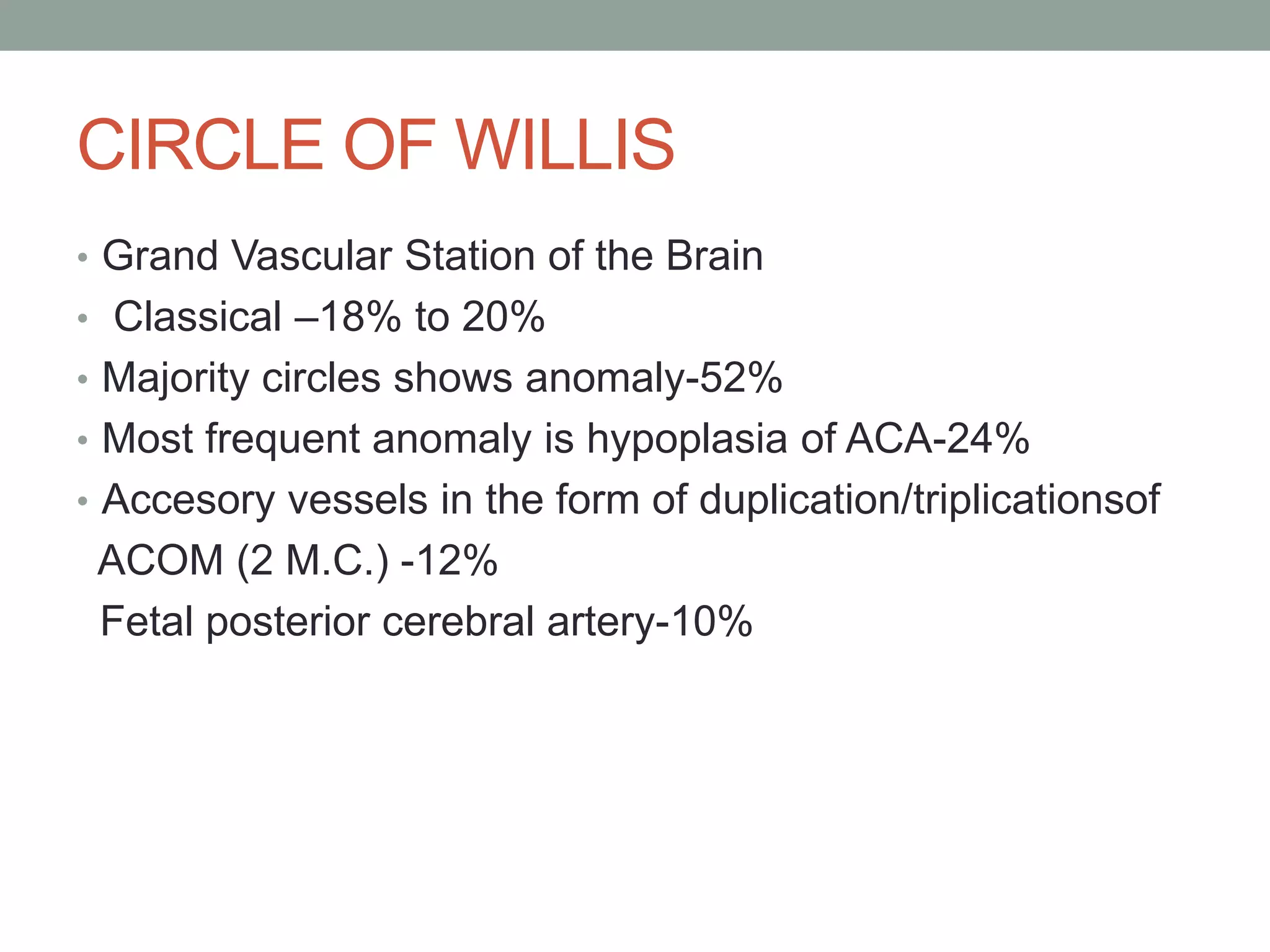
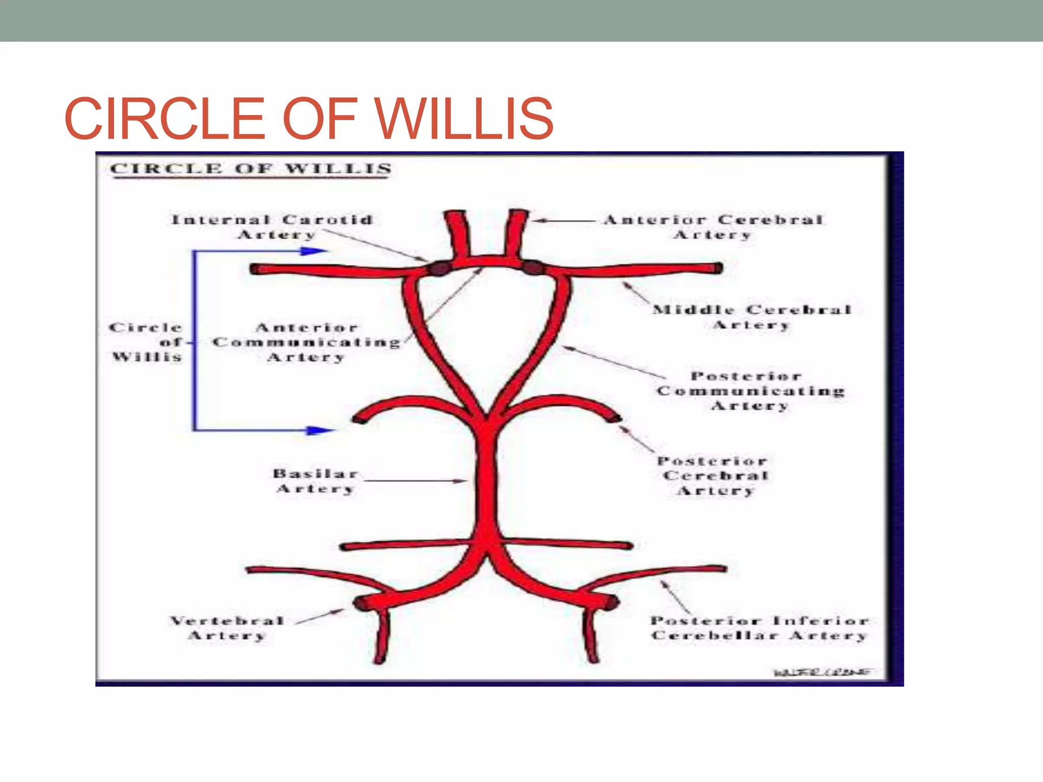
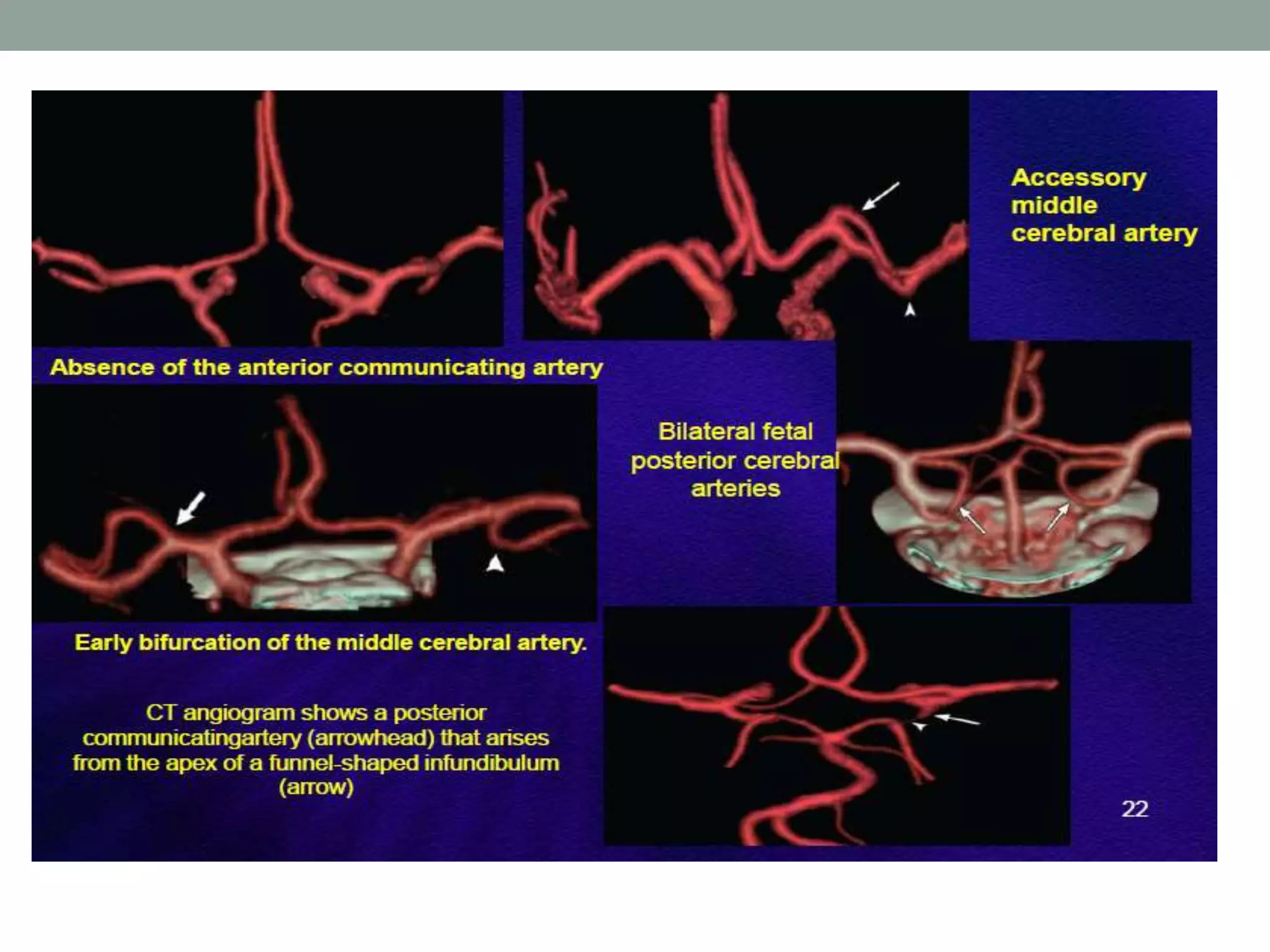
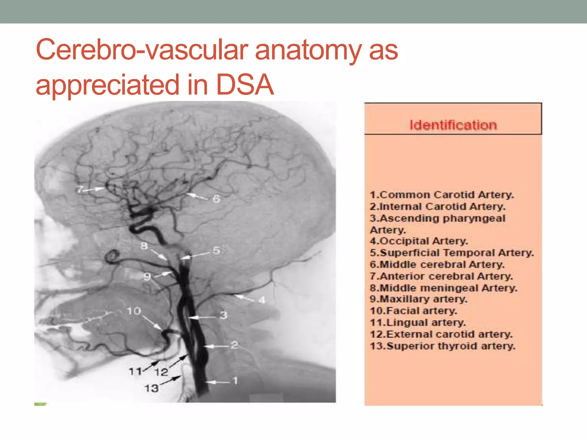
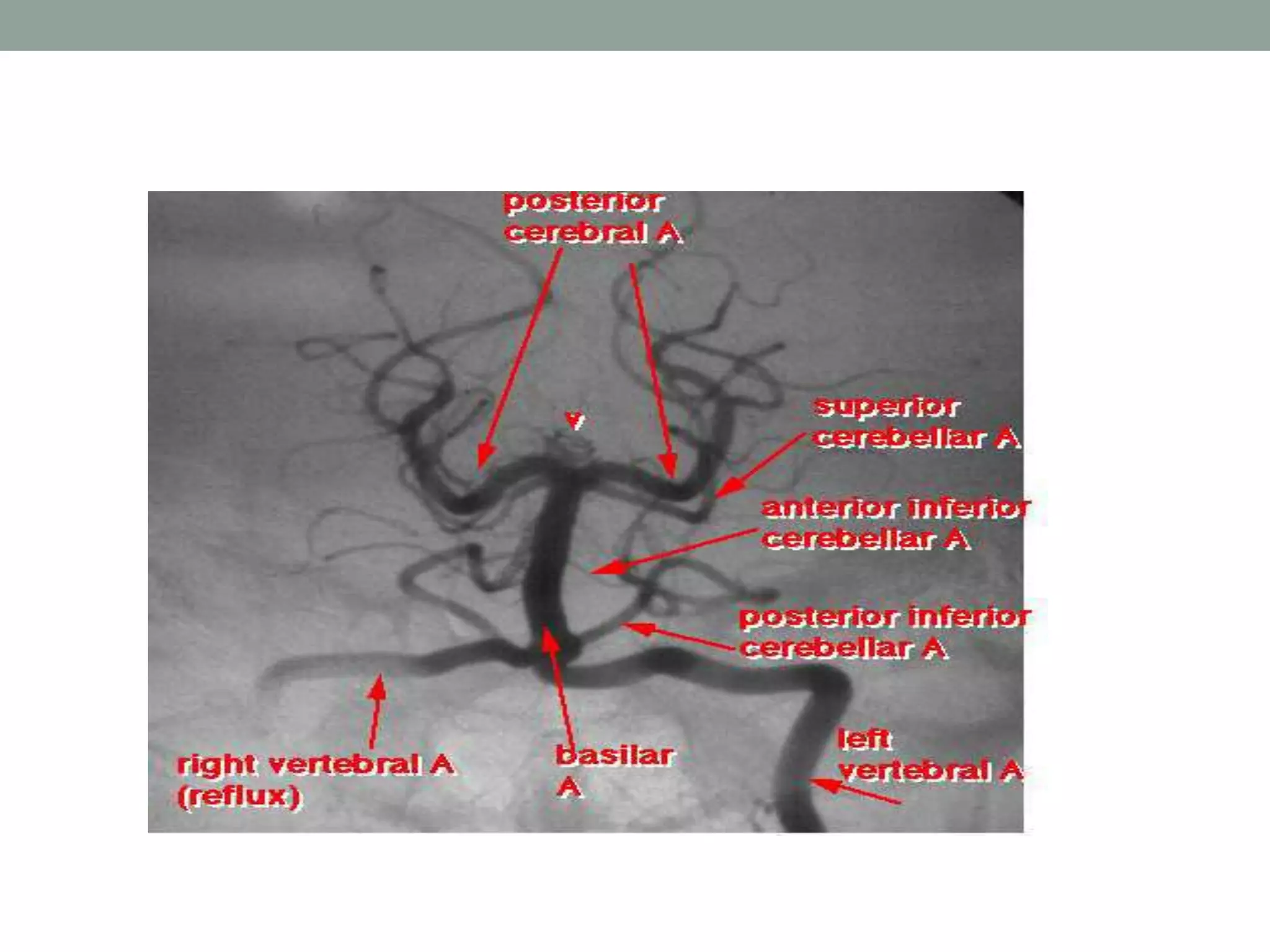
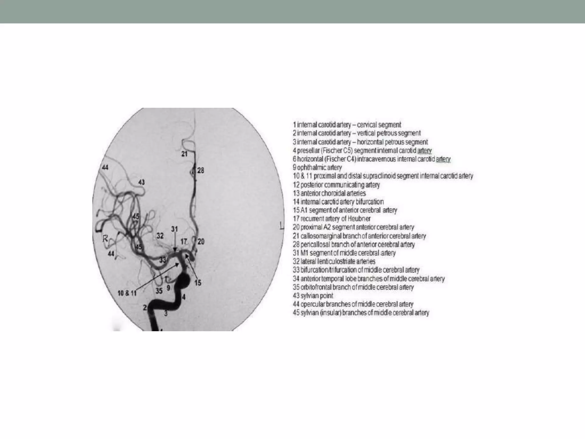
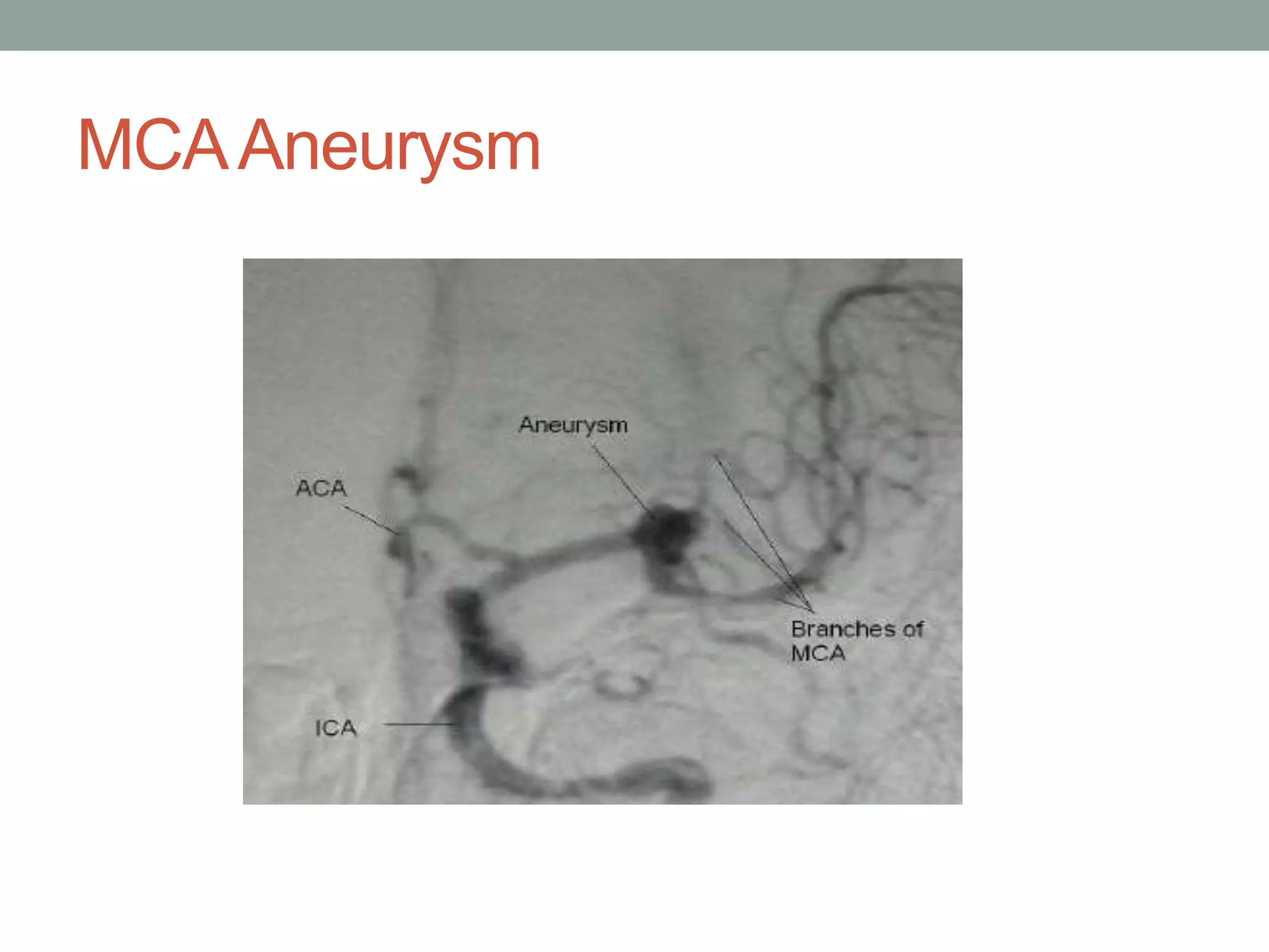
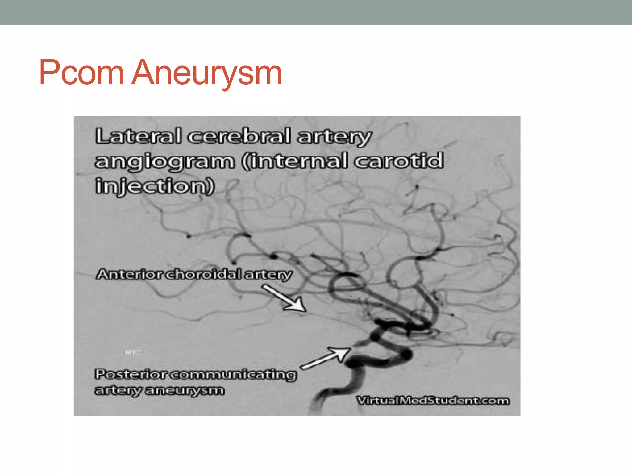
Digital subtraction angiography (DSA) is the gold standard for evaluating the cerebral vasculature. It involves injecting iodinated contrast material into arteries and using subtraction techniques to visualize vessels. The normal anatomy includes the circle of Willis and branches of major arteries. Variants are common. DSA is used to diagnose conditions like aneurysms and arteriovenous malformations. Newer digital systems provide 3D reconstruction and less radiation exposure compared to older techniques. DSA remains an important tool for interventional procedures and treatment planning of complex vascular lesions of the brain.
Cerebral angiography - medical information martinshaji
Cerebral angiography is an X-ray examination method with the help of which the state of blood vessels, their functional abilities, roundabout blood circulation, the presence of deviations and the size of pathological zones are determined. i hope this will be useful among the medical professionals.
please comment
thank u
Starting with the Definition, Coverage of field, Seldinger technique, Instruments used in IR we move forward into the embolization Techniques and applications, IR procedures in hepatobiliary system, Portal hypertension, Varicose veins
and lastly RFA for bone tumors like ostoid osteoma
SELDINGER TECHNIQUE & INTERVENTIONAL RADIOLOGYRiyas M K
its a basic introduction about Seldinger technique and Intervetional radiology.In interventional radiology, procedures generally start with the Seldinger technique to access the vasculature, using a needle through which a guidewire is inserted, followed by navigation of catheters within the vessels.
Basics of Coronary Angiography Hewad Gulzai.pptxHewad Gulzai
Basics of Coronary Angiography for beginners, MD, DNB, DM students, Nurses, cathlab technicians, physicians and other healthcare members .
hope you will learn something from this ppt. 😀
Carotid artery disease is commonly seen in association with atherosclerosis and complicate the situation. clearcut guidelines with necessary surgical details are provided in presentations.
CONCEPT OF NODOPATHIES AND PARANODOPATHIES.pptxNeurologyKota
emergence of autoimmune neuropathies and role of nodal and paranodal regions in their pathophysiology.
Peripheral neuropathies are traditionally categorized into demyelinating or axonal.
dysfunction at nodal/paranodal region key for better understanding of patients with immune mediated neuropathies.
antibodies targeting node and paranode of myelinated nerves have been increasingly detected in patients with immune mediated neuropathies.
have clinical phenotype similar common inflammatory neuropathies like Guillain Barre syndrome and chronic inflammatory demyelinating polyradiculoneuropathy
they respond poorly to conventional first line immunotherapies like IVIG
This presentation briefs out the approach of dementia assessment in line with consideration of recent advances. Now the pattern of assessment has evolved towards examining each individual domain rather than lobar assessment.
This presentation contains information about Dementia in Young onset. Also it describes the etiologies, clinical feature of common YOD & their management.
Entrapment Syndromes of Lower Limb.pptxNeurologyKota
This presentation contains information about the various Entrapment syndromes of Lower limb in descending order of topography. It also contains information about etiology, clinical features and management of each of these entrapment syndromes with special emphasis on electrodiagnostic confirmation.
Couples presenting to the infertility clinic- Do they really have infertility...Sujoy Dasgupta
Dr Sujoy Dasgupta presented the study on "Couples presenting to the infertility clinic- Do they really have infertility? – The unexplored stories of non-consummation" in the 13th Congress of the Asia Pacific Initiative on Reproduction (ASPIRE 2024) at Manila on 24 May, 2024.
These simplified slides by Dr. Sidra Arshad present an overview of the non-respiratory functions of the respiratory tract.
Learning objectives:
1. Enlist the non-respiratory functions of the respiratory tract
2. Briefly explain how these functions are carried out
3. Discuss the significance of dead space
4. Differentiate between minute ventilation and alveolar ventilation
5. Describe the cough and sneeze reflexes
Study Resources:
1. Chapter 39, Guyton and Hall Textbook of Medical Physiology, 14th edition
2. Chapter 34, Ganong’s Review of Medical Physiology, 26th edition
3. Chapter 17, Human Physiology by Lauralee Sherwood, 9th edition
4. Non-respiratory functions of the lungs https://academic.oup.com/bjaed/article/13/3/98/278874
Report Back from SGO 2024: What’s the Latest in Cervical Cancer?bkling
Are you curious about what’s new in cervical cancer research or unsure what the findings mean? Join Dr. Emily Ko, a gynecologic oncologist at Penn Medicine, to learn about the latest updates from the Society of Gynecologic Oncology (SGO) 2024 Annual Meeting on Women’s Cancer. Dr. Ko will discuss what the research presented at the conference means for you and answer your questions about the new developments.
These lecture slides, by Dr Sidra Arshad, offer a quick overview of physiological basis of a normal electrocardiogram.
Learning objectives:
1. Define an electrocardiogram (ECG) and electrocardiography
2. Describe how dipoles generated by the heart produce the waveforms of the ECG
3. Describe the components of a normal electrocardiogram of a typical bipolar leads (limb II)
4. Differentiate between intervals and segments
5. Enlist some common indications for obtaining an ECG
Study Resources:
1. Chapter 11, Guyton and Hall Textbook of Medical Physiology, 14th edition
2. Chapter 9, Human Physiology - From Cells to Systems, Lauralee Sherwood, 9th edition
3. Chapter 29, Ganong’s Review of Medical Physiology, 26th edition
4. Electrocardiogram, StatPearls - https://www.ncbi.nlm.nih.gov/books/NBK549803/
5. ECG in Medical Practice by ABM Abdullah, 4th edition
6. ECG Basics, http://www.nataliescasebook.com/tag/e-c-g-basics
HOT NEW PRODUCT! BIG SALES FAST SHIPPING NOW FROM CHINA!! EU KU DB BK substit...GL Anaacs
Contact us if you are interested:
Email / Skype : kefaya1771@gmail.com
Threema: PXHY5PDH
New BATCH Ku !!! MUCH IN DEMAND FAST SALE EVERY BATCH HAPPY GOOD EFFECT BIG BATCH !
Contact me on Threema or skype to start big business!!
Hot-sale products:
NEW HOT EUTYLONE WHITE CRYSTAL!!
5cl-adba precursor (semi finished )
5cl-adba raw materials
ADBB precursor (semi finished )
ADBB raw materials
APVP powder
5fadb/4f-adb
Jwh018 / Jwh210
Eutylone crystal
Protonitazene (hydrochloride) CAS: 119276-01-6
Flubrotizolam CAS: 57801-95-3
Metonitazene CAS: 14680-51-4
Payment terms: Western Union,MoneyGram,Bitcoin or USDT.
Deliver Time: Usually 7-15days
Shipping method: FedEx, TNT, DHL,UPS etc.Our deliveries are 100% safe, fast, reliable and discreet.
Samples will be sent for your evaluation!If you are interested in, please contact me, let's talk details.
We specializes in exporting high quality Research chemical, medical intermediate, Pharmaceutical chemicals and so on. Products are exported to USA, Canada, France, Korea, Japan,Russia, Southeast Asia and other countries.
Lung Cancer: Artificial Intelligence, Synergetics, Complex System Analysis, S...Oleg Kshivets
RESULTS: Overall life span (LS) was 2252.1±1742.5 days and cumulative 5-year survival (5YS) reached 73.2%, 10 years – 64.8%, 20 years – 42.5%. 513 LCP lived more than 5 years (LS=3124.6±1525.6 days), 148 LCP – more than 10 years (LS=5054.4±1504.1 days).199 LCP died because of LC (LS=562.7±374.5 days). 5YS of LCP after bi/lobectomies was significantly superior in comparison with LCP after pneumonectomies (78.1% vs.63.7%, P=0.00001 by log-rank test). AT significantly improved 5YS (66.3% vs. 34.8%) (P=0.00000 by log-rank test) only for LCP with N1-2. Cox modeling displayed that 5YS of LCP significantly depended on: phase transition (PT) early-invasive LC in terms of synergetics, PT N0—N12, cell ratio factors (ratio between cancer cells- CC and blood cells subpopulations), G1-3, histology, glucose, AT, blood cell circuit, prothrombin index, heparin tolerance, recalcification time (P=0.000-0.038). Neural networks, genetic algorithm selection and bootstrap simulation revealed relationships between 5YS and PT early-invasive LC (rank=1), PT N0—N12 (rank=2), thrombocytes/CC (3), erythrocytes/CC (4), eosinophils/CC (5), healthy cells/CC (6), lymphocytes/CC (7), segmented neutrophils/CC (8), stick neutrophils/CC (9), monocytes/CC (10); leucocytes/CC (11). Correct prediction of 5YS was 100% by neural networks computing (area under ROC curve=1.0; error=0.0).
CONCLUSIONS: 5YS of LCP after radical procedures significantly depended on: 1) PT early-invasive cancer; 2) PT N0--N12; 3) cell ratio factors; 4) blood cell circuit; 5) biochemical factors; 6) hemostasis system; 7) AT; 8) LC characteristics; 9) LC cell dynamics; 10) surgery type: lobectomy/pneumonectomy; 11) anthropometric data. Optimal diagnosis and treatment strategies for LC are: 1) screening and early detection of LC; 2) availability of experienced thoracic surgeons because of complexity of radical procedures; 3) aggressive en block surgery and adequate lymph node dissection for completeness; 4) precise prediction; 5) adjuvant chemoimmunoradiotherapy for LCP with unfavorable prognosis.
Tom Selleck Health: A Comprehensive Look at the Iconic Actor’s Wellness Journeygreendigital
Tom Selleck, an enduring figure in Hollywood. has captivated audiences for decades with his rugged charm, iconic moustache. and memorable roles in television and film. From his breakout role as Thomas Magnum in Magnum P.I. to his current portrayal of Frank Reagan in Blue Bloods. Selleck's career has spanned over 50 years. But beyond his professional achievements. fans have often been curious about Tom Selleck Health. especially as he has aged in the public eye.
Follow us on: Pinterest
Introduction
Many have been interested in Tom Selleck health. not only because of his enduring presence on screen but also because of the challenges. and lifestyle choices he has faced and made over the years. This article delves into the various aspects of Tom Selleck health. exploring his fitness regimen, diet, mental health. and the challenges he has encountered as he ages. We'll look at how he maintains his well-being. the health issues he has faced, and his approach to ageing .
Early Life and Career
Childhood and Athletic Beginnings
Tom Selleck was born on January 29, 1945, in Detroit, Michigan, and grew up in Sherman Oaks, California. From an early age, he was involved in sports, particularly basketball. which played a significant role in his physical development. His athletic pursuits continued into college. where he attended the University of Southern California (USC) on a basketball scholarship. This early involvement in sports laid a strong foundation for his physical health and disciplined lifestyle.
Transition to Acting
Selleck's transition from an athlete to an actor came with its physical demands. His first significant role in "Magnum P.I." required him to perform various stunts and maintain a fit appearance. This role, which he played from 1980 to 1988. necessitated a rigorous fitness routine to meet the show's demands. setting the stage for his long-term commitment to health and wellness.
Fitness Regimen
Workout Routine
Tom Selleck health and fitness regimen has evolved. adapting to his changing roles and age. During his "Magnum, P.I." days. Selleck's workouts were intense and focused on building and maintaining muscle mass. His routine included weightlifting, cardiovascular exercises. and specific training for the stunts he performed on the show.
Selleck adjusted his fitness routine as he aged to suit his body's needs. Today, his workouts focus on maintaining flexibility, strength, and cardiovascular health. He incorporates low-impact exercises such as swimming, walking, and light weightlifting. This balanced approach helps him stay fit without putting undue strain on his joints and muscles.
Importance of Flexibility and Mobility
In recent years, Selleck has emphasized the importance of flexibility and mobility in his fitness regimen. Understanding the natural decline in muscle mass and joint flexibility with age. he includes stretching and yoga in his routine. These practices help prevent injuries, improve posture, and maintain mobilit
Prix Galien International 2024 Forum ProgramLevi Shapiro
June 20, 2024, Prix Galien International and Jerusalem Ethics Forum in ROME. Detailed agenda including panels:
- ADVANCES IN CARDIOLOGY: A NEW PARADIGM IS COMING
- WOMEN’S HEALTH: FERTILITY PRESERVATION
- WHAT’S NEW IN THE TREATMENT OF INFECTIOUS,
ONCOLOGICAL AND INFLAMMATORY SKIN DISEASES?
- ARTIFICIAL INTELLIGENCE AND ETHICS
- GENE THERAPY
- BEYOND BORDERS: GLOBAL INITIATIVES FOR DEMOCRATIZING LIFE SCIENCE TECHNOLOGIES AND PROMOTING ACCESS TO HEALTHCARE
- ETHICAL CHALLENGES IN LIFE SCIENCES
- Prix Galien International Awards Ceremony
TEST BANK for Operations Management, 14th Edition by William J. Stevenson, Ve...kevinkariuki227
TEST BANK for Operations Management, 14th Edition by William J. Stevenson, Verified Chapters 1 - 19, Complete Newest Version.pdf
4. CIRCLE OF WILLIS
• Grand Vascular Station of the Brain
• Classical –18% to 20%
• Majority circles shows anomaly-52%
• Most frequent anomaly is hypoplasia of ACA-24%
• Accesory vessels in the form of duplication/triplicationsof
ACOM (2 M.C.) -12%
Fetal posterior cerebral artery-10%
9. MIDDLE CEREBRALARTERY
• Larger terminal branch of ICA
• Run laterally in stem of lateral sulcus
• Curves on superolateral surface
• Runs backwards in depth of posterior ramus of lateral
sulcus
10. • M1 segment =horizontal segment from origin to its
bifurcation (it is in sylvian fissure)
• M2 segment =lacunar segment -in the insula loops over
insula—laterally to exit from sylvian fissure
• M3 segment = opercular branch-from sylvian fissure &
ramify over cerebral cortex
• Anomalies of MCA are uncommon
13. • 2 major terminal br of PCA—
•
• parieto occipital art & calcarine art
14. POSTERIOR FOSSA
• Vertebral artery
• Basilar artrery
• Vertebral arteries
• Originate from the subclavian arteries
• Left VA is dominant in 60% cases
16. BASILAR ARTERY
• Right and left VA unite to form basilar artery
• Courses infront of pons (Prepontine cistern)
terminates in the interpeduncular cistern
• 3cm in length,1.5 to 4mm in width
18. Normal VARIANTS
• Fenestrations and duplications
• Variants of the circle of Willis
• Persistent carotid-basilar anastomoses
• Anomalies identified in the skull base
23. Imaging Techniques
• Vascular structures of brain can be imaged by 4
means:
• 1. DSA: gold standard
• Invasive and risk of nephrotoxic contrast,ionising
radiation
• 2. Vascular ultrasound: least invasive, can be
done bedside, cost effective.
• Best choice for imaging vessels close to skin
surface.
24. • Drawback: limited anatomic coverage, deep
vessels cant be imaged, operator dependent,
requires skill.
25. • 3 CT angio: main drawbacks are contrast use and
radiation exposure,
• calcifications are overestimated.
• Preferred for aorta and coronaries
• 4 MRA : non invasive, no radiation exposure.
• Preferred for carotids and intracranial vessels as MRI
brain can also be obtained
• widely used in neurological disorders
26. • ANGIO?
• ‘ANGIO means blood vessel’
• SUBTRACTION?
• It is simply a technique by which bone
structures images are subtracted or canceled
out from a film of bones plus opacified vessels,
“leaving an unobscured image of the vessels”
27. DSA
• Acquisition of digital fluoroscopic images
combined with injection of contrast material
and real time subtraction of pre- and post
contrast images to perform angiography is
referred to as digital subtraction angiography
28. HISTORY
Portuguese neurologist Egas Moniz,( Nobel
Prize winner 1949), in 1927 developed the
technique of contrast x-ray cerebral angiography
to diagnose diseases,
such as tumors and arteriovenous
malformations.
29. • Idea of subtraction images was first proposed
by the Dutch radiologist Ziedses des Plantes in
the 1935, when he was able to produce
subtracted images using plain films.
30. HISTORICAL DEVELOPMENT
• CONVENTIONAL SUBTRACTION TECHNIQUE:
• Photographic method used to eliminate
unwanted images.
• No addition of information; only purpose to
make diagnostically important information
31. • Developed in 1970s, University of Wisconsin,
University of Arizona, University of Kiel.
• USA
• Commercial systems introduced in 1980.
32. • 3 conditions:
• SCOUT FILM
• ANGIOGRAM FILM-CONTRAST
• NO MOTION OF HEAD
33. PRINCIPAL
• Principles of subtraction are based on the
following:
• Scout film shows the structural details of the
skull and the adjacent soft tissue.
• Angiogram film shows exactly the same
anatomic details, if the patient does not move,
plus the opacified blood vessels.
34. • If all the information in the scout film could be
subtracted from the angiogram film, only the
opacified vessel pattern would remain visible.
37. WHY DSA?
• Digital subtraction angiography (DSA) was developed
to improve vessel contrast.
• Technique that uses a computer to subtract two
images, obtained before and after contrast media is
injected into the vessels of interest.
• Anatomical structures that are the same in the two
images can be removed and the resulting image
shows the vessels only
38. • Modern DSA systems are based on digital
fluoroscopy/fluorography systems, which are
equipped with special software and display
facilities
39. • Image before the contrast agent is
administered is called the mask image.
•
• Once the contrast is administered, a sequence
of images are taken by a television camera in
analog form, which is then digitised by
computer
40. • DSA processor has two separate image
memories, one for the mask and the other for
the images with contrast medium.
• These two image memories are subtracted
from one another arithmetically, and the result
goes to an image processing and display unit.
45. Contra indications
• No absolute contraindication.
• Poor renal reserve.
• Deranged coagulogram.
• Allergic to contrast media
46. Contrast Media
• Blood vessels are not normally seen in an x-ray
image, because of low tissue contrast.
• To increase image contrast, contrast agents,
which are dense fluids with elements of high
atomic numbers, such as iodine, are injected into
a blood vessel during angiography. Because of its
higher density and high atomic number, iodine
absorbs photons more than blood and tissue.
47. Creates detailed images of the blood vessels in
real time.
• First contrast media used for intravascular
injection were called high-osmolar contrast
media (HOCM).
48. • High osmolarity caused adverse effects such as
pain, endothelial damage, thrombosis, and
increased pressure in the pulmonary circulation.
• Low-osmolar contrast media (LOCM) were first
developed in the 1970's reduce side effects.
• Major risks of modern iodine contrast media is an
allergic reaction to iodine
49. • Non ionic Iso-osmolar contrast media.
• 30-40% dilution with normal saline.
• 50 ml of diluted contrast media is enough to do
a standard cerebral angiogram with total 8
projections.
• Approx. 5-8 ml diluted contrast / injection.
54. PREPARATION
• Nil orally 4-6 hrs.
• On trolley
• In hospital gown
• Groin shave
• Records
• Should be well
hydrated.
• Should void before
procedure.
• Peripheral pulses
marked.
• I.V line in place.
• Informed consent
55. PROCEDURE
• Gaining arterial access.
• Selective arterial catheterization.
• Image acquisition.
• Closure of arterial access.
• Post processing
• Hard copy
56. • Patients may be sedated to reduce anxiety.
• Monitor of vitals
• Local anesthetic is usually used in the area
where the catheter is to be inserted,
• Most common femoral artery
57. PROCEDURE
• Small incision given, medicut is inserted into the artery,
• Fluoroscopy is used to guide the needle to the proper
position .
• Needle is then removed after placing guide wire in the
artery and vascular sheath is inserted over the guide wire
.
• Catheter is then inserted along the guide wire through the
sheath
58. • When the catheter is in the correct position, the wire is
pulled out and dye is injected through the catheter.
• Images are acquired during contrast injection.
• Injections can be made directly into the artery of interest
(selective arteriography)
60. POSTPROCEDURAL CARE
• After the catheter is removed compression is applied to
the puncture site
• Bed rest for a minimum of 4 hours
• During rest patient is monitored and vital sign like
peripheral pulse like distal to Puncture are regularly
• Extremity is also checked for warmth, color, numbness to
ensure circulation has not been disrupted.
72. Digital subtraction angiography based
advances
• Better visualization and less radiation exposure
are key tenets in the development of digital
subtraction angiography
• Complexity of disease types increases and
more technically challenging, cutting-edge
procedures are performed
73. • Digital subtraction angiography systems with flat
panel : ADVANTAGES-
• High spatial resolution, wide dynamic range,
square field of view, and real-time imaging
capabilities with no geometric distortion—all of
which may be used for improve image quality,
reduced patient exposure to radiation
74. • Hatakeyama et al have shown that two-dimensional (2D)
and three-dimensional (3D)digital subtraction angiography
using the flat panel detector system of the direct
conversion type, with low radiation dose, is superior in
image quality for visualizing small intracranial vessels with
significantly decreased radiation exposure compared with
digital subtraction angiography with the conventional
image intensifier television system
• Hatakeyama Y, Kakeda S, Korogi Y, et al. Intracranial 2D and 3DDSA with flat panel detector of the
direct conversion type: initial experience. Eur Radiol 2006;16:2594 –2602.
77. • Allura Xper FD20/10 biplane flat detector
system with integrated 3D for intricate
neurovascular procedures.
• 3D- reconstructions
• Xper CT
• SPECTRA BEAM
• 3D Roadmapping
• multi-modality information integration
78.
79. • Redefines image clarity and captures
information at a resolution four times greater
than conventional X-ray systems.
80. Xper CT
• With XperCT clinicians can access CT-like imaging right
on the angio system so can assess soft tissue, bone
structure and other body structures before, during or after
an interventional procedure.
• XperCT reconstruction is created from rotational
acquisition performed on the Allura Xper system.
• This reconstruction can be overlaid with the 3D vascular
image.
81. • 3D soft tissue imaging supports diagnosis
planning, interventions and treatment follow-up
• XperCT can be combined with Allura 3D-RA
images to visualize soft tissue and vascular
anatomy on one image.
82. Spectra Beam
• Basically a selectable copper beam filtration
• Combination of Spectra Beam with the MRC-
tube allows increased X-ray output with better
filtration of soft radiation.
• Reduces patient X-ray dose for cardiac and
vascular applications, while maintaining the same
image quality.
83. Disadvantage of DSA
• Limited spatial resolution
• Artifacts
• Small visual field
• Not a good technique for neoplasm
84. Conclusion
• Despite recent advances in CT angiography and
MR angiography, DSA remains the standard
imaging technique for evaluation of the
cerebral vasculature .
• 3D reconstruction of the dataset acquired
during rotational DSA represents the latest
development in the neurovascular imaging .
85. • 3D-DSA has taken a prominent role in
treatment planning by enabling better
appreciation of the morphology of complex
vascular lesions before endovascular or surgical
management.
• Superior in the performance of sophisticated
tasks such as aneurysm volume measurement
86. • On the other hand, inability of 3D-DSA to
simultaneously image osseous and vascular
structures is noted as a weakness of this
technique compared with CT angiography .
87. Refrences-
• The Principle of Digital Subtraction Angiography and Radiological Protection;
K. OKAMOTO, . IT, K. SAKAI, S. YOSHIMURA ; Interventional
Neuroradiology 6 (Suppl1): 25-31, 2000
• Advances in Interventional Neuroimaging; Vallabh Janardhan and Adnan I.
Qureshi; Vol. 4, 414–419, July 2007 c The American Society for Experimental
NeuroTherapeutics,
• Intracranial Vascular Stenosis and Occlusive Disease: Evaluation with CT
Angiography, MR Angiography, and Digital Subtraction Angiography; Bash et
al; AJNR Am J Neuroradiol 26:1012–1021, May 2005
• Uptodate.com
• Digital substraction Angiography ;John hopkins Medical university
• Gray’s text book of anatomy
• Teachme anatomy .com