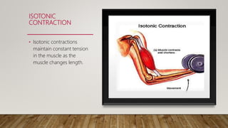
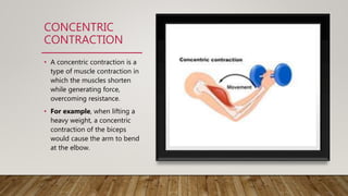
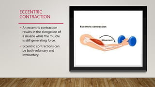
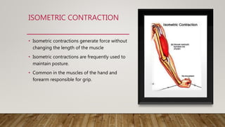
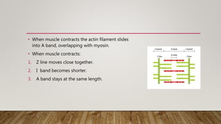
TYPES OF MUSCLE CONTRACTION
AND MECHANISM OF
CONTRACTION
DR. JASJYOT KAUR SABHARWAL (PT)
J
TWO TYPES OF CONTRACTIONS
• Isotonic Contraction
• Isometric contraction
ISOTONIC
CONTRACTION
• Isotonic contractions
maintain constant tension
in the muscle as the
muscle changes length.
TWO TYPE OF ISOTONIC CONTRACTION
Concentric Contraction
Isometric Contraction
CONCENTRIC
CONTRACTION
• A concentric contraction is a
type of muscle contraction in
which the muscles shorten
while generating force,
overcoming resistance.
• For example, when lifting a
heavy weight, a concentric
contraction of the biceps
would cause the arm to bend
at the elbow.
ECCENTRIC
CONTRACTION
• An eccentric contraction
results in the elongation of
a muscle while the muscle
is still generating force.
• Eccentric contractions can
be both voluntary and
involuntary.
ISOMETRIC CONTRACTION
• Isometric contractions generate force without
changing the length of the muscle
• Isometric contractions are frequently used to
maintain posture.
• Common in the muscles of the hand and
forearm responsible for grip.
SLIDING
FILAMENT
THEORY
Muscle contraction occurs by sliding filament
mechanism whereby the sarcomere shorten ( the
Z-line come close together) by the action of actin
filament sliding over the myosin filament.
The force behind the muscle contraction is the
ratchet movement of myosin head towards centre
of sarcomere.
Electron microscopy shows that muscle is
composed of 2 contractile proteins:
•Thin filament actin attached to Z line
•Thick filament myosin, found in A band
• When muscle contracts the actin filament slides
into A band, overlapping with myosin.
• When muscle contracts:
1. Z line moves close together.
2. I band becomes shorter.
3. A band stays at the same length.
• The process of a muscle contraction can be divided:
1. A nerve impulse arrives at the neuromuscular junction, which causes a release
of a chemical called Acetylcholine (Ach) into the synaptic cleft. The presence of
Acetylcholine causes Calcium (Ca+) to be released from the sarcoplasmic
reticulum.
2. In the presence of high concentrations of Ca+, the Ca+ binds to Troponin
changing its shape. This moves Tropomyosin from the active site of the Actin to
expose uncovered sites. Therefore, Myosin filaments can now attach to the
Actin forming a cross-bridge.
3. The cross bridge binds to the active sites. Energy from ATP is used to break the
bond straighten the cross bridge and form new bond with another active site.
4. This process of muscular contraction can last for as long as there is enough ATP
and Ca+ stores. Once the impulse stops the Ca+ is pumped back to the
Sarcoplasmic Reticulum and the Actin returns to its resting position causing the
muscle to lengthen and relax.
THANKYOU

Muscle contraction ppt

  • 1.
    TYPES OF MUSCLECONTRACTION AND MECHANISM OF CONTRACTION DR. JASJYOT KAUR SABHARWAL (PT) J
  • 2.
    TWO TYPES OFCONTRACTIONS • Isotonic Contraction • Isometric contraction
  • 3.
    ISOTONIC CONTRACTION • Isotonic contractions maintainconstant tension in the muscle as the muscle changes length.
  • 4.
    TWO TYPE OFISOTONIC CONTRACTION Concentric Contraction Isometric Contraction
  • 5.
    CONCENTRIC CONTRACTION • A concentriccontraction is a type of muscle contraction in which the muscles shorten while generating force, overcoming resistance. • For example, when lifting a heavy weight, a concentric contraction of the biceps would cause the arm to bend at the elbow.
  • 6.
    ECCENTRIC CONTRACTION • An eccentriccontraction results in the elongation of a muscle while the muscle is still generating force. • Eccentric contractions can be both voluntary and involuntary.
  • 7.
    ISOMETRIC CONTRACTION • Isometriccontractions generate force without changing the length of the muscle • Isometric contractions are frequently used to maintain posture. • Common in the muscles of the hand and forearm responsible for grip.
  • 8.
    SLIDING FILAMENT THEORY Muscle contraction occursby sliding filament mechanism whereby the sarcomere shorten ( the Z-line come close together) by the action of actin filament sliding over the myosin filament. The force behind the muscle contraction is the ratchet movement of myosin head towards centre of sarcomere. Electron microscopy shows that muscle is composed of 2 contractile proteins: •Thin filament actin attached to Z line •Thick filament myosin, found in A band
  • 9.
    • When musclecontracts the actin filament slides into A band, overlapping with myosin. • When muscle contracts: 1. Z line moves close together. 2. I band becomes shorter. 3. A band stays at the same length.
  • 10.
    • The processof a muscle contraction can be divided: 1. A nerve impulse arrives at the neuromuscular junction, which causes a release of a chemical called Acetylcholine (Ach) into the synaptic cleft. The presence of Acetylcholine causes Calcium (Ca+) to be released from the sarcoplasmic reticulum. 2. In the presence of high concentrations of Ca+, the Ca+ binds to Troponin changing its shape. This moves Tropomyosin from the active site of the Actin to expose uncovered sites. Therefore, Myosin filaments can now attach to the Actin forming a cross-bridge.
  • 11.
    3. The crossbridge binds to the active sites. Energy from ATP is used to break the bond straighten the cross bridge and form new bond with another active site. 4. This process of muscular contraction can last for as long as there is enough ATP and Ca+ stores. Once the impulse stops the Ca+ is pumped back to the Sarcoplasmic Reticulum and the Actin returns to its resting position causing the muscle to lengthen and relax.
  • 12.