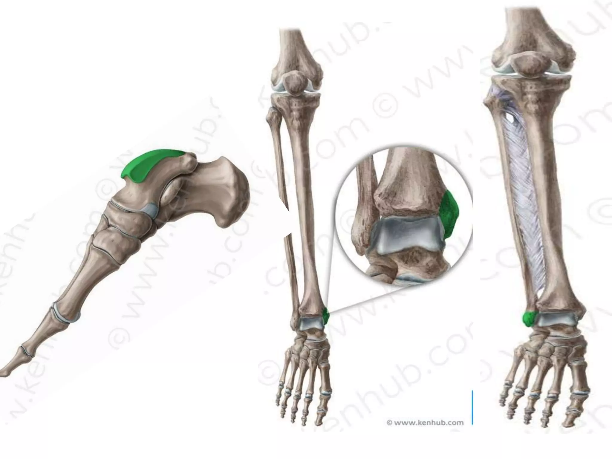
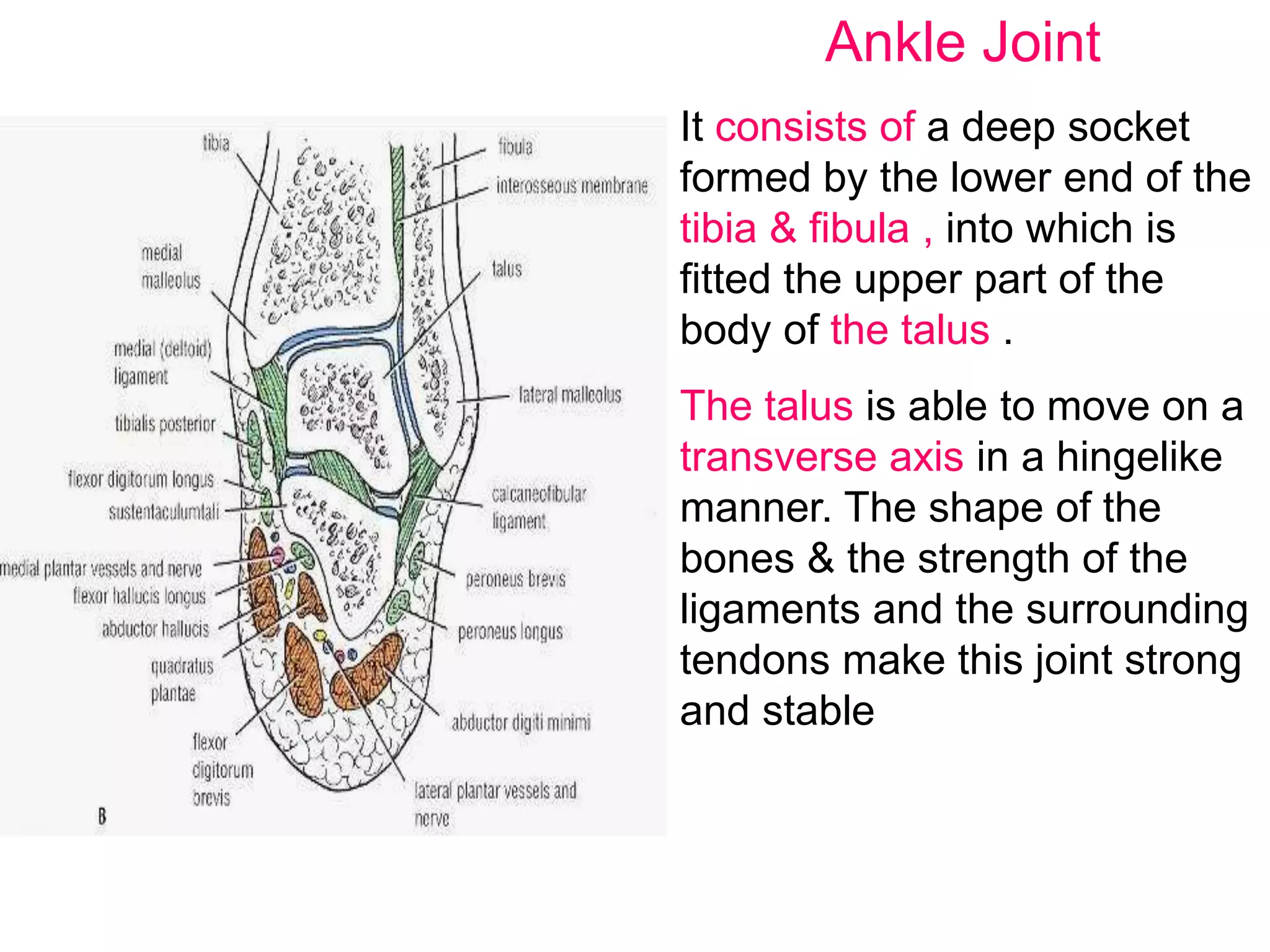
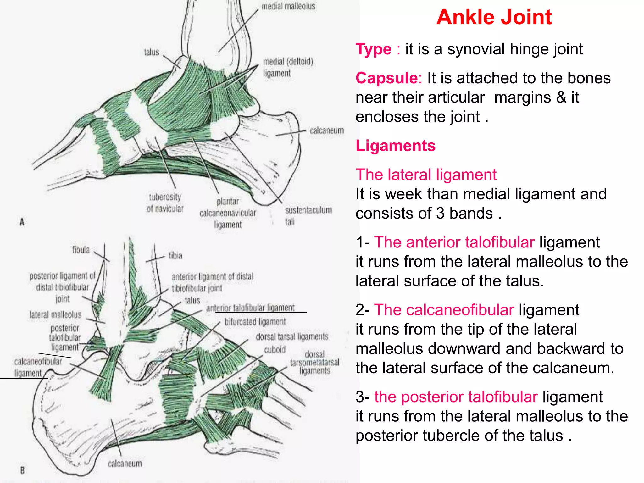
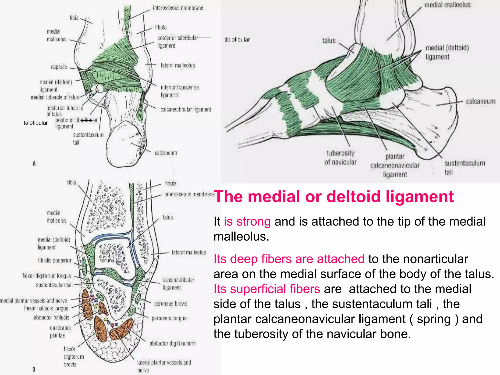
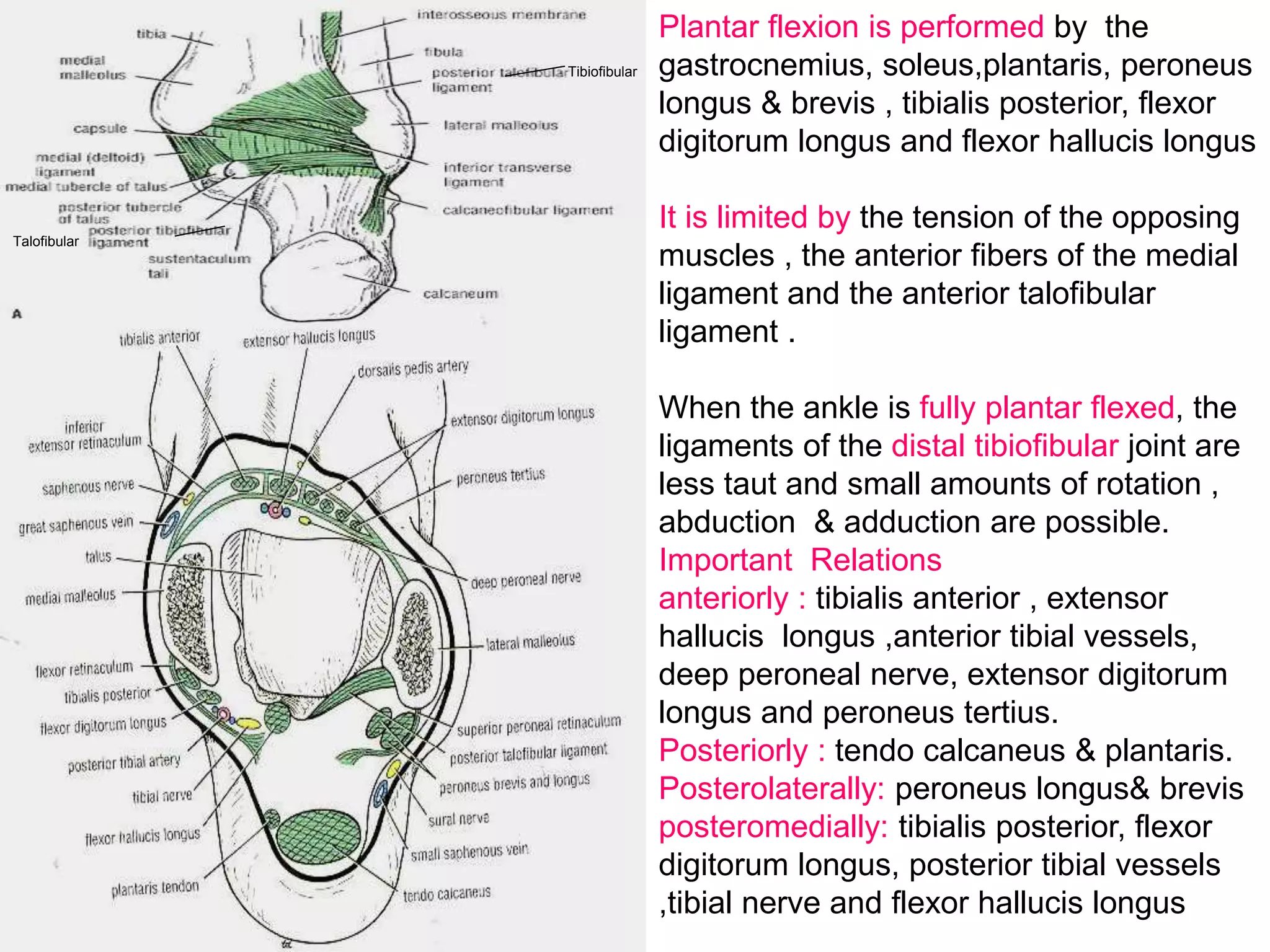
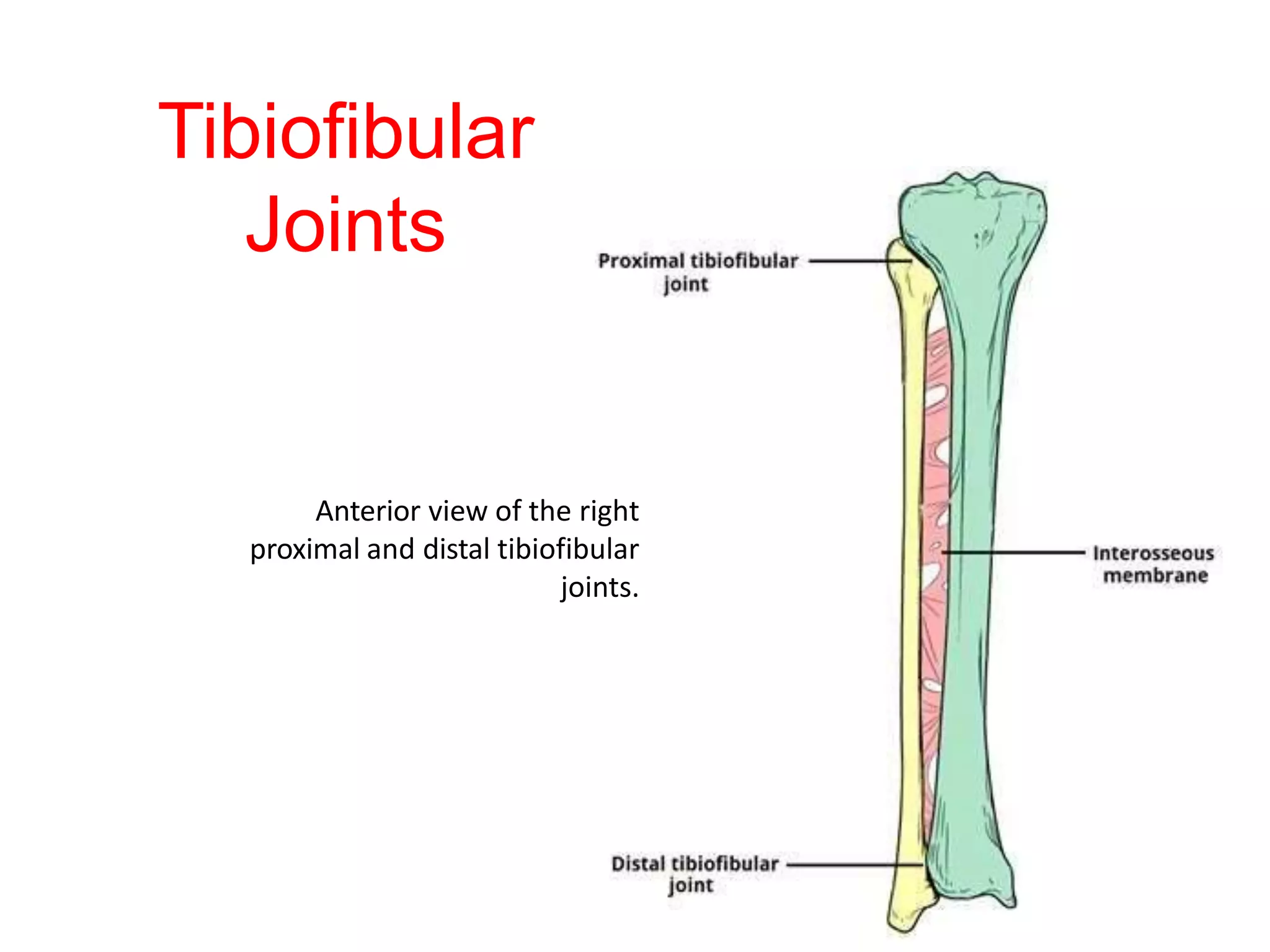
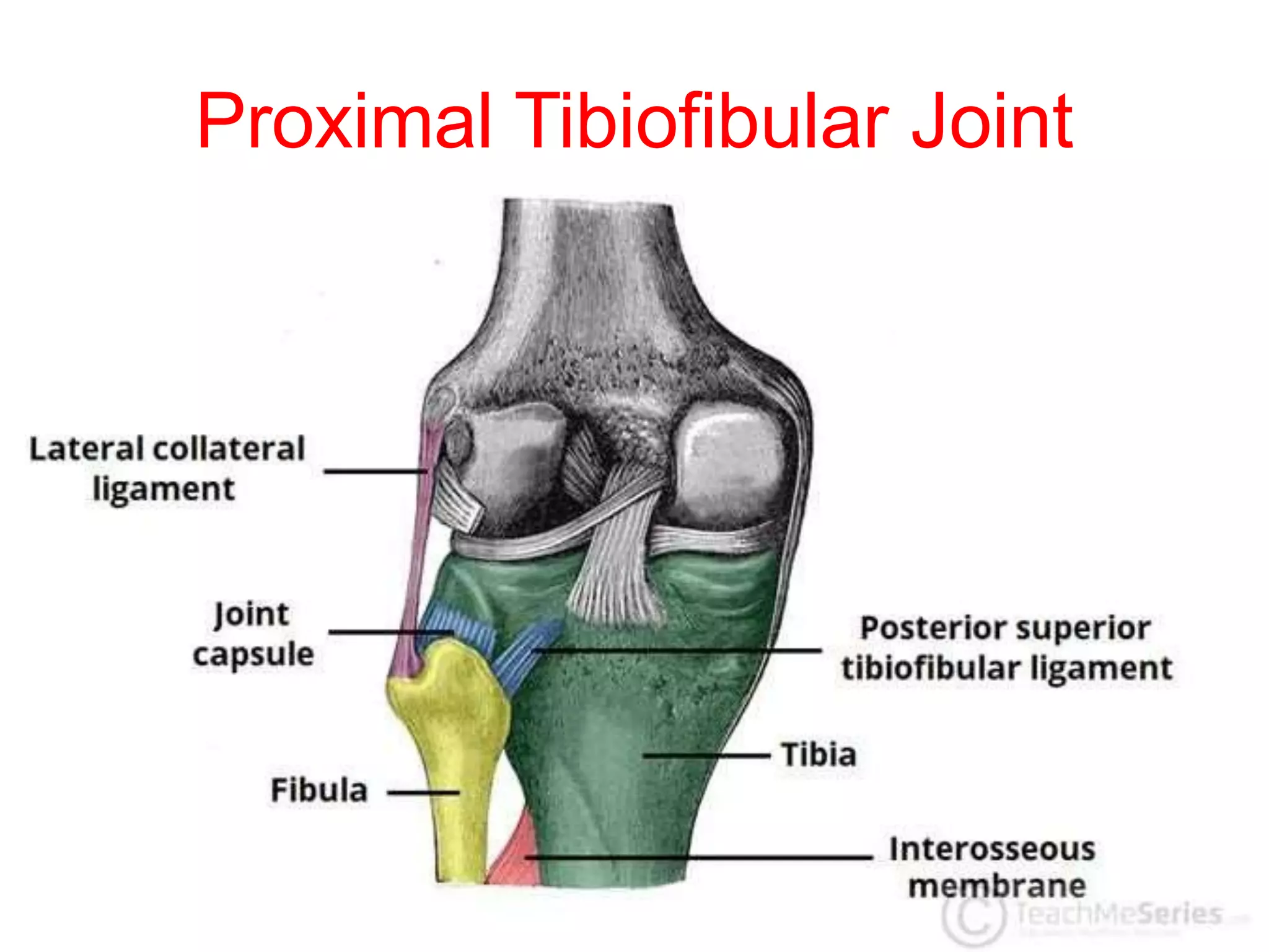
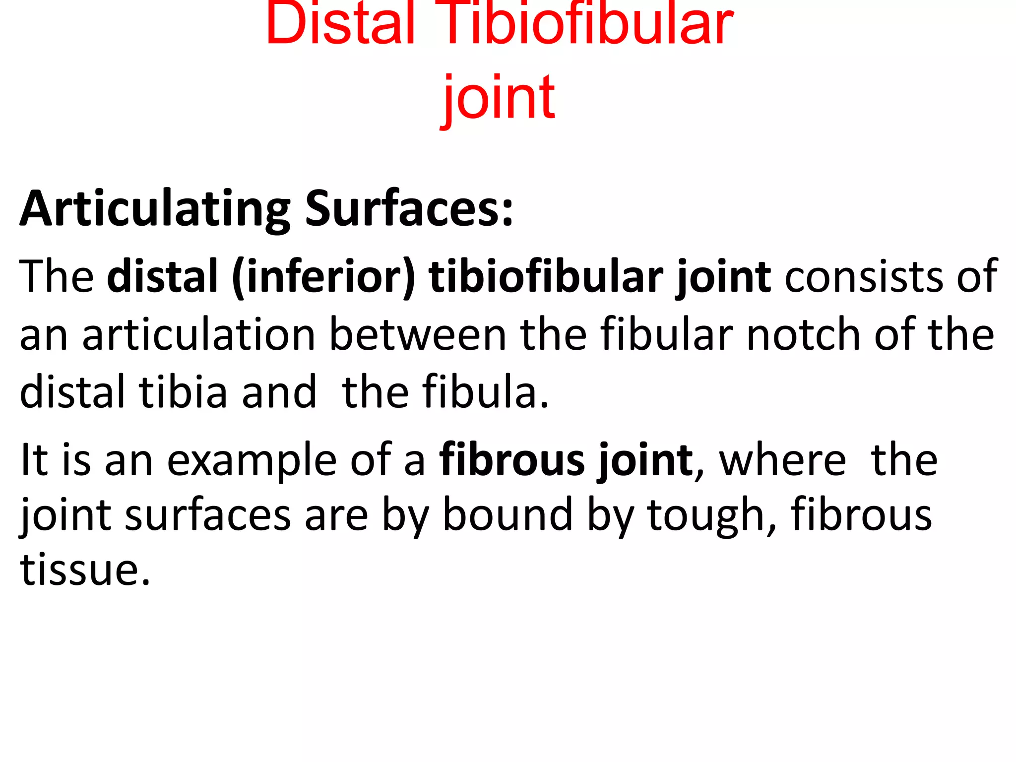
The tibiofibular joints are a set of articulations that unite the tibia and fibula. These two bones of the leg are connected via three junctions; The superior (proximal) tibiofibular joint - between the superior ends of tibia and fibula. The inferior (distal) tibiofibular joint - between their inferior ends.
Escort Service Call Girls In Sarita Vihar,, 99530°56974 Delhi NCR
Ankle and Tibiofibular Joint.pptx
1. Joints of Lower Limb
Tibio-Fibular Joint & Ankle Joint
Dr Mathew Joseph
MBBS, MD(AIIMS), BCC(Palliative Medicine)
Assistant Professor
Department of Anatomy
Amala Institute of Medical Sciences, Thrissur
6. Ankle Joint
It consists of a deep socket
formed by the lower end of the
tibia & fibula , into which is
fitted the upper part of the
body of the talus .
The talus is able to move on a
transverse axis in a hingelike
manner. The shape of the
bones & the strength of the
ligaments and the surrounding
tendons make this joint strong
and stable
7. Ankle Joint
•
Articulation
•
It is between the lower end of the tibia & the 2
malleoli and the body of the talus.
•
The articular surfaces are covered by hyaline
cartilage.
•
The inferior transverse tibiofibular ligament ,
which runs between the lateral malleolus and
the posterior border of the lower end of the tibia
,deepens the socket into which the body of the
talus fits.
8. Ankle Joint
Type : it is a synovial hinge joint
Capsule: It is attached to the bones
near their articular margins & it
encloses the joint .
Ligaments
The lateral ligament
It is week than medial ligament and
consists of 3 bands .
1- The anterior talofibular ligament
it runs from the lateral malleolus to the
lateral surface of the talus.
2- The calcaneofibular ligament
it runs from the tip of the lateral
malleolus downward and backward to
the lateral surface of the calcaneum.
3- the posterior talofibular ligament
it runs from the lateral malleolus to the
posterior tubercle of the talus .
9.
10. The medial or deltoid ligament
It is strong and is attached to the tip of the medial
malleolus.
Its deep fibers are attached to the nonarticular
area on the medial surface of the body of the talus.
Its superficial fibers are attached to the medial
side of the talus , the sustentaculum tali , the
plantar calcaneonavicular ligament ( spring ) and
the tuberosity of the navicular bone.
tibiofibular
talofibular
11.
12.
13.
14. Ankle Joint
Synovial Membrane : It lines the capsule.
Nerve Supply : Deep peroneal & tibial nerves
Movements: Dorsiflexion & plantar flexion .
N. B. inversion & eversion tack place at the
tarsal joints .
Dorsiflexion is performed by the tibialis
anterior , extensor hallucis longus, extensor
digitorum longus, and peroneus tertius.
It is limited by the tension of the
tendocalcaneus , the posterior fibers of the
medial ligament and the calcaneofibular
ligament.
During dorsiflexion the wider anterior part of
the articular surface of the talus is forced
between the medial & lateral malleolus,
causing them to separate slightly & tighten
the ligaments of the distal tibiofibular joint.
This arrangement increases the stability of
the ankle joint when the foot is in the initial
position for major thrusting movements in
running , jumping and walk .
15. Plantar flexion is performed by the
gastrocnemius, soleus,plantaris, peroneus
longus & brevis , tibialis posterior, flexor
digitorum longus and flexor hallucis longus
It is limited by the tension of the opposing
muscles , the anterior fibers of the medial
ligament and the anterior talofibular
ligament .
When the ankle is fully plantar flexed, the
ligaments of the distal tibiofibular joint are
less taut and small amounts of rotation ,
abduction & adduction are possible.
Important Relations
anteriorly : tibialis anterior , extensor
hallucis longus ,anterior tibial vessels,
deep peroneal nerve, extensor digitorum
longus and peroneus tertius.
Posteriorly : tendo calcaneus & plantaris.
Posterolaterally: peroneus longus& brevis
posteromedially: tibialis posterior, flexor
digitorum longus, posterior tibial vessels
,tibial nerve and flexor hallucis longus
Tibiofibular
Talofibular
16.
17. The Tibiofibular Joints
TFJs are a set of articulations that unite tibia and fibula.
These two bones of the leg are connected via three junctions;
The superior (proximal) tibiofibular joint - between the superior
ends of tibia and fibula
The inferior (distal) tibiofibular joint - between their inferior
ends
The interosseous membrane of leg (middle) - connects their
shafts
18.
19. Tibiofibular Joints
The proximal and distal tibiofibular joints
refer to two articulations between the tibia
and fibula of the leg.
These joints have minimal function in terms
of movement but play a greater role in
stability and weight-bearing.
21. Proximal Tibiofibular Joint
Articulating Surfaces:
The proximal tibiofibular joint is formed by an
articulation between the head of the fibula
and the lateral condyle of the tibia.
It is a plane type synovial joint; where the
bones to glide over one another to create
movement.
23. Proximal Tibiofibular Joint
The joint capsule receives additional support from:
Anterior and posterior superior tibiofibular
ligaments – span between the fibular head and
lateral tibial condyle
Lateral collateral ligament of the knee joint
–
Biceps femoris – provides reinforcement as
it inserts onto the fibular head.
24. Proximal Tibiofibular
Joint
Neurovascular Supply:
The arterial supply to the proximal tibiofibular joint is
via the inferior genicular arteries and the anterior
tibial recurrent arteries.
The joint is innervated by branches of
the common fibular nerve and the nerve to the
popliteus (a branch of the tibial nerve).
26. Distal Tibiofibular
joint
Articulating Surfaces:
The distal (inferior) tibiofibular joint consists of
an articulation between the fibular notch of the
distal tibia and the fibula.
It is an example of a fibrous joint, where the
joint surfaces are by bound by tough, fibrous
tissue.
27. Distal Tibiofibular
joint
Supporting Structures:
–
The distal tibiofibular joint is supported by:
Interosseous membrane – a fibrous structure
spanning the length of the tibia and fibula.
Anterior and posterior inferior tibiofibular
ligaments
Inferior transverse tibiofibular ligament – a
continuation of the posterior inferior tibiofibular
ligament.
– As it is a fibrous joint, the distal tibiofibular joint does not
have a joint capsule (only synovial joints have a joint
capsule).
28. Distal Tibiofibular
joint
A syndesmosis is a fibrous joint between two bones
and linked by ligaments and a strong membrane. [
The distal tibiofibular syndesmosis is a syndesmotic
joint.
It is formed between the distal tibia and fibula and it
is attached by
interosseous ligament (IOL),
anterior-inferior tibiofibular ligament (AITFL),
posterior-inferior tibiofibular ligament
The (PITFL)the transverse tibiofibular ligament (TTFL).
29. Distal Tibiofibular
joint
The left distal tibiofibular joint, supported by
the interosseous membrane and the anterior
inferior tibiofibular ligament. The posterior
ligaments are not visible in this illustration.
30. Distal Tibiofibular
joint
Neurovascular Supply:
Arterial supply to the distal tibiofibular joint
is via branches of the fibular artery and the
anterior and posterior tibial arteries.
The nerve supply is derived from
the deep peroneal and tibial nerves.
31. INTEROSSEUS MEMBRANE
OF LEG
(middle tibiofibular ligament)
This ligament extends through the fibula and tibia's
interosseous borders and separates the muscles in
the back of the leg from the muscles located in the
front of the leg.
It is made of an aponeurotic lamina, which is a thin
layer of oblique, tendon-like fibers.
Most of the fibers run laterally and
downwards while the others run in an
opposite direction.
32. INTEROSSEUS MEMBRANE of leg
(middle tibiofibular ligament)
The ligament thins out at the lower portion, but is
broader in the upper half.
The upper portion of the interosseous membrane of leg does
not reach the tibiofibular joint, but does create a large
concave border that allows the anterior tibial vessels to pass
through to the front of the leg.
The
lower part of the interosseous membrane of leg there
is an opening so that the anterior peroneal vessels can
pass through.
In addition to the two main openings for the passage of
vessels, there are also numerous openings so that small
vessels can pass through.
33. INTEROSSEUS MEMBRANE of leg
(middle tibiofibular ligament)
Function:
The inferior segment assists in
stabilising the tibiofibular syndesmosis.
34. LIGAMENTS DESCRIPTION PROXIMAL
ATTACHMENT
DISTAL ATTACHMENT ROLE / FUNCTION
Anterior-inferior
tibiofibular ligament
(AITFL)
Trapezoid shape (the
tibial insertion is
wider)
The ligament runs
obliquely
Weaker than the
PITFL
20% intra-articular
Anterior tubercle of the
distal tibia
Anterior surface of the
distal fibula at the
lateral malleolus
One of the primary
stabilisers
Limits excessive:
external rotation of
the foot on the leg
distal fibular motion
on the tibia
Posterior or posterior-
inferior tibiofibular
ligament
(PITFL)
Strong compact
ligament
Known as the
Superficial
component of the
PITFL
Posterior edge of the
lateral malleolus
Posterior tibial
tubercle
One of the primary
stabilisers Limits
excessive:
external rotation of
the foot on the leg
distal fibular motion
on the tibia
Transverse ligament
or the Transverse
tibiofibular ligament
(TTFL)
Cone shaped
Also known as the
Deep component of
the PTIFL
Proximal area of the
malleolar fossa
Posterior edge of the
tibia -- directly posterior
to the cartilaginous
covering of the inferior
tibial articular surface
and may extent up to
the medial malleolus
Forms a true labrum
Provides talocrural joint
stability.
Prevents Posterior
translation
Interosseus ligament
or the interosseous
tibiofibular ligament
(IOL)
Thickened portion of
the distal
interosseous
membrane
Dense mass of short
fibers with adipose
tissue and small
branching vessels
from the peroneal
artery
Span between the tibia
and fibula
The most proximal
fibres attach to the
apex of the incisura
tibialis on the tibia
Most distal fibres attach
to the anterior tubercle
of the tibia and
descends straight to the
talocrural joint of the
fibula
The length of the fibres
increase from proximal
to distal
One of the primary
stabilisers
Buffer to neutralise forces
during weight bearing as it
transfers some of the axial
compressive load to the
fibula
'Spring' action - allowing
for minor separation
between the distal tibia
and fibula during
dorsiflexion. Allowing
slight wedging of the talus
in the mortise
35.
36.
37.
38. Tarsal joints
1- Subtalar joint
It is between the inferior surface of the body
of the talus and the facet on the middle of
the upper surface of the calcaneum.
2- Talocalcaneonavicular joint
It is between the rounded head of the talus ,
the upper surface of the sustentaculum tali
and the posterior concave surface of the
navicular bone.
3- Calcaneocuboid joint
It is between the anterior end of the
calcaneum & the posterior surface of the
cuboid .
The talocalcaneonavicular & calcaneo-
cuboid joints are together referred as
midtarsal or transverse tarsal joints .
N.B. The inversion & eversion movements
take place at the 3 previous joints .