AML
----WILLIAMS Hematology 9e
----HARRISON’s 19e
Acute Myelogenous Leukemia
A clonal, malignant disease of hematopoietic tissues that is
characterized by
1.Accumulation of abnormal (leukemic) blast cells, principally in the
marrow and
2.Impaired production of normal blood cells
Result of a sequence of somatic mutations in a primitive multipotential
hematopoietic cell
Etiology
Environmental factors :
Radiation
Benzene
Alkylating agents
Topoisomerase II inhibitors
Tobacco smoke RR-1.5-2.0
Alpha emitter-Thorium
Obesity-APL ? Leptin ,Adiponectin
Etiology
Acquired diseases :
Clonal myeloid diseases –
CML
Primary myelofibrosis
ET
PV
PNH
genomic instability and the acquisition of additional mutations
Etiology
Sibling with AML 1.DNA Repair defects
Congenital 2.Susceptible gene favouring 2nd mutation
Bloom syndrome1 3.Tumour suppressor
Congenital agranulocytosis (Kostmann syndrome) defect
Chronic thrombocytopenia with chromosome 21q 22.12 microdeletion
Diamond-Blackfan syndrome
Down syndrome
Dyskeratosis congenita3
Familial platelet disorder2
Fanconi anemia1
MonoMAC and Emberger syndromes (GATA2 mutations)
Neurofibromatosis
Werner syndrome (progeria)
MOLECULAR PATHOGENESIS
The Leukemia Stem Cell
Series of somatic mutations in a primitive hematopoietic multipotential
progenitor cell
Bulk of AML cases arise from one of two predominant CD34+ cell
populations:
CD34+CD45RA+CD38–CD90– (multipotential myeloid progenitor)
or
CD34+CD38+CD45RA+CD110+ (granulocyte-monocyte progenitor).
MOLECULAR PATHOGENESIS
Preleukemic Stem Cells
Accumulation of genetic and epigenetic changes in normal
pluripotential HSC
AML progresses from such cells carrying founder mutations
Thought to form a reservoir after therapy that can lead to relapse
HSC with DNMT3A,TET2,IDH1&2-promote self-renewal and block
differentiation of stem and progenitor cells.
MOLECULAR PATHOGENESIS
Somatic Mutations
mutant protein product often is a transcription factor or an element in
the transcription pathway-disrupts the regulatory sequences
controlling growth rate or survival of blood cell progenitors & their
differentiation and maturation
core binding factor (CBF)- {CBF-β & RUNX1}-10% AML
retinoic acid receptor-α (RAR-α)
HOX family
mixed-lineage leukemia (MLL),
MOLECULAR PATHOGENESIS
Somatic Mutations continued
primary mutations are not sufficient to cause AML
Additional activating mutations
Fms-like tyrosine kinase (FLT)3
KIT
N-RAS and K-RAS are required to induce a proliferative advantage in the
affected primitive cell
Other proto oncogene mutations that occur in leukemic cells involve
FES, FOS, GATA-1, JUN B, MPL, MYC, p53, PU.1, RB, WT1, WNT, NPM1,
CEPBA (CCAAT-enhancer binding protein A),
MOLECULAR PATHOGENESIS
Somatic Mutations continued
Significance
PML-RARa or CEPBA double mutations-very favorable OS-83%
RUNX1-RUNX1T1, CBFB-MYH11,NPM1 without FLT3-ITD OS-62.6%,
MLL-PTD or RUNX1, or ASXL1 mutation OS-22%
and very unfavorable TP53 mutation (OS at 3 years, 0%).
Classification
IHC
LAB
Blood:
Anemia with Retic count 0.5-2.0
TLC -< 5*109 in 50% at diagnosis
ANC-<1*109
Platelets <50*109 in 50% at diagnosis
Auer Rods
Faggot Cells
Bone Marrow:
Blast cells around 3-95% at diagnosis
WHO criteria more >/= 20% blasts except APL
DIFFERENTIAL DIAGNOSIS
In adults the term Pseudoleukemia has been applied to circumstances
that mimic the marrow appearance of promyelocytic leukemia.
Recovery from drug-induced or Pseudomonas aeruginosa–induced
agranulocytosis is characterized by a striking cohort of promyelocytes in
the marrow, which upon inspection of the marrow aspirate or biopsy
mimics promyelocytic leukemia
The promyelocytes in Pseudoleukemia contain a prominent
paranuclear clear (Golgi) zone not covered with granules; and
promyelocytes do not have Auer rods
TREATMENT-Preparation of patient
Pretreatment laboratory examination
blood cell counts
cytochemistry analysis
immunophenotyping of leukemic cells from blood or marrow,
marrow examination including cytogenetic and molecular analyses to include
FLT3 ITD, NPM-1, CEBPα, and KIT mutation status.
Herpes simplex virus and cytomegalovirus serotyping may be helpful if
transplantation is a consideration.
HIV and hepatitis serology(sos)
patients should have a baseline cardiac scan to determine ejection fraction
prior to administration of an anthracycline antibiotic.
TREATMENT-Preparation of patient
A peripherally inserted central catheter or a tunneled central venous
catheter should be placed.
Circulation facilitates administration of chemotherapy, blood
components, antibiotics, and other intravenous fluids and medications
TREATMENT-Preparation of patient
Therapy for hyperuricemia is required
(1) the pretreatment uric acid level is greater than 7 mg/dL,
(2) the marrow is packed with blast cells or
(3) the blood blast cell count is moderately or markedly elevated
Allopurinol 300 mg/day orally
discontinued after the risk of acute hyperuricosuria or tumor lysis has
passed (usually 4 to 7 days)
Recombinant urate oxidase (rasburicase) can be used to prevent urate-
induced nephropathy, recommended dose is 0.2 mg/kg daily for 5 to 7
days i.v..
TREATMENT-INDUCTION
Goal of induction therapy
achievement of complete remission
<2 percent blasts in the marrow,
a neutrophil count greater than 1000/μL and
a platelet count greater than 100,000/μL
TREATMENT-INDUCTION
Current standard induction treatment for non-APL AML involves drug
regimens with two or more agents that include an anthracycline
antibiotic or an anthraquinone and cytarabine
Remission rates 55-90%
Age
Antecedant chemotherapy and clonal myeloid disorder
TREATMENT-INDUCTION
standard induction Regimen the “7 plus 3” regimen
Cytarabine -100 mg/m2 daily by continuous infusion on
days 1 through 7 and
Daunorubicin at 45 to 90 mg/m2 on
days 1 through 3
Idarubicin 12 mg/m2 gives better complete remission rates in younger
adults than does daunorubicin 45 mg/m2, each given for 3 days
----WILLIAMS Hematology 9e
TREATMENT-INDUCTION
Age <60 years
Cytarabine -100 to 200 mg/m2 daily by continuous infusion on
days 1 through 7 or 2gm/m2 i.v. q12h for 6 days and
Daunorubicin at 60 to 90 mg/m2 on
days 1 through 3
High dose Cytarabine associated with higher remission rates..
Toxicity of High dose cytarabine-Pulmonary,Cerebellar(ocassionally
irreversible)
----HARRISON’s 19e
TREATMENT-INDUCTION
Timed Sequential Therapy
Mitoxantrone i.v. on days 1 to 3,
Etoposide i.v. on days 8 to 10 along with
Cytarabine i.v. on days 1 to 3 and 8 to 10 resulted in a complete
remission in 60 percent of patients
TREATMENT-INDUCTION
Novel and Molecular Targeting agents
For patients with FLT3ITD + trials with tyrosine kinase inhibitors are
ongoing.
Patients with CBF + may benefit from combination of Gemtuzumab
ozogamicin, a monoclonal CD33 antibody linked to the cytotoxic agent
calicheamicin, with induction and consolidation chemotherapies
TREATMENT
Very old patients or patients with comorbid conditions who are unfit
for intensive regimens
single-agent therapies with clofarabine or
Hypomethylating agents (i.e., 5-azacitidine or decitabine)
TREATMENT-INDUCTION
Special Considerations during Induction Therapy :
Hyperleukocytosis
Patients with blast counts greater than 100 × 109/L require prompt
treatment to prevent serious complications of hyperleukocytosis:
intracranial hemorrhage or pulmonary insufficiency.
Hydration should be administered promptly to maintain urine flow
greater than 100 mL/h/m2
Cytoreduction therapy can be initiated with hydroxyurea 1.5 to 2.5 g orally
every 6 hours (total dose 6 to 10 g/day) for approximately 36 hours
TREATMENT-INDUCTION
Special Considerations during Induction Therapy :
Antibiotic Therapy
Pancytopenia is worsened or induced shortly after treatment is
instituted. Absolute neutrophil counts less than 100/μL (0.1 × 10⁹/L)
are expected and are a sign of effective drug action.
The patient usually becomes febrile (>38°C), often with associated
rigors.
Centers use prophylactic antibacterial, antifungal, and/or antiviral
antibiotics
TREATMENT-INDUCTION
Special Considerations during Induction Therapy :
Component Transfusion Therapy
Red cell transfusions should be used to keep the hemoglobin level greater
than 7.0 g/dL, or higher in special cases (e.g., symptomatic coronary artery
disease)
Platelet transfusions should be used for hemorrhagic manifestations related
to thrombocytopenia and prophylactically if necessary to maintain the
platelet count between 5 × 10⁹/L and 10 × 109/L
All red cell and platelet products should be depleted of leukocytes, and all
products, including granulocytes for transfusions, should be irradiated to
prevent transfusion-associated graft-versus-host disease (GVHD) in this
immunosuppressed population
TREATMENT-INDUCTION
Special Considerations during Induction Therapy :
Management of Central Nervous System Disease
Prophylactic therapy usually not indicated but examination of spinal fluid
after remission should be considered in
(1) monocytic subtypes
(2) cases with extramedullary disease
(3) cases with inversion 16 and t(8;21) cytogenetics
(4) CD7- and CD56-positive (neural-cell adhesion molecule)
immunophenotypes and
(5) patients who present with very high blood blast cell counts
TREATMENT-INDUCTION
Special Considerations during Induction Therapy :
Management of Central Nervous System Disease
In these situations, the risk of meningeal leukemia or a brain myeloid
sarcoma is more but prophylactic intrathecal chemotherapy is not
recommended if high-dose cytarabine is used for consolidation
Treatment of meningeal leukemia can include high-dose intravenous
cytarabine (which penetrates the blood–brain barrier), intrathecal
methotrexate, intrathecal cytarabine, cranial radiation, or chemotherapy and
radiation in combination
If CNS leukemia is present, intrathecal therapy is often given twice/week
until blasts are cleared & then once/week for 4-6 weeks.
TREATMENT-INDUCTION
Special Considerations during Induction Therapy :
Management of Nonleukemic Myeloid Sarcoma:
Myeloid sarcoma may be the presenting finding in approximately 1% of
patients with AML. Such patients should receive intensive AML
induction therapy
Intensive therapy results in a longer nonleukemic period than patients
who have undergone surgical resection or resection followed by local
irradiation
TREATMENT
POSTREMISSION THERAPY
Postremission therapy is intended to prolong remission duration and
overall survival but no consensus exists regarding the best approach
Intensive consolidation therapy after remission results in a somewhat
longer remission duration
Currently transplantation is recommended for all but good-prognosis
patients (CBF leukemias or those with NPM1 mutation without a FLT3
mutation).
TREATMENT
POSTREMISSION THERAPY
For patients who do not receive high-dose chemotherapy with
autologous or allogeneic transplantation in first remission,
consolidation chemotherapy regimens containing high-dose cytarabine
provide better results
RAS mutations are associated with benefit from high-dose cytarabine
therapy.Patients with CBF leukemias such as t(8;21) also have
particularly favorable responses to repetitive cycles of high-dose
cytarabine Relapse Rate-19%
TREATMENT
NOVEL CHEMOTHERAPY
Epigenetic Modulation
Methylation of DNA at critical sites cause transcriptional inactivation of
genes or chromosomal instability.
In AML aberrant methylation especially preferential methylation of
chromosome 11 has been described.
Presumptive demethylating agents such as 5-azacytidine or decitabine,
silencing mediated by histone deacetylation is a target for histone
deacetylases-Depsipeptide, vorinostat promote histone acetylation and
gene transcription in RUNX1-positive leukemic
TREATMENT
NOVEL CHEMOTHERAPY
Antibodies to CD33
Gemtuzumab ozogamicin is a recombinant humanized anti-CD33
monoclonal IgG4 antibody conjugated to cytotoxin calicheamicin
Rapidly internalized causes subsequent cell apoptosis.
Hyperbilirubinemia and transaminase elevations can occur.
TREATMENT
NOVEL CHEMOTHERAPY
Therapies Targeted to Signal Transduction Mediators:
FLT3 Inhibitors:Crenolanib, Quizartinib, midostaurin
Kit Tyrosine Kinase Inhibitors: Imatinib Mesylate, Dasatinib(Kit+CBF)
Nuclear Factor-Kappa B Inhibitors:Bortezomib
Prenylation Inhibitors:lovastatin, Simvastatin
TREATMENT-APL
INDUCTION:
ATRA combined with an anthracycline-Idarubicin or with arsenic
trioxide during induction treatment for most benefit and to prevent
drug resistance
Typical induction regimen ATRA 45 mg/m2 daily in divided doses with
idarubicin at standard induction doses (e.g., 12 mg/m2 on days 1 to 3)
MOA:ATRA overcome the recruitment of histone deacetylase activity by
PMLRAR-α fusion gene through interference with a nuclear corepressor
TREATMENT-APL
Arsenic Trioxide :
Trigger apoptosis of APL cells at high concentrations & maturation at
low concentrations. The presence of PML-RAR-α is important for the
response. Apoptosis may occur through induction of activation of
caspase-1 and caspase-3. also may function through NF-κB inhibition
0.06 to 0.12 mg/kg body weight per day until leukemic cells were
eliminated from the marrow-induced remission within 12 to 89 days
S.E: retinoic acid–like syndrome,Torsades pointes
TREATMENT-APL
Maintenance Therapy :
patients should be in a molecular remission i.e.,PCR- negative for PML-
RAR-α.
Best results were achieved when ATRA was combined with 6-
mercaptopurine and methotrexate
Maintenance is usually recommended for 2 years
During maintenance, PCR monitoring on blood samples is
recommended
TREATMENT-APL
Differentiation Syndrome
A rapid increase in the total blood leukocyte count to as high as 80 ×
109/L in the first several weeks of therapy
median time of onset is 11 days
Fever, weight gain, dependent edema, pleural or pericardial effusion
Respiratory distress is the key feature
Once respiratory distress is evident, the patient should receive
dexamethasone 10 mg IV every 12 hours for several days
TREATMENT-APL
Treatment of Coagulopathy :
Requires use of fresh-frozen plasma, platelet replacement, and
fibrinogen replacement
Targeted levels platelet counts 30 to 50 × 109 ,fibrinogen levels 1.5 g/L
AML PROGNOSIS
CR & CRPlatelets (CRp)
Initial remission rates now approach
90 % in children
70 % in young adults
60 % in middle-aged patients and
40 % in older patients
AML PROGNOSIS

Acute myeloid leukemia

  • 1.
  • 2.
    Acute Myelogenous Leukemia Aclonal, malignant disease of hematopoietic tissues that is characterized by 1.Accumulation of abnormal (leukemic) blast cells, principally in the marrow and 2.Impaired production of normal blood cells Result of a sequence of somatic mutations in a primitive multipotential hematopoietic cell
  • 4.
    Etiology Environmental factors : Radiation Benzene Alkylatingagents Topoisomerase II inhibitors Tobacco smoke RR-1.5-2.0 Alpha emitter-Thorium Obesity-APL ? Leptin ,Adiponectin
  • 5.
    Etiology Acquired diseases : Clonalmyeloid diseases – CML Primary myelofibrosis ET PV PNH genomic instability and the acquisition of additional mutations
  • 6.
    Etiology 
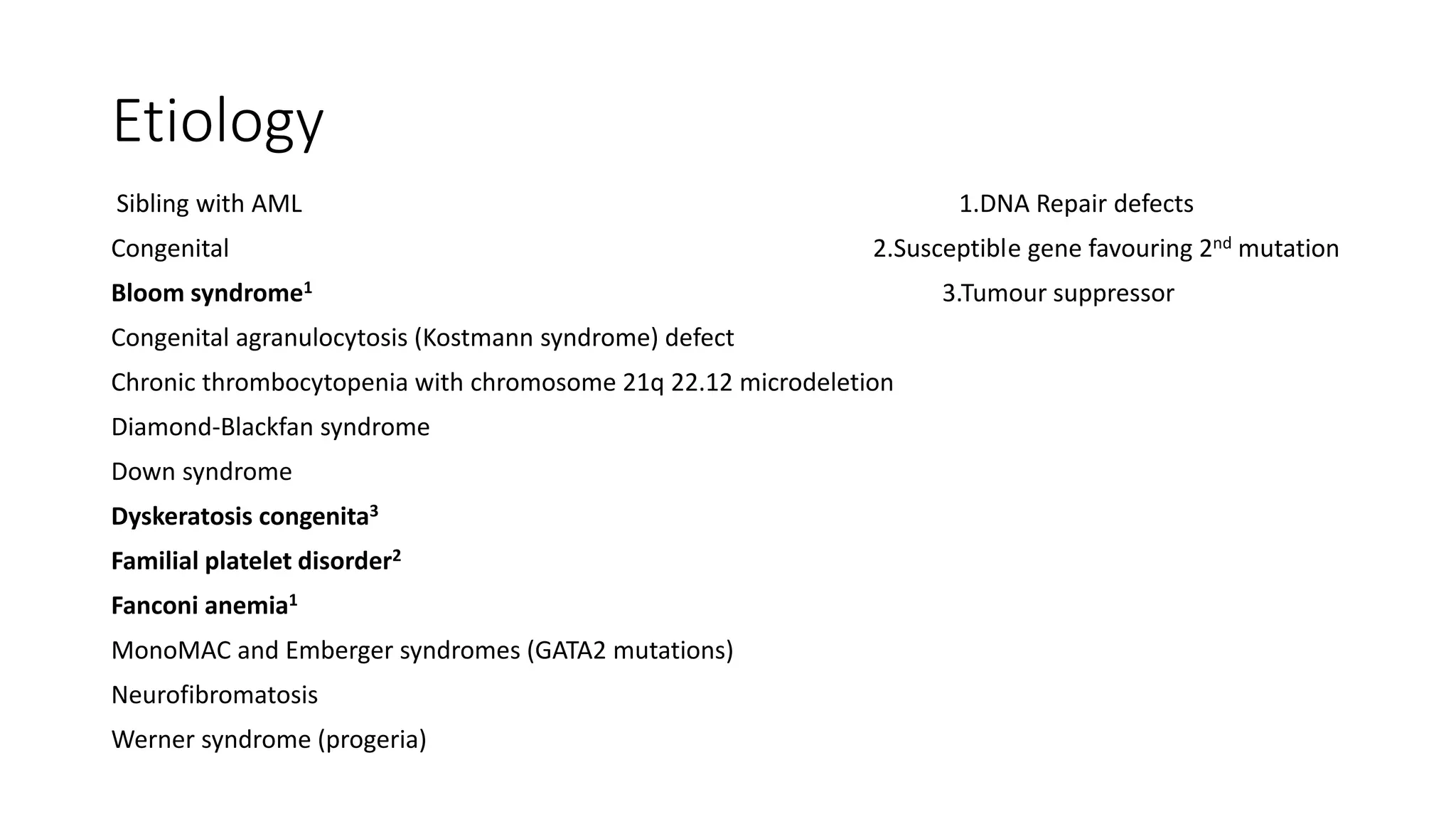
Sibling with AML1.DNA Repair defects Congenital 2.Susceptible gene favouring 2nd mutation Bloom syndrome1 3.Tumour suppressor Congenital agranulocytosis (Kostmann syndrome) defect Chronic thrombocytopenia with chromosome 21q 22.12 microdeletion Diamond-Blackfan syndrome Down syndrome Dyskeratosis congenita3 Familial platelet disorder2 Fanconi anemia1 MonoMAC and Emberger syndromes (GATA2 mutations) Neurofibromatosis Werner syndrome (progeria)
  • 7.
    MOLECULAR PATHOGENESIS The LeukemiaStem Cell Series of somatic mutations in a primitive hematopoietic multipotential progenitor cell Bulk of AML cases arise from one of two predominant CD34+ cell populations: CD34+CD45RA+CD38–CD90– (multipotential myeloid progenitor) or CD34+CD38+CD45RA+CD110+ (granulocyte-monocyte progenitor).
  • 8.
    MOLECULAR PATHOGENESIS Preleukemic StemCells Accumulation of genetic and epigenetic changes in normal pluripotential HSC AML progresses from such cells carrying founder mutations Thought to form a reservoir after therapy that can lead to relapse HSC with DNMT3A,TET2,IDH1&2-promote self-renewal and block differentiation of stem and progenitor cells.
  • 9.
    MOLECULAR PATHOGENESIS Somatic Mutations mutantprotein product often is a transcription factor or an element in the transcription pathway-disrupts the regulatory sequences controlling growth rate or survival of blood cell progenitors & their differentiation and maturation core binding factor (CBF)- {CBF-β & RUNX1}-10% AML retinoic acid receptor-α (RAR-α) HOX family mixed-lineage leukemia (MLL),
  • 10.
    MOLECULAR PATHOGENESIS Somatic Mutationscontinued primary mutations are not sufficient to cause AML Additional activating mutations Fms-like tyrosine kinase (FLT)3 KIT N-RAS and K-RAS are required to induce a proliferative advantage in the affected primitive cell Other proto oncogene mutations that occur in leukemic cells involve FES, FOS, GATA-1, JUN B, MPL, MYC, p53, PU.1, RB, WT1, WNT, NPM1, CEPBA (CCAAT-enhancer binding protein A),
  • 11.
    MOLECULAR PATHOGENESIS Somatic Mutationscontinued Significance PML-RARa or CEPBA double mutations-very favorable OS-83% RUNX1-RUNX1T1, CBFB-MYH11,NPM1 without FLT3-ITD OS-62.6%, MLL-PTD or RUNX1, or ASXL1 mutation OS-22% and very unfavorable TP53 mutation (OS at 3 years, 0%).
  • 12.
  • 14.
  • 15.
    LAB Blood: Anemia with Reticcount 0.5-2.0 TLC -< 5*109 in 50% at diagnosis ANC-<1*109 Platelets <50*109 in 50% at diagnosis Auer Rods Faggot Cells Bone Marrow: Blast cells around 3-95% at diagnosis WHO criteria more >/= 20% blasts except APL
  • 16.
    DIFFERENTIAL DIAGNOSIS In adultsthe term Pseudoleukemia has been applied to circumstances that mimic the marrow appearance of promyelocytic leukemia. Recovery from drug-induced or Pseudomonas aeruginosa–induced agranulocytosis is characterized by a striking cohort of promyelocytes in the marrow, which upon inspection of the marrow aspirate or biopsy mimics promyelocytic leukemia The promyelocytes in Pseudoleukemia contain a prominent paranuclear clear (Golgi) zone not covered with granules; and promyelocytes do not have Auer rods
  • 17.
    TREATMENT-Preparation of patient Pretreatmentlaboratory examination blood cell counts cytochemistry analysis immunophenotyping of leukemic cells from blood or marrow, marrow examination including cytogenetic and molecular analyses to include FLT3 ITD, NPM-1, CEBPα, and KIT mutation status. Herpes simplex virus and cytomegalovirus serotyping may be helpful if transplantation is a consideration. HIV and hepatitis serology(sos) patients should have a baseline cardiac scan to determine ejection fraction prior to administration of an anthracycline antibiotic.
  • 18.
    TREATMENT-Preparation of patient Aperipherally inserted central catheter or a tunneled central venous catheter should be placed. Circulation facilitates administration of chemotherapy, blood components, antibiotics, and other intravenous fluids and medications
  • 19.
    TREATMENT-Preparation of patient Therapyfor hyperuricemia is required (1) the pretreatment uric acid level is greater than 7 mg/dL, (2) the marrow is packed with blast cells or (3) the blood blast cell count is moderately or markedly elevated Allopurinol 300 mg/day orally discontinued after the risk of acute hyperuricosuria or tumor lysis has passed (usually 4 to 7 days) Recombinant urate oxidase (rasburicase) can be used to prevent urate- induced nephropathy, recommended dose is 0.2 mg/kg daily for 5 to 7 days i.v..
  • 20.
    TREATMENT-INDUCTION Goal of inductiontherapy achievement of complete remission <2 percent blasts in the marrow, a neutrophil count greater than 1000/μL and a platelet count greater than 100,000/μL
  • 21.
    TREATMENT-INDUCTION Current standard inductiontreatment for non-APL AML involves drug regimens with two or more agents that include an anthracycline antibiotic or an anthraquinone and cytarabine Remission rates 55-90% Age Antecedant chemotherapy and clonal myeloid disorder
  • 22.
    TREATMENT-INDUCTION standard induction Regimenthe “7 plus 3” regimen Cytarabine -100 mg/m2 daily by continuous infusion on days 1 through 7 and Daunorubicin at 45 to 90 mg/m2 on days 1 through 3 Idarubicin 12 mg/m2 gives better complete remission rates in younger adults than does daunorubicin 45 mg/m2, each given for 3 days ----WILLIAMS Hematology 9e
  • 23.
    TREATMENT-INDUCTION Age <60 years Cytarabine-100 to 200 mg/m2 daily by continuous infusion on days 1 through 7 or 2gm/m2 i.v. q12h for 6 days and Daunorubicin at 60 to 90 mg/m2 on days 1 through 3 High dose Cytarabine associated with higher remission rates.. Toxicity of High dose cytarabine-Pulmonary,Cerebellar(ocassionally irreversible) ----HARRISON’s 19e
  • 24.
    TREATMENT-INDUCTION Timed Sequential Therapy Mitoxantronei.v. on days 1 to 3, Etoposide i.v. on days 8 to 10 along with Cytarabine i.v. on days 1 to 3 and 8 to 10 resulted in a complete remission in 60 percent of patients
  • 25.
    TREATMENT-INDUCTION Novel and MolecularTargeting agents For patients with FLT3ITD + trials with tyrosine kinase inhibitors are ongoing. Patients with CBF + may benefit from combination of Gemtuzumab ozogamicin, a monoclonal CD33 antibody linked to the cytotoxic agent calicheamicin, with induction and consolidation chemotherapies
  • 26.
    TREATMENT Very old patientsor patients with comorbid conditions who are unfit for intensive regimens single-agent therapies with clofarabine or Hypomethylating agents (i.e., 5-azacitidine or decitabine)
  • 27.
    TREATMENT-INDUCTION Special Considerations duringInduction Therapy : Hyperleukocytosis Patients with blast counts greater than 100 × 109/L require prompt treatment to prevent serious complications of hyperleukocytosis: intracranial hemorrhage or pulmonary insufficiency. Hydration should be administered promptly to maintain urine flow greater than 100 mL/h/m2 Cytoreduction therapy can be initiated with hydroxyurea 1.5 to 2.5 g orally every 6 hours (total dose 6 to 10 g/day) for approximately 36 hours
  • 28.
    TREATMENT-INDUCTION Special Considerations duringInduction Therapy : Antibiotic Therapy Pancytopenia is worsened or induced shortly after treatment is instituted. Absolute neutrophil counts less than 100/μL (0.1 × 10⁹/L) are expected and are a sign of effective drug action. The patient usually becomes febrile (>38°C), often with associated rigors. Centers use prophylactic antibacterial, antifungal, and/or antiviral antibiotics
  • 29.
    TREATMENT-INDUCTION Special Considerations duringInduction Therapy : Component Transfusion Therapy Red cell transfusions should be used to keep the hemoglobin level greater than 7.0 g/dL, or higher in special cases (e.g., symptomatic coronary artery disease) Platelet transfusions should be used for hemorrhagic manifestations related to thrombocytopenia and prophylactically if necessary to maintain the platelet count between 5 × 10⁹/L and 10 × 109/L All red cell and platelet products should be depleted of leukocytes, and all products, including granulocytes for transfusions, should be irradiated to prevent transfusion-associated graft-versus-host disease (GVHD) in this immunosuppressed population
  • 30.
    TREATMENT-INDUCTION Special Considerations duringInduction Therapy : Management of Central Nervous System Disease Prophylactic therapy usually not indicated but examination of spinal fluid after remission should be considered in (1) monocytic subtypes (2) cases with extramedullary disease (3) cases with inversion 16 and t(8;21) cytogenetics (4) CD7- and CD56-positive (neural-cell adhesion molecule) immunophenotypes and (5) patients who present with very high blood blast cell counts
  • 31.
    TREATMENT-INDUCTION Special Considerations duringInduction Therapy : Management of Central Nervous System Disease In these situations, the risk of meningeal leukemia or a brain myeloid sarcoma is more but prophylactic intrathecal chemotherapy is not recommended if high-dose cytarabine is used for consolidation Treatment of meningeal leukemia can include high-dose intravenous cytarabine (which penetrates the blood–brain barrier), intrathecal methotrexate, intrathecal cytarabine, cranial radiation, or chemotherapy and radiation in combination If CNS leukemia is present, intrathecal therapy is often given twice/week until blasts are cleared & then once/week for 4-6 weeks.
  • 32.
    TREATMENT-INDUCTION Special Considerations duringInduction Therapy : Management of Nonleukemic Myeloid Sarcoma: Myeloid sarcoma may be the presenting finding in approximately 1% of patients with AML. Such patients should receive intensive AML induction therapy Intensive therapy results in a longer nonleukemic period than patients who have undergone surgical resection or resection followed by local irradiation
  • 33.
    TREATMENT POSTREMISSION THERAPY Postremission therapyis intended to prolong remission duration and overall survival but no consensus exists regarding the best approach Intensive consolidation therapy after remission results in a somewhat longer remission duration Currently transplantation is recommended for all but good-prognosis patients (CBF leukemias or those with NPM1 mutation without a FLT3 mutation).
  • 34.
    TREATMENT POSTREMISSION THERAPY For patientswho do not receive high-dose chemotherapy with autologous or allogeneic transplantation in first remission, consolidation chemotherapy regimens containing high-dose cytarabine provide better results RAS mutations are associated with benefit from high-dose cytarabine therapy.Patients with CBF leukemias such as t(8;21) also have particularly favorable responses to repetitive cycles of high-dose cytarabine Relapse Rate-19%
  • 35.
    TREATMENT NOVEL CHEMOTHERAPY Epigenetic Modulation Methylationof DNA at critical sites cause transcriptional inactivation of genes or chromosomal instability. In AML aberrant methylation especially preferential methylation of chromosome 11 has been described. Presumptive demethylating agents such as 5-azacytidine or decitabine, silencing mediated by histone deacetylation is a target for histone deacetylases-Depsipeptide, vorinostat promote histone acetylation and gene transcription in RUNX1-positive leukemic
  • 36.
    TREATMENT NOVEL CHEMOTHERAPY Antibodies toCD33 Gemtuzumab ozogamicin is a recombinant humanized anti-CD33 monoclonal IgG4 antibody conjugated to cytotoxin calicheamicin Rapidly internalized causes subsequent cell apoptosis. Hyperbilirubinemia and transaminase elevations can occur.
  • 37.
    TREATMENT NOVEL CHEMOTHERAPY Therapies Targetedto Signal Transduction Mediators: FLT3 Inhibitors:Crenolanib, Quizartinib, midostaurin Kit Tyrosine Kinase Inhibitors: Imatinib Mesylate, Dasatinib(Kit+CBF) Nuclear Factor-Kappa B Inhibitors:Bortezomib Prenylation Inhibitors:lovastatin, Simvastatin
  • 38.
    TREATMENT-APL INDUCTION: ATRA combined withan anthracycline-Idarubicin or with arsenic trioxide during induction treatment for most benefit and to prevent drug resistance Typical induction regimen ATRA 45 mg/m2 daily in divided doses with idarubicin at standard induction doses (e.g., 12 mg/m2 on days 1 to 3) MOA:ATRA overcome the recruitment of histone deacetylase activity by PMLRAR-α fusion gene through interference with a nuclear corepressor
  • 39.
    TREATMENT-APL Arsenic Trioxide : Triggerapoptosis of APL cells at high concentrations & maturation at low concentrations. The presence of PML-RAR-α is important for the response. Apoptosis may occur through induction of activation of caspase-1 and caspase-3. also may function through NF-κB inhibition 0.06 to 0.12 mg/kg body weight per day until leukemic cells were eliminated from the marrow-induced remission within 12 to 89 days S.E: retinoic acid–like syndrome,Torsades pointes
  • 40.
    TREATMENT-APL Maintenance Therapy : patientsshould be in a molecular remission i.e.,PCR- negative for PML- RAR-α. Best results were achieved when ATRA was combined with 6- mercaptopurine and methotrexate Maintenance is usually recommended for 2 years During maintenance, PCR monitoring on blood samples is recommended
  • 41.
    TREATMENT-APL Differentiation Syndrome A rapidincrease in the total blood leukocyte count to as high as 80 × 109/L in the first several weeks of therapy median time of onset is 11 days Fever, weight gain, dependent edema, pleural or pericardial effusion Respiratory distress is the key feature Once respiratory distress is evident, the patient should receive dexamethasone 10 mg IV every 12 hours for several days
  • 42.
    TREATMENT-APL Treatment of Coagulopathy: Requires use of fresh-frozen plasma, platelet replacement, and fibrinogen replacement Targeted levels platelet counts 30 to 50 × 109 ,fibrinogen levels 1.5 g/L
  • 43.
    AML PROGNOSIS CR &CRPlatelets (CRp) Initial remission rates now approach 90 % in children 70 % in young adults 60 % in middle-aged patients and 40 % in older patients
  • 44.