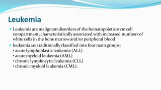
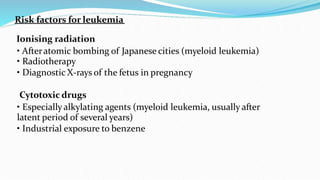
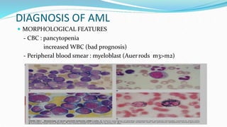
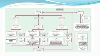
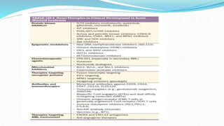
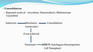
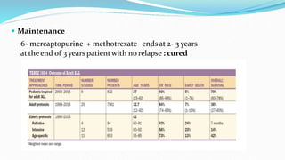
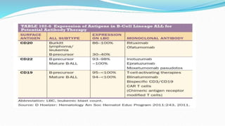
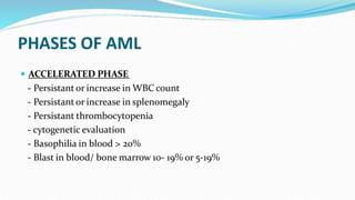
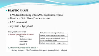
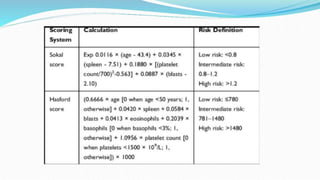
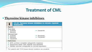
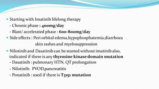
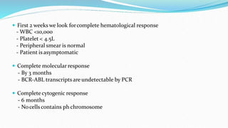
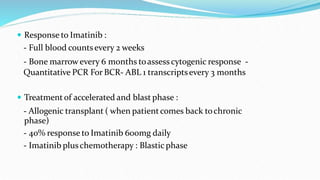
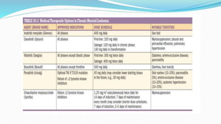
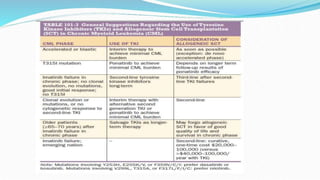
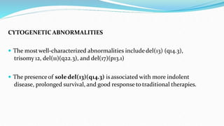
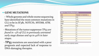
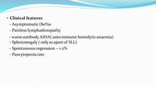
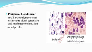
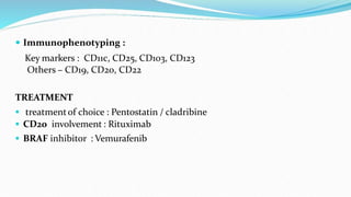
DIAGNOSIS AND MANAGEMENT
Presented by - Dr PRIYA A
Guided by – Dr Daruka sir
Leukemia
 Leukemiaare malignantdisordersof the hematopoieticstemcell
compartment, characteristicallyassociated with increased numbers of
whitecells in the bone marrowand/or peripheral blood
 leukemiaare traditionallyclassified into four main groups:
• acute lymphoblastic leukemia (ALL)
• acute myeloid leukemia (AML)
• chronic lymphocytic leukemia (CLL)
• chronic myeloid leukemia (CML).
Risk factors for leukemia
Ionising radiation
• Afteratomic bombing of Japanese cities (myeloid leukemia)
• Radiotherapy
• Diagnostic X-rays of the fetus in pregnancy
Cytotoxic drugs
• Especiallyalkylating agents (myeloid leukemia, usually after
latent period of several years)
• Industrial exposure to benzene
Retroviruses
• Adult T-cell leukemia/lymphoma (ATLL) caused by human T-cell
lymphotropicvirus 1(HTLV-1, mostprevalent in Japan, the Caribbean and
someareas of Central and South America and Africa
Genetic
• Identical twin of patients with leukemia
• Down’ssyndromeand certain othergenetic disorders
Immunological
• Immunedeficiency states (e.g. hypogammaglobulinaemia A)
Acute leukemia
• There is a failure of cell maturation in acute leukemia.
• Proliferation of cells that do not mature leads toan accumulation of
primitivecells that take up more and more marrow spaceat the
expenseof the normal hematopoieticelements.
• Acute myeloid leukemia (AML) is about four times more common
than acute lymphoblastic leukemia (ALL) in adults.
• In children, the proportions are reversed, the lymphoblastic variety
being morecommon.
ACUTE MYELOID LEUKEMIA(AML)
 Mostcommon leukemia in india
 Morecommon in males>females, averageage >65years
 >20% marrow filled with myeloblast
 Lossof differentiation of myeloblast
 Pancytopenia
 Lymphadenopathy,hepatosplenomegaly: uncommon
 Onsetwithin 2-3 months
 Hyperuricemia
 Skin involvementseen
DIAGNOSIS OF AML
 MORPHOLOGICAL FEATURES
- CBC : pancytopenia
increased WBC (bad prognosis)
- Peripheral blood smear : myeloblast (Auerrods m3>m2)
 CYTOCHEMISTRY
Positive for Myeloperoxidaseand sudan black
 IMMUNOPHENOTYPING
Toconfirm the diagnosis of AML
 Myeloblast – CD11b,CD13,CD15,CD33,CD117,HLA-DR
 Myelomonocyte - CD11b,CD13,CD14,CD15,CD32,CD33,HLA-DR
 Erythroid – spectrin,ABH ambigens, glycophonin,carbonic anhydrase 1,
HLA-DR
 Promyelocytic – CD13,CD33
 Monocytic – CD11b,11C,CD13,CD14,CD33,CD65,HLA-DR
 Megakaryoblastic – CD34,CD41,CD42,CD61,anti-von Willibrand factor
 CYTOGENETICS
- Translocation( 15;17) : PML –RAR alpha best prognosis
- t ( 8;21 ) : RUN –X1 – RUN X1 good prognosisexcept for m2
- t (16;16 ) : associatewith inversion 16
good prognosisexcept for m4
- any translocationsother than ( 16;16 , 8;21, 15;17 ) have bad prognosis
 MOLECULAR GENETICS
- Nucleophosmin Gene mutation(M/C) : good prognosis
- CEBPA( cancerenhancer binding protein) : good prognosis
- FLT3 – ITD : bad prognosis
BLOOD PICTURE IN AML
 Anaemia
 Thrombocytopenia
 Platelet <50000/microliter
 TLC < 5000/ microliter in 50%
 10% havecounts > 1,00,000/microliter
BONE MARROW PICTURE IN AML
 3-95% leukemic blast cells
 >20% is thearbitrarycut off
 Mpoand Sudan black positive
 Auerrods : m3>m2
 Decrease in granulopoiesis,erythropoiesis,megakaryopoiesis
 Marrow fibrosis is minimal
TREATMENT FOR AML




The first decision must be whetheror not togive specific treatment to
attempt toachieve remission.
This is generally aggressive, has numerousside-effects, and may not be
appropriate for theveryelderlyor patients with serious comorbidities In
these patients, supportive treatmentcan effectconsiderable
improvement in well-being.
Low-intensitychemotherapy, such as low-dosecytosinearabinosideor,
recently, azacitidine, is frequentlyused in elderlyand more frail patients
butonly induces remission in less than 20% of patients
 Specific therapy
- Ideally, whenever possible, patientswith acute leukemiashould be
treated within a clinical trial.
- The aim of treatment is todestroy the leukaemiccloneof cells without
destroying the residual normal stemcell compartment from which
repopulationof the haematopoietic tissueswill occur.
- Patientshould be prepared as follows before the specific therapy
1. Existing infections identified and treated (e.g. urinary tract
infections, oral candidiasis, dental, gingival and skin infections)
2. Anaemiacorrected by red cell concentratetransfusion
3. Thrombocytopenic bleeding controlled by platelet transfusions
4 . If possible, central venouscatheter (e.g. Hickman line) inserted to
facilitateaccess to the circulation fordeliveryof chemotherapy,
fluids, blood productsand othersupportivedrugs
5. Tumourlysis risk assessed and prevention started: fluidswith
allopurinol or rasburicase
6 .Therapeutic regimen carefullyexplained to the patientand informed
consentobtained
7. Consideration of entry intoclinical trial
Drugs commonly used in the treatmentof acute myeloid
leukemia
Induction Phase : Daunorubicin (IV)
Cytarabine (IV)
Etoposide (IV and oral)
Gentuzumabozogamicin (IV) All-
trans retinoicacid (ATRA) (oral)
Arsenic trioxide (ATO)
 CONSOLIDATION PHASE : Cytarabine (IV)
Amsacrine (IV)
Mitoxantrone (IV)
 RELAPSE PHASE : Fludarabine
Cytarabine
Arsenic trioxide (ATO)
Idarubicin
 Aggressive front line chemotherapy in fit patients
- < 40yrs,withoutcomorbid condition
- 7 + 3 days regimen
7 – cytarabine ( 100-200mg /m2/day)
3 – daunorubicin (60-90mg/day)
- from day 7 : period of strictvigilant monitoring
- from day 14 : bone marrowstudy
look forremission (molecular> morphological)
 Morphological remission
 Blasts - <5%
 Platelet - >100000
 Absoluteeosinophilic count >100
 Molecularremission : minimal residual disease is not detected
on flow cytometry
- If there is a morphological remission – Consolidate
- If tehre is no morphological remission – Repeat 7+ 3 regime
ACUTE LYMPHOBLASTIC LEUKEMIA
 In acute lymphoblastic leukemia (ALL), the malignantclonearises
from hematopoietic progenitors in the bone marrowor lymphatic
system resulting in an increaseof immature nonfunctioning leukemic
cells.
 Infiltration of bone marrow leads toanemia, granulocytopenia, and
thrombocytopeniawith the clinical manifestationsof fatigue,
weakness, infection, and hemorrhages.
 These symptomsare moreoften the reason a patient first seeks
medical advice rather than consequences of tumor bulk, such as
lymph nodeenlargement, hepatosplenomegalycaused by leukemic
infiltration, orsymptomsof the central nervoussystem
 ALL is the most frequent neoplasticdisease in children with an
early peak at the ageof 3–4 years
 Patientswith some rare congenital chromosomal abnormalities havea
higher risk of developmentof acute leukemia; e.g., Klinefelter’s
syndrome, Fanconi’sanemia, Bloom’ssyndrome, ataxia telangiectasia,
and neurofibromatosis.
 There is a twentyfold increased incidenceof leukemia in patientswith
Down syndrome, in whom ALL is increased in childhood orAML atan
olderage.
 Mostcommon typeof ALL is Pre B cell typewhich has a good
prognosis rest other like Pro B cell type, Pro T cell,Pre T cell type has
bad prognosis
Acute Lymphoid Leukemia - Diagnosis
 Bood counts = low
 Peripheral Smear = lymphoblast
 Bone marrow = > 20% blastcells
if the countsare high = very bad prognosis
Cytological
 Stains : PAS ( Periodic Acid Schiff ) positive
 Acid phosphatase positive (only forT - Cell all)
Immunophenotyping
 Early pre 6-Cell ALL : CD11,CD19,CD79a,TdT Positive
 If associated with tumor : CO20, CD22 also present
 Pro 6 - Cell ALL CD10 absent
Cytogenetics
1) Children – t(12:21) – good prognosis
2) Adults - t (9:22), t(4:11),t(1:19) has got poor prognosis and poor
response
Moleculargenetics
 NOTCH-1 and HOX-11 seen in T cell ALL has verygood prognosis
Acute lymphoid leukemia -Treatment
 Curerate – Adults – 40-50%
Children – 90%
 Induction
- Steroids + vincristine/ Daunorubicin +/- L-Asperginase( may increase
the risk of pancreatities,thrombosis)
- High dose Methotrexate : good CNS penetration
- Accompanied by intrathecal methotrexate : Prevents CNS relapse
 Consolidation
- Repeated cycles of vincristine, Daunorubicin, Methotrexate
Cytarabine
Induction Remission Consolidation
(molecular)
if notachieved
Treatment AHSCT( Autologous Hematopoietic
Cell Transplant)
 Maintenance
6- mercaptopurine + methotrexate ends at 2- 3 years
at the end of 3 years patientwith no relapse : cured
• Chimeric Antigen Receptor (CAR) Tcells
- The adoptive transferof CAR-modified Tcells directed against CD19 is a
promising approach to the treatmentof CD19+ childhood oradult ALL.
• CRISPR geneeditting technique
- The CRISPER gene editing technique is used in humans to remove specfic
genes toallow the immune system to be more activated against cancer.
- Recently,this CRISPER technology has used to insert genes that allow
immunecells toattack cancer cells,potentially leaving normal cells
unharmed and increasing the effectiveness of immunotherapy




CHRONIC MYELOID leukemia
Chronic myeloid leukemia (CML) is a myeloproliferativestemcell
disorderresulting in proliferation of all haematopoietic lineages but
manifesting predominantly in the granulocytic series. Maturation
of cells proceeds fairly normally.
The disease occurschiefly between the ages of 30 and 80 years, with a
peak incidenceat 55 years.
The defining characteristicof CML is the chromosomeabnormality
known as the Philadelphia (Ph) chromosome




The break on chromosome 22 occurs in the breakpointclusterregion
(BCR).
The fragment from chromosome 9 that joins the BCR carries the abl
oncogene, which forms a fusion gene with the remains of the BCR.
This BCR ABL fusion gene codes for a 210 kDa protein with tyrosine
kinase activity, which playsa causative role in thedisease as an
oncogene, influencing cellularproliferation, differentiation and
survival.
In some patients in whom conventional chromosomal analysisdoes not
detecta Ph chromosome, the BCR ABL gene product is detectable by
moleculartechniques.
Thedisease has three phases:
• A chronic phase :
- in which the disease is responsive to treatmentand is easilycontrolled, which
used to last 3–5 years
- With the introduction of imatinib therapy, this phase has been prolonged to
encompass a normal lifeexpectancy in many patients.
• An accelerated phase (not always seen) :
- in which disease control becomes more difficult
Blast crises :
- In this phase the diseasewill transforms intoan acute leukemia,either
Myeloblast(70%) or Lymphoblastic (30%), which is relatively
refractory to treatment
- This is the causeof death in majorityof patients, therefore survival is
dictated by the timing of blastcrisis,which cannot be predicted.
Clinical features
 Symptomsat presentation may include lethargy, weight loss,abdominal
discomfort, goutand sweating, butabout 25% of patientsare asymptomatic
atdiagnosis.
 Splenomegaly is present in 90%; in about 10%, theenlargement is massive,
extending toover 15 cm below the costal margin.
 A friction rub may be heard in cases of splenic infarction.
 Hepatomegaly occurs in about 50%.
 Lymphadenopathy is unusual.
DIAGNOSIS OF CML
 Blood picture :-
- decreased hemoglobin
- mild anisopoikilocytosis
- WBC count >25000/microliter
- 50% havecount >1,00,000/microliter
- cytochemicallyabnormal WBC: LOW LAP
- Absoluteeosinophil count is increased
- Thrombocytosis : hemorrhage and thrombosisare
very rare
- Basophilia/ tryptase raised
- Wright geimsa stain showing bone marrow hypercellularity with
myeloid hyperplasia
 Bone marrow
- marked hypercellularity
- predominantgranulopoiesis ( 30:1)
- decreased erythropoiesis
- Dwarf megakaryocytes increased
- Sea blue histiocytes/ pseudogaucher
cells
 Moleculardiagnosis
- 95% will have philadelphiachromosome
G banding : sent in T lymphocytes
- 5% can still have BCR-ABL transcripts detectable by PCR
- FISH: is specific for ( 9:22) translocation
- Deletion of 20q, trisomy 8, del 3q, double philadelphiachromosome
has Bad prognosis
 Chemical findings
uricacid - high
LDH - high
histamine - high
angiogenic factor - high
PHASES OF AML
 ACCELERATED PHASE
- Persistant or increase in WBC count
- Persistant or increase in splenomegaly
- Persistant thrombocytopenia
- cytogenetic evaluation
- Basophilia in blood > 20%
- Blast in blood/ bone marrow 10- 19% or 5-19%
 BLASTIC PHASE
- CML transforming into AML,myeloid sarcoma
- Blast > 20% in blood/bone marrow
- LAP increased
- myeloid > lymphoid
Treatment of CML
 Thyrosine kinase inhibitors
 Starting with Imatinib lifelong therapy
- Chronic phase : 400mg/day
- Blast/ accelerated phase : 600-800mg/day
 Side effects : Peri orbital edema,hypophosphatemia,diarrhoea
skin rashes and myelosuppression
 Nilotiniband Dasatinibcan be started without imatinib also,
indicated if there is any thyrosine kinasedomain mutation
- Dasatinib : pulmonary HTN, QT prolongation
- Nilotinib: PVOD,pancreatitis
- Ponatinib : used if there is T3151 mutation
 First 2 weeks we look forcomplete hematological response
- WBC <10,000
- Platelet < 4.5L
- Peripheral smear is normal
- Patient is asymptomatic
 Complete molecular response
- By 3 months
- BCR-ABL transcripts are undetectable by PCR
 Complete cytogenic response
- 6 months
- Nocells contains ph chromosome
 Response to Imatinib :
- Full blood countsevery 2 weeks
- Bone marrowevery 6 months toassess cytogenic response -
Quantitative PCR For BCR- ABL 1 transcriptsevery 3 months
 Treatment of accelerated and blast phase :
- Allogenic transplant ( when patientcomes back tochronic
phase)
- 40% response to Imatinib 600mg daily
- Imatinib pluschemotherapy : Blastic phase
CHRONIC LYMPHOCYTIC LEUKEMIA
 Chronic lymphocytic leukemia (CLL) is a monoclonal proliferation of
mature B lymphocytesdefined byan absolute numberof malignant cells
in the blood (5 × 10⁹/mL).
 The presence of malignant B cells under this count in the blood without
nodal, spleen, or liver involvementand absentcytopenias is a precursorof
this disease called monoclonal B-cell lymphocytosis (MBL) with ~1–2%
chance per year of progressing to overt CLL.
 CLL is a heterogeneousdisease in termsof natural history, with some
patients presenting asymptomaticallyand neverrequiring therapy,
• Whereas others presentwith symptomaticdisease, require multiple
lines of therapy, and eventuallydie of theirdisease


CLL is primarilya disease of olderadults, with a median age atdiagnosis
of 71 years
The male:female ratio is 2:1; however, as patients age, the ratio becomes
more even, and over the age of 80, the incidence is equal between men
and women
 The morphology, immunophenotype, and gene expression pattern of
CLL cells are that of a mature B cell, and so it has been presumed that
the initiating cell is a mature lymphocyte, perhaps memory B
 Monoclonal B cell count > 5000cells/microliter– CLL
 Monoclonal B cell count < 5000cells/microliter – monoclonal B cell
Lymphocytosis
- Bone marrow not involved, perpheral blood is involved
 CD markers ; CD19,CD20,CD21,CD22,CD79a,CD79b,S1gm
 On mature B cell CD5,CD23 is stronglyexpressed
CYTOGENETIC ABNORMALITIES
 The mostwell-characterized abnormalities includedel(13) (q14.3),
trisomy 12, del(11)(q22.3), and del(17)(p13.1)
 The presence of soledel(13)(q14.3) isassociated with more indolent
disease, prolonged survival, and good response to traditional therapies.
 GENE MUTATIONS
- Wholegenome and whole exome sequencing
have identified the most common mutations in
CLL to be in SF3B1, NOTCH1, MYD88, ATM,
and TP53
- Mutations of the tumorsuppressor TP53 are
found in ~5% of CLL in previously untreated
early stage disease and up to 40% in later
stages.
- TP53 mutations are associated with a poor
prognosis and expected lack of response to
DNA-damaging therapies.
 Clinical features
- Asymptomatic (80%0
- Painless lymphadenopathy
- warm antibody AIHA( auto immune hemolyticanaemia)
- Splenomegaly ( onlyas apartof SLL)
- Spontaneous regression – 1-2%
- Pancytopenia rare
 Peripheral blood smear
- small, mature lymphocytes
with scanty bluish cytoplasm
and moderatecondensation
- smudgecells
 Fish – cytogenetics in CLL
1. 13q deletion assosiated with mutated Ig heavychain –Good
prognosis
2. Trisomy 12,Deletion 11q, Deletion 17p – Bad prognosis
 Coombs test - Todetectwarm antibody AIHA
Treatment of CLL
 First lineof treatment
FCR regime : Fludarabine, Cyclophosphamide,Rituximab
BR regime : Bendamustine,Rituximab
< 60 year FCR regime, >60 year BR regime
 AntiapoptoticTherapies
- BCL2 is anotherpromising target in CLL.
- Venetoclax is an orally bioavailable, selective BCL2 inhibitor.
- It is currently Food and Drug Administration (FDA) approved for
marketing in patients with relapsed or refractory CLL who have the
del(17)(p13.1).
Immune Therapies




Current immune therapies include allogenic stem cell transplantation,
chimericantigen receptor(CAR) T-cell therapy, and oral
immunomodulatory agents such as lenalidomide
Stem cell transplantation is currently considered theonly standard curative
approach to CLL.
Because most CLL patients areolderand many havesignificant comobidity
myeloablative transplants results in extensive morbidityand mortality, making
them prohibitive in many individuals
Reduced intensityconditioning (RIC) allogeneic transplants have been
successfully incorporated into the treatmentof patients up to ~75 years in age but
still havea ≥50% frequencyof chronic graft-versus-hostdisease.
HAIRY CELL leukemia






This is a rarechronic B-cell lymphoproliferativedisorder.
The male-to-femaleratio is 6 : 1 and the median age at diagnosis is 50 years.
Presenting symptomsare general ill health and recurrent infections.
Splenomegalyoccurs in 90% but lymph node enlargement is unusual.
Severe neutropenia, monocytopenia and the characteristic hairycells in the
blood and bone marrow are typical.
These cells usually havea B-lymphocyte immunotype but theyalso
characteristicallyexpress CD25 and CD103.
Recently, all patients with hairycell leukemia have been found to havea
mutation in the BRAF gene.
 Immunophenotyping :
Key markers : CD11c, CD25, CD103, CD123
Others – CD19, CD20, CD22
TREATMENT
 treatmentof choice : Pentostatin / cladribine
 CD20 involvement : Rituximab
 BRAF inhibitor : Vemurafenib
THANK YOU

leukemia1-230103123650-8c41bba7.pptx

  • 1.
    DIAGNOSIS AND MANAGEMENT Presentedby - Dr PRIYA A Guided by – Dr Daruka sir
  • 2.
    Leukemia  Leukemiaare malignantdisordersofthe hematopoieticstemcell compartment, characteristicallyassociated with increased numbers of whitecells in the bone marrowand/or peripheral blood  leukemiaare traditionallyclassified into four main groups: • acute lymphoblastic leukemia (ALL) • acute myeloid leukemia (AML) • chronic lymphocytic leukemia (CLL) • chronic myeloid leukemia (CML).
  • 3.
    Risk factors forleukemia Ionising radiation • Afteratomic bombing of Japanese cities (myeloid leukemia) • Radiotherapy • Diagnostic X-rays of the fetus in pregnancy Cytotoxic drugs • Especiallyalkylating agents (myeloid leukemia, usually after latent period of several years) • Industrial exposure to benzene
  • 4.
    Retroviruses • Adult T-cellleukemia/lymphoma (ATLL) caused by human T-cell lymphotropicvirus 1(HTLV-1, mostprevalent in Japan, the Caribbean and someareas of Central and South America and Africa Genetic • Identical twin of patients with leukemia • Down’ssyndromeand certain othergenetic disorders Immunological • Immunedeficiency states (e.g. hypogammaglobulinaemia A)
  • 5.
    Acute leukemia • Thereis a failure of cell maturation in acute leukemia. • Proliferation of cells that do not mature leads toan accumulation of primitivecells that take up more and more marrow spaceat the expenseof the normal hematopoieticelements. • Acute myeloid leukemia (AML) is about four times more common than acute lymphoblastic leukemia (ALL) in adults. • In children, the proportions are reversed, the lymphoblastic variety being morecommon.
  • 6.
    ACUTE MYELOID LEUKEMIA(AML) Mostcommon leukemia in india  Morecommon in males>females, averageage >65years  >20% marrow filled with myeloblast  Lossof differentiation of myeloblast  Pancytopenia  Lymphadenopathy,hepatosplenomegaly: uncommon  Onsetwithin 2-3 months  Hyperuricemia  Skin involvementseen
  • 11.
    DIAGNOSIS OF AML MORPHOLOGICAL FEATURES - CBC : pancytopenia increased WBC (bad prognosis) - Peripheral blood smear : myeloblast (Auerrods m3>m2)
  • 12.
     CYTOCHEMISTRY Positive forMyeloperoxidaseand sudan black  IMMUNOPHENOTYPING Toconfirm the diagnosis of AML  Myeloblast – CD11b,CD13,CD15,CD33,CD117,HLA-DR  Myelomonocyte - CD11b,CD13,CD14,CD15,CD32,CD33,HLA-DR  Erythroid – spectrin,ABH ambigens, glycophonin,carbonic anhydrase 1, HLA-DR  Promyelocytic – CD13,CD33  Monocytic – CD11b,11C,CD13,CD14,CD33,CD65,HLA-DR  Megakaryoblastic – CD34,CD41,CD42,CD61,anti-von Willibrand factor
  • 13.
     CYTOGENETICS - Translocation(15;17) : PML –RAR alpha best prognosis - t ( 8;21 ) : RUN –X1 – RUN X1 good prognosisexcept for m2 - t (16;16 ) : associatewith inversion 16 good prognosisexcept for m4 - any translocationsother than ( 16;16 , 8;21, 15;17 ) have bad prognosis  MOLECULAR GENETICS - Nucleophosmin Gene mutation(M/C) : good prognosis - CEBPA( cancerenhancer binding protein) : good prognosis - FLT3 – ITD : bad prognosis
  • 14.
    BLOOD PICTURE INAML  Anaemia  Thrombocytopenia  Platelet <50000/microliter  TLC < 5000/ microliter in 50%  10% havecounts > 1,00,000/microliter BONE MARROW PICTURE IN AML  3-95% leukemic blast cells  >20% is thearbitrarycut off  Mpoand Sudan black positive  Auerrods : m3>m2  Decrease in granulopoiesis,erythropoiesis,megakaryopoiesis  Marrow fibrosis is minimal
  • 15.
    TREATMENT FOR AML     Thefirst decision must be whetheror not togive specific treatment to attempt toachieve remission. This is generally aggressive, has numerousside-effects, and may not be appropriate for theveryelderlyor patients with serious comorbidities In these patients, supportive treatmentcan effectconsiderable improvement in well-being. Low-intensitychemotherapy, such as low-dosecytosinearabinosideor, recently, azacitidine, is frequentlyused in elderlyand more frail patients butonly induces remission in less than 20% of patients
  • 16.
     Specific therapy -Ideally, whenever possible, patientswith acute leukemiashould be treated within a clinical trial. - The aim of treatment is todestroy the leukaemiccloneof cells without destroying the residual normal stemcell compartment from which repopulationof the haematopoietic tissueswill occur. - Patientshould be prepared as follows before the specific therapy 1. Existing infections identified and treated (e.g. urinary tract infections, oral candidiasis, dental, gingival and skin infections) 2. Anaemiacorrected by red cell concentratetransfusion
  • 17.
    3. Thrombocytopenic bleedingcontrolled by platelet transfusions 4 . If possible, central venouscatheter (e.g. Hickman line) inserted to facilitateaccess to the circulation fordeliveryof chemotherapy, fluids, blood productsand othersupportivedrugs 5. Tumourlysis risk assessed and prevention started: fluidswith allopurinol or rasburicase 6 .Therapeutic regimen carefullyexplained to the patientand informed consentobtained 7. Consideration of entry intoclinical trial
  • 18.
    Drugs commonly usedin the treatmentof acute myeloid leukemia Induction Phase : Daunorubicin (IV) Cytarabine (IV) Etoposide (IV and oral) Gentuzumabozogamicin (IV) All- trans retinoicacid (ATRA) (oral) Arsenic trioxide (ATO)  CONSOLIDATION PHASE : Cytarabine (IV) Amsacrine (IV) Mitoxantrone (IV)
  • 19.
     RELAPSE PHASE: Fludarabine Cytarabine Arsenic trioxide (ATO) Idarubicin  Aggressive front line chemotherapy in fit patients - < 40yrs,withoutcomorbid condition - 7 + 3 days regimen 7 – cytarabine ( 100-200mg /m2/day) 3 – daunorubicin (60-90mg/day) - from day 7 : period of strictvigilant monitoring - from day 14 : bone marrowstudy look forremission (molecular> morphological)
  • 20.
     Morphological remission Blasts - <5%  Platelet - >100000  Absoluteeosinophilic count >100  Molecularremission : minimal residual disease is not detected on flow cytometry - If there is a morphological remission – Consolidate - If tehre is no morphological remission – Repeat 7+ 3 regime
  • 24.
    ACUTE LYMPHOBLASTIC LEUKEMIA In acute lymphoblastic leukemia (ALL), the malignantclonearises from hematopoietic progenitors in the bone marrowor lymphatic system resulting in an increaseof immature nonfunctioning leukemic cells.  Infiltration of bone marrow leads toanemia, granulocytopenia, and thrombocytopeniawith the clinical manifestationsof fatigue, weakness, infection, and hemorrhages.
  • 25.
     These symptomsaremoreoften the reason a patient first seeks medical advice rather than consequences of tumor bulk, such as lymph nodeenlargement, hepatosplenomegalycaused by leukemic infiltration, orsymptomsof the central nervoussystem  ALL is the most frequent neoplasticdisease in children with an early peak at the ageof 3–4 years
  • 26.
     Patientswith somerare congenital chromosomal abnormalities havea higher risk of developmentof acute leukemia; e.g., Klinefelter’s syndrome, Fanconi’sanemia, Bloom’ssyndrome, ataxia telangiectasia, and neurofibromatosis.  There is a twentyfold increased incidenceof leukemia in patientswith Down syndrome, in whom ALL is increased in childhood orAML atan olderage.  Mostcommon typeof ALL is Pre B cell typewhich has a good prognosis rest other like Pro B cell type, Pro T cell,Pre T cell type has bad prognosis
  • 27.
    Acute Lymphoid Leukemia- Diagnosis  Bood counts = low  Peripheral Smear = lymphoblast  Bone marrow = > 20% blastcells if the countsare high = very bad prognosis
  • 28.
    Cytological  Stains :PAS ( Periodic Acid Schiff ) positive  Acid phosphatase positive (only forT - Cell all) Immunophenotyping  Early pre 6-Cell ALL : CD11,CD19,CD79a,TdT Positive  If associated with tumor : CO20, CD22 also present  Pro 6 - Cell ALL CD10 absent Cytogenetics 1) Children – t(12:21) – good prognosis 2) Adults - t (9:22), t(4:11),t(1:19) has got poor prognosis and poor response
  • 29.
    Moleculargenetics  NOTCH-1 andHOX-11 seen in T cell ALL has verygood prognosis
  • 32.
    Acute lymphoid leukemia-Treatment  Curerate – Adults – 40-50% Children – 90%  Induction - Steroids + vincristine/ Daunorubicin +/- L-Asperginase( may increase the risk of pancreatities,thrombosis) - High dose Methotrexate : good CNS penetration - Accompanied by intrathecal methotrexate : Prevents CNS relapse
  • 33.
     Consolidation - Repeatedcycles of vincristine, Daunorubicin, Methotrexate Cytarabine Induction Remission Consolidation (molecular) if notachieved Treatment AHSCT( Autologous Hematopoietic Cell Transplant)
  • 34.
     Maintenance 6- mercaptopurine+ methotrexate ends at 2- 3 years at the end of 3 years patientwith no relapse : cured
  • 36.
    • Chimeric AntigenReceptor (CAR) Tcells - The adoptive transferof CAR-modified Tcells directed against CD19 is a promising approach to the treatmentof CD19+ childhood oradult ALL. • CRISPR geneeditting technique - The CRISPER gene editing technique is used in humans to remove specfic genes toallow the immune system to be more activated against cancer. - Recently,this CRISPER technology has used to insert genes that allow immunecells toattack cancer cells,potentially leaving normal cells unharmed and increasing the effectiveness of immunotherapy
  • 37.
        CHRONIC MYELOID leukemia Chronicmyeloid leukemia (CML) is a myeloproliferativestemcell disorderresulting in proliferation of all haematopoietic lineages but manifesting predominantly in the granulocytic series. Maturation of cells proceeds fairly normally. The disease occurschiefly between the ages of 30 and 80 years, with a peak incidenceat 55 years. The defining characteristicof CML is the chromosomeabnormality known as the Philadelphia (Ph) chromosome
  • 38.
        The break onchromosome 22 occurs in the breakpointclusterregion (BCR). The fragment from chromosome 9 that joins the BCR carries the abl oncogene, which forms a fusion gene with the remains of the BCR. This BCR ABL fusion gene codes for a 210 kDa protein with tyrosine kinase activity, which playsa causative role in thedisease as an oncogene, influencing cellularproliferation, differentiation and survival. In some patients in whom conventional chromosomal analysisdoes not detecta Ph chromosome, the BCR ABL gene product is detectable by moleculartechniques.
  • 39.
    Thedisease has threephases: • A chronic phase : - in which the disease is responsive to treatmentand is easilycontrolled, which used to last 3–5 years - With the introduction of imatinib therapy, this phase has been prolonged to encompass a normal lifeexpectancy in many patients. • An accelerated phase (not always seen) : - in which disease control becomes more difficult
  • 40.
    Blast crises : -In this phase the diseasewill transforms intoan acute leukemia,either Myeloblast(70%) or Lymphoblastic (30%), which is relatively refractory to treatment - This is the causeof death in majorityof patients, therefore survival is dictated by the timing of blastcrisis,which cannot be predicted.
  • 41.
    Clinical features  Symptomsatpresentation may include lethargy, weight loss,abdominal discomfort, goutand sweating, butabout 25% of patientsare asymptomatic atdiagnosis.  Splenomegaly is present in 90%; in about 10%, theenlargement is massive, extending toover 15 cm below the costal margin.  A friction rub may be heard in cases of splenic infarction.  Hepatomegaly occurs in about 50%.  Lymphadenopathy is unusual.
  • 42.
    DIAGNOSIS OF CML Blood picture :- - decreased hemoglobin - mild anisopoikilocytosis - WBC count >25000/microliter - 50% havecount >1,00,000/microliter - cytochemicallyabnormal WBC: LOW LAP - Absoluteeosinophil count is increased - Thrombocytosis : hemorrhage and thrombosisare very rare - Basophilia/ tryptase raised
  • 43.
    - Wright geimsastain showing bone marrow hypercellularity with myeloid hyperplasia  Bone marrow - marked hypercellularity - predominantgranulopoiesis ( 30:1) - decreased erythropoiesis - Dwarf megakaryocytes increased - Sea blue histiocytes/ pseudogaucher cells
  • 44.
     Moleculardiagnosis - 95%will have philadelphiachromosome G banding : sent in T lymphocytes - 5% can still have BCR-ABL transcripts detectable by PCR - FISH: is specific for ( 9:22) translocation - Deletion of 20q, trisomy 8, del 3q, double philadelphiachromosome has Bad prognosis  Chemical findings uricacid - high LDH - high histamine - high angiogenic factor - high
  • 45.
    PHASES OF AML ACCELERATED PHASE - Persistant or increase in WBC count - Persistant or increase in splenomegaly - Persistant thrombocytopenia - cytogenetic evaluation - Basophilia in blood > 20% - Blast in blood/ bone marrow 10- 19% or 5-19%
  • 46.
     BLASTIC PHASE -CML transforming into AML,myeloid sarcoma - Blast > 20% in blood/bone marrow - LAP increased - myeloid > lymphoid
  • 48.
    Treatment of CML Thyrosine kinase inhibitors
  • 49.
     Starting withImatinib lifelong therapy - Chronic phase : 400mg/day - Blast/ accelerated phase : 600-800mg/day  Side effects : Peri orbital edema,hypophosphatemia,diarrhoea skin rashes and myelosuppression  Nilotiniband Dasatinibcan be started without imatinib also, indicated if there is any thyrosine kinasedomain mutation - Dasatinib : pulmonary HTN, QT prolongation - Nilotinib: PVOD,pancreatitis - Ponatinib : used if there is T3151 mutation
  • 50.
     First 2weeks we look forcomplete hematological response - WBC <10,000 - Platelet < 4.5L - Peripheral smear is normal - Patient is asymptomatic  Complete molecular response - By 3 months - BCR-ABL transcripts are undetectable by PCR  Complete cytogenic response - 6 months - Nocells contains ph chromosome
  • 51.
     Response toImatinib : - Full blood countsevery 2 weeks - Bone marrowevery 6 months toassess cytogenic response - Quantitative PCR For BCR- ABL 1 transcriptsevery 3 months  Treatment of accelerated and blast phase : - Allogenic transplant ( when patientcomes back tochronic phase) - 40% response to Imatinib 600mg daily - Imatinib pluschemotherapy : Blastic phase
  • 54.
    CHRONIC LYMPHOCYTIC LEUKEMIA Chronic lymphocytic leukemia (CLL) is a monoclonal proliferation of mature B lymphocytesdefined byan absolute numberof malignant cells in the blood (5 × 10⁹/mL).  The presence of malignant B cells under this count in the blood without nodal, spleen, or liver involvementand absentcytopenias is a precursorof this disease called monoclonal B-cell lymphocytosis (MBL) with ~1–2% chance per year of progressing to overt CLL.
  • 55.
     CLL isa heterogeneousdisease in termsof natural history, with some patients presenting asymptomaticallyand neverrequiring therapy, • Whereas others presentwith symptomaticdisease, require multiple lines of therapy, and eventuallydie of theirdisease   CLL is primarilya disease of olderadults, with a median age atdiagnosis of 71 years The male:female ratio is 2:1; however, as patients age, the ratio becomes more even, and over the age of 80, the incidence is equal between men and women
  • 56.
     The morphology,immunophenotype, and gene expression pattern of CLL cells are that of a mature B cell, and so it has been presumed that the initiating cell is a mature lymphocyte, perhaps memory B  Monoclonal B cell count > 5000cells/microliter– CLL  Monoclonal B cell count < 5000cells/microliter – monoclonal B cell Lymphocytosis - Bone marrow not involved, perpheral blood is involved  CD markers ; CD19,CD20,CD21,CD22,CD79a,CD79b,S1gm  On mature B cell CD5,CD23 is stronglyexpressed
  • 57.
    CYTOGENETIC ABNORMALITIES  Themostwell-characterized abnormalities includedel(13) (q14.3), trisomy 12, del(11)(q22.3), and del(17)(p13.1)  The presence of soledel(13)(q14.3) isassociated with more indolent disease, prolonged survival, and good response to traditional therapies.
  • 58.
     GENE MUTATIONS -Wholegenome and whole exome sequencing have identified the most common mutations in CLL to be in SF3B1, NOTCH1, MYD88, ATM, and TP53 - Mutations of the tumorsuppressor TP53 are found in ~5% of CLL in previously untreated early stage disease and up to 40% in later stages. - TP53 mutations are associated with a poor prognosis and expected lack of response to DNA-damaging therapies.
  • 59.
     Clinical features -Asymptomatic (80%0 - Painless lymphadenopathy - warm antibody AIHA( auto immune hemolyticanaemia) - Splenomegaly ( onlyas apartof SLL) - Spontaneous regression – 1-2% - Pancytopenia rare
  • 60.
     Peripheral bloodsmear - small, mature lymphocytes with scanty bluish cytoplasm and moderatecondensation - smudgecells
  • 61.
     Fish –cytogenetics in CLL 1. 13q deletion assosiated with mutated Ig heavychain –Good prognosis 2. Trisomy 12,Deletion 11q, Deletion 17p – Bad prognosis  Coombs test - Todetectwarm antibody AIHA
  • 63.
  • 64.
     First lineoftreatment FCR regime : Fludarabine, Cyclophosphamide,Rituximab BR regime : Bendamustine,Rituximab < 60 year FCR regime, >60 year BR regime  AntiapoptoticTherapies - BCL2 is anotherpromising target in CLL. - Venetoclax is an orally bioavailable, selective BCL2 inhibitor. - It is currently Food and Drug Administration (FDA) approved for marketing in patients with relapsed or refractory CLL who have the del(17)(p13.1).
  • 65.
    Immune Therapies     Current immunetherapies include allogenic stem cell transplantation, chimericantigen receptor(CAR) T-cell therapy, and oral immunomodulatory agents such as lenalidomide Stem cell transplantation is currently considered theonly standard curative approach to CLL. Because most CLL patients areolderand many havesignificant comobidity myeloablative transplants results in extensive morbidityand mortality, making them prohibitive in many individuals Reduced intensityconditioning (RIC) allogeneic transplants have been successfully incorporated into the treatmentof patients up to ~75 years in age but still havea ≥50% frequencyof chronic graft-versus-hostdisease.
  • 67.
    HAIRY CELL leukemia       Thisis a rarechronic B-cell lymphoproliferativedisorder. The male-to-femaleratio is 6 : 1 and the median age at diagnosis is 50 years. Presenting symptomsare general ill health and recurrent infections. Splenomegalyoccurs in 90% but lymph node enlargement is unusual. Severe neutropenia, monocytopenia and the characteristic hairycells in the blood and bone marrow are typical. These cells usually havea B-lymphocyte immunotype but theyalso characteristicallyexpress CD25 and CD103. Recently, all patients with hairycell leukemia have been found to havea mutation in the BRAF gene.
  • 69.
     Immunophenotyping : Keymarkers : CD11c, CD25, CD103, CD123 Others – CD19, CD20, CD22 TREATMENT  treatmentof choice : Pentostatin / cladribine  CD20 involvement : Rituximab  BRAF inhibitor : Vemurafenib
  • 70.